What Are Wisdom Teeth?
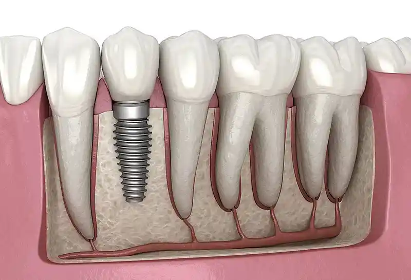
Wisdom teeth, referred to in dental literature as “third molars” or wisdom teeth, are the last elements of the masticatory system to evolve, positioned at the furthest terminals of the lower and upper dental arches. The historical narrowing of human phylogeny and anthropological jaw morphology has pushed these teeth into the status of vestigial organs that are mostly non-functional and subject to space limitations in modern humans. In normal anatomical variations, a total of four third molars develop in the mouth, one in each jaw quadrant. Although the basic tissue structure of these teeth is the same as other molars, the fact that their eruption period coincides with the standstill phase of jaw skeletal development makes them highly susceptible to producing pathology.
- Molar Morphology: Hard tissue anatomy with a wide chewing surface, multiple roots, and high calcification density.
- Anatomical Terminal Position: A cramped placement geometry limited by the anterior border of the ramus in the lower jaw and the tuber region in the upper jaw.
- Evolutionary Jaw Narrowing: The biology of the alveolar bone arch shrinking to an extent that leaves no room for these teeth as a result of soft food dietary habits.
When Do Wisdom Teeth Erupt?
The eruption schedule of the third molars into the oral cavity is shaped by the individual’s skeletal maturation and root calcification hierarchy rather than the chronological age scale. While the appearance of the crown portions of these teeth within the bone begins at ages 12–14 in general biological literature, the stage where root development is completed and punctures the mucosal boundary corresponds to the period between the ages of 17 and 25. Since this period is the young adulthood phase and symbolizes the age of mental maturation, it is popularly called “wisdom tooth”. Teeth that are not visible in the mouth despite exceeding these age limits are registered as impacted.
- Active Eruption Turn: Chronologically the 17–25 age range (the phase when skeletal development comes to an end).
- Root Development Rate: The complete closure of the root apices is generally completed within the 22–24 age limits.
- Mucosal Barrier Dam: The frequency of remaining semi-impacted by failing to completely overcome the thick attached gingival barrier even if it filters through the bone.
In Which Situations Do Wisdom Teeth Cause Problems?
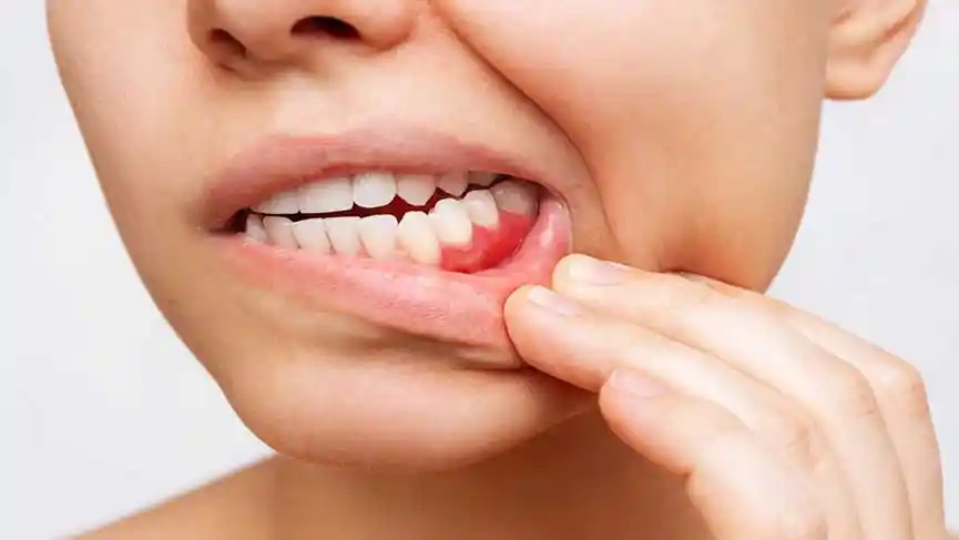
Third molar segments turn into active pathological mechanisms that radically sabotage oral homeostasis when they cannot find their own tight seal corridor in the jaw arch and are forced to erupt at faulty angles. The fact that only a portion of the tooth as small as a fingernail opens into the oral environment while the rest of the body remains under the mucosa creates an insidious pocket for bacteria to leak into. Since these blind pockets contain micro-grooves that toothbrush bristles cannot physically fit into, food debris ferments inside, producing chronic infection foci, unbearable jaw locking, and irreversible resorption in adjacent teeth.
The main variations in which wisdom teeth cause clinical problems in oral hard and soft tissues are as follows:
- When anaerobic microbes breeding under the semi-impacted gum trigger acute “pericoronitis” (inflammation of the gum hood) abscesses,
- When it leans horizontally (mesioangular) against the adjacent second molar tooth, insidiously initiating interproximal decay on the root surface of that tooth,
- When the embryonic follicle sac surrounding the tooth accumulates fluid over time and evolves into “dentigerous cyst” pathologies that melt the bone from the inside,
- When it produces chronic referred pain radiating to the ear, temple, and lower jawbone due to the mechanical compression pressure it creates in the corner of the jaw (ramus region).
In Whom Should Wisdom Teeth Be Extracted?
This medical surgical intervention is biologically indicated for all patients whose third molars are registered as poorly positioned in oral clinical and radiological scans, whose pericoronitis inflammation attacks become chronic, who threaten adjacent gingival health, and who carry a risk of orthodontic relapse. The extraction decision is not made blindly; the passive benefit provided by the tooth to the oral ecosystem and the active tissue destruction coefficient it produces are rationally weighed on the surgical scale. The criteria for planning wisdom tooth extraction according to clinical condition groups are detailed in the medical matrix below:
| Clinical Tooth Condition / Position | Degree of Surgical Intervention Necessity | Expected Medical and Cellular Outcome of the Operation |
|---|---|---|
| Horizontally Impacted and Root-Resorbing Case | Absolutely Mandatory / Urgent | The root destruction of the second molar is halted, and the insidious cyst formation chain is broken from the root. |
| Semi-Impacted and Frequently Abscessed (Pericoronitis) Tooth | Mandatory Indication | The mucosal pocket, which is a bacterial harbor, is eliminated, ending bad breath and jaw locking. |
| Completely Bone-Impacted, Non-Pathological Standing Tooth | Passive Follow-up / Postponable | If there is no pressure on surrounding tissues and nerves, it can be left in place with 6-month periodic radiological monitoring. |
| Completely Erupted, Opposing Tooth Present, Functional Tooth | No Extraction Required / Contraindicated | The tooth, which participates in chewing mechanics and whose hygiene is maintained, is preserved within organic integrity. |
What Is an Impacted Wisdom Tooth?
An impacted wisdom tooth is a third molar pathology that has failed to erupt into the oral cavity in its normal anatomical position despite exceeding the chronological eruption timeline of 25 years, remaining under the jawbone integrity (osteotomy line) or thick attached mucosal cover. The degree of impaction is clinically registered under two main headings: In “completely bone-impacted” cases, all borders of the tooth are covered by the alveolar bone network, and its contact with oral fluids is zero. In “semi-impacted” cases, the crown portion of the tooth has emerged from the bone but has not entirely overcome the thick gum cover (operculum) over it, leaving a leaky corridor open to the oral bacterial flora; it is these semi-impacted variations that produce the most aggressive abscesses and stubborn decays in clinical practices.
- Mucosal Impaction: The condition where the tooth passes the bone border and remains hidden only under the gum tissue.
- Bony Impaction: The tooth remaining impacted and integrating with the skeleton in the deep layers of the alveolar cortical bone.
- Positional Classification: The tooth taking a vertical, horizontal, or angled (mesio/distoangular) position according to its standing angle in the jaw.
How Are Wisdom Teeth Evaluated?
The medical and surgical evaluation of third molar segments is an analytical examination phase that determines the three-dimensional spatial relationship of the tooth with the bone, nerves, and adjacent soft tissues using rational parameters, rather than carrying out random guesses. In the evaluation, the clinician centers the “Pell and Gregory” and “Winter” classification indices. In line with these registered charts, how far behind the mandibular ramus bone the tooth is impacted and at which milimetric vertical level it is hidden relative to the occlusal plane is mapped with mathematical certainty.
- Path of Entry Axis Analysis: The phase of determining with which movement angle (vector) the tooth will be removed from the line during extraction.
- Periodontal Pocket Scanning: Measuring the depth of bone pockets formed behind the second molar tooth with the help of periodontal probes.
- Mucosa Biotype Registration: Inspecting the thickness of the gum in the area to ensure leak-proof suturing after surgery.
How Is the Oral Examination Performed Before Wisdom Tooth Extraction?
The clinical oral examination performed before the operation schedule is finalized is the most vital pre-operative check-up phase that determines the infection load of the surgical field and the patient’s anatomical limitations. In the dental chair, the physician first measures the patient’s maximum mouth opening distance (degree of trismus); limited mouth opening is a factor that narrows the surgical field of view. Then, the color of the mucosa in the wisdom tooth area, the edema coefficient, and whether pus comes out upon palpation are meticulously inspected to rule out the presence of an active acute inflammation; because making an incision in an actively abscessed tissue invites the infection to spread to deep facial spaces.
- Trismus Inspection: Determining whether the temporomandibular joint (TMJ) can tolerate remaining open for a long time during the operation.
- Operculum Examination: Scanning the bacterial plaque density under the tissue flap covering the semi-impacted tooth using staining tablets.
- Referred Pain Mapping: Registering the neurological connection of the patient’s head, ear, and temple aches with the wisdom tooth pressure lines.
Why Are Digital Imaging Methods Important in Wisdom Teeth?
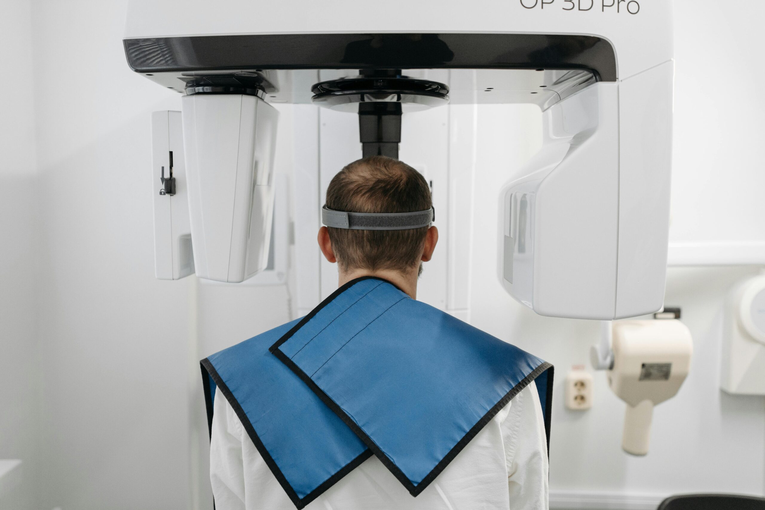
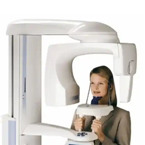
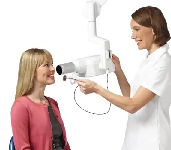
Digital 3D imaging systems in wisdom tooth surgery register surgical safety at the highest note by completely eliminating the depth blindness and superimposed illusion risks brought by two-dimensional classical X-rays. In particular, the relationship of the lower wisdom tooth roots with the “Inferior Alveolar Nerve” canal, which is the main sensory nerve line of the lower jaw, carries a vital risk. Thanks to CBCT 3D dental tomography scanning, the surgeon examines whether the nerve canal passes between the roots, underneath them, or from the lateral wall on a 1:1 scale in three axes; this digital accuracy prevents blind shots from being fired during surgery, definitively breaking the complication chain of permanent lower lip numbness and loss of sensation (paresthesia).
- Registration of Root Curvatures: Radiological clarity to prevent hook-shaped (dilacerated) micro root tips from breaking.
- Bone Density Mapping: Calibrating the handpiece speed during the operation according to the rigidity coefficient of the cortical alveolar bone surrounding the tooth.
- Sinus Floor Scanning: Scanning the anatomical boundary lines of the upper wisdom tooth roots with the maxillary sinus floor membrane (Schneiderian membrane).
How Is Wisdom Tooth Extraction Performed?
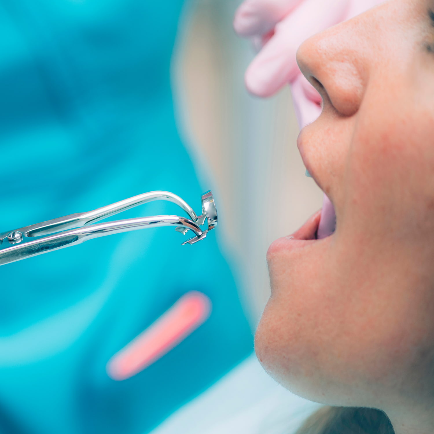
The extraction procedure for wisdom teeth that are completely erupted, visible to the naked eye in the intraoral ecosystem, but must be sacrificed due to decay or lack of space, is a practical micro-surgical step carried out within the framework of conventional “Simple Extraction” protocols. After pain transmission lines are locked with computer-controlled digital anesthesia, the marginal boundaries of the gum adhering to the tooth are loosened milimetrically with special periodontal luxator instruments. Then, the periodontal ligament fibers surrounding the root are slowly torn using rotational expansion movements (luxation forces); the tooth is removed as a whole from the line by applying minimum mechanical pressure to the bone socket, and the bed is closed with a sterile gauze pad.
- Ligament Separation: Milimetric cutting of the fiber bridges connecting the tooth root to the bone using luxator tips.
- Socket Integrity Preservation: Vertical axis-directed forceps maneuvers to prevent fracturing the alveolar outer cortical bone wall during extraction.
- Bleeding Control: Applying mechanical pressure to initiate a full and healthy blood clot organization at the base of the socket.
How Is Impacted Wisdom Tooth Surgery Applied?
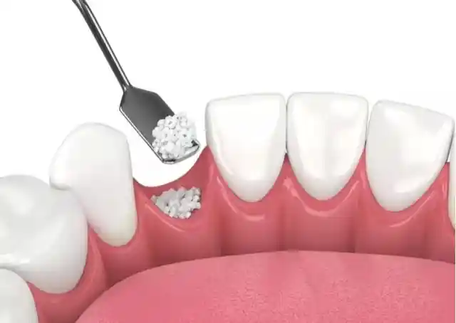
Surgical removal of impacted or semi-impacted third molars is a highly technical, focus-driven jaw surgery operation performed under local anesthesia block in polyclinic operating rooms subject to full sterilization rules. In the first phase, a smooth incision line is drawn with the help of a scalpel, taking into account the topographical projection boundaries of the tooth within the bone, and the mucosal flap is lifted off the bone membrane (periosteum) to expose the bone. Then, the upper cortical bone layer blocking the exit path of the tooth is shaved with micron precision using water-cooled surgical burs or ultrasonic piezo tips (osteotomy). To avoid applying elevator leverage pressure to the surrounding bone, the crown and root bodies of the tooth are divided into pieces with micro-blades (separation technique), picked out from the socket with tweezers, and removed; the socket is washed with antiseptic solutions and closed tension-free with primary sutures.
- Atraumatic Bone Window: Shaving the minimal-sized bone corridor that will allow access to the tooth with precise burs.
- Tooth Separation: Dividing the tooth into two or three pieces according to its geometric axis; this avoids major bone losses.
- Socket Toilet (Curettage): Scraping and removing root tip granulation tissues and follicle remnants, ensuring a clean bed registration.
How Long Does a Wisdom Tooth Extraction Take?
The active chairside session duration of wisdom tooth operations varies depending on the depth of the tooth’s impaction within the bone, the asymmetry of the root count, and the surgical technique to be applied (simple or separated extraction). Following anesthesia absorption, the simple extraction stages of a completely erupted, straight-rooted wisdom tooth are usually completed in a remarkably short time frame of just 5 to 10 minutes. However, in advanced horizontally impacted lower wisdom surgeries involving bone shaving, tooth division, and milimetric suturing steps, the operation time is meticulously shaped between 20 to 45 minutes per session on average.
- Simple Extraction Cycle: 5–10 Minutes (A practical timeline that does not tire the patient and does not interrupt the social calendar).
- Completely Impacted Surgical Cycle: 20–45 Minutes (Meticulous craftsmanship duration covering tissue loosening, bone filing, and suture closure steps).
What Anesthesia Methods Are Used in Wisdom Tooth Extraction?
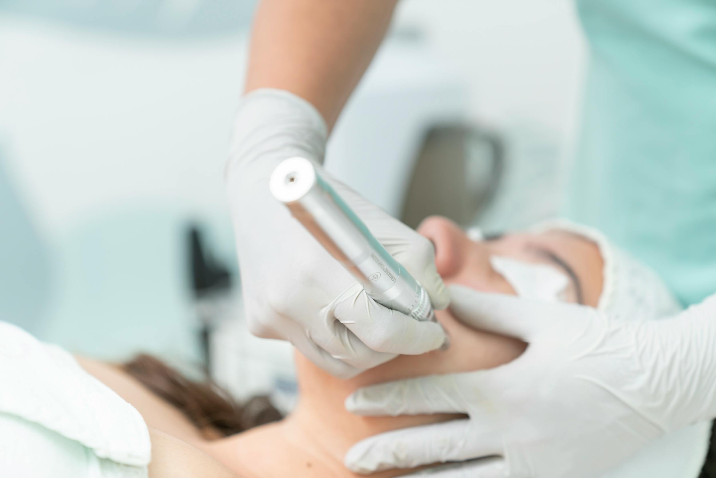
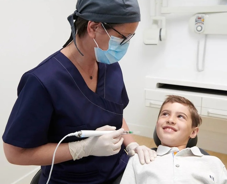
Anesthesia technologies with high vascular safety are deployed in modern oral surgery to eliminate the patient’s psychological anxiety level at the moment of surgery and to render pain transmission lines completely numb. In standard cases, painless local anesthetic solutions infiltrated at a speed suitable for tissue resistance with the help of computer-controlled digital anesthesia devices (Wand technology) are the primary rule; this completely eliminates that distressing pressure sting during injection. However, in children or adult cases with the highest level of needle phobia or a history of panic attacks, sedation (semi-sleep) or general anesthesia (narcosis) infrastructures are successfully integrated to manage the process in a full relaxation mode.
- Mandibular Regional Block: The main nerve locking technique that completely numbs the tongue and lip half in lower jaw wisdom tooth surgeries.
- Local Infiltration: Performing local mucosal infiltration in upper jaw wisdom segments by utilizing the porous structure of the bone.
How Does the Healing Process Proceed After Wisdom Tooth Extraction?
The tissue recovery phase after the operation is a completely natural, predictable, and linear repair cycle initiated by the body to close the wound site. Forming a temporary micro-edema (swelling) in the cheek area during the first 48 hours following the surgery due to intra-bone stimulation and mucosal loosening steps is a completely normal physiological wound repair reflex given by the extracellular matrix. The healing process flows smoothly; on the 7th day, the mucosal sutures close tightly, the cheek swelling completely subsides, and the complete weaving of the intra-bone cavity with new cortical bone trabeculae requires an average ossification schedule of 4 months.
- First 24 Hours (Clot Shield): The organization of a red blood clot inside the socket; it acts as a living biological blanket for bone-forming cells to proliferate.
- 1st Week Turning Point: The period when soft tissue margins unite, and surgical sutures are painlessly removed from the line during clinical control.
What Should Be Considered After Wisdom Tooth Extraction?
The care discipline that the patient will follow in the home environment after the procedure is of vital importance to make the success of the surgical session permanent, protect that vital clot integrity within the socket, and zero out infection risks. Since the extraction bed temporarily shifts into an extremely delicate phase against external stimulants and intraoral pressure waves during the first hours, it is a medical necessity for the patient to maintain homeostasis balance.
The aspects that must be meticulously followed at home during the critical period after the surgery are as follows:
- The sterile gauze pad placed over the extraction cavity must be bitten uncompromisingly for at least 30-45 minutes to establish the clot pressure that will stop the bleeding, and then discarded in one go.
- For the first 24 hours following the operation, the mouth should not be vacuumed and spit out in order not to displace the clot and revive bleeding; saliva should be swallowed instead (spitting triggers bleeding).
- Hot baths, saunas, Turkish baths, and heavy sports activities that will dilate capillary walls and increase the risk of oozing and cheek swelling should be postponed for the first 48 hours.
- Smoking, tobacco products, and alcohol consumption must be completely cut off for at least 3 days so as not to paralyze the micro-cell repair mechanisms at the wound site.
How Should Oral Care Be Performed After Wisdom Tooth Extraction?
Post-session home hygiene processes must contain a delicate balance that will limit the intraoral pathogenic bacterial population to prevent socket infections (alveolitis) but will also not batter the newly formed sensitive clot shield. On the day of extraction, the mouth should not be rinsed forcefully with water; starting from the next day, while normal teeth are cleaned as standard, the surgical area is entrusted to chemical protection.
- Antiseptic Mouthwash Shield: Performing gentle rinsing morning and evening with chlorhexidine-based medical mouthwashes without touching the toothbrush to the wound site, starting 24 hours after the surgery.
- Gentle Sweeping Motion: Utilizing ultra soft brushes manufactured specifically for sensitive surgical periods to clean the adjacent teeth in that area starting from the 48th hour.
How Do Wisdom Teeth Affect the Health of Other Teeth?
The faulty positions and eruption anomalies of wisdom teeth are a source of chronic risk that is not limited only to the local rear region but endangers the health of the entire tooth series in the jaw arch with a sinsi chain reaction. Especially a wisdom tooth standing mesioangularly (tilted forward) applies continuous mechanical pressure to the cervical region of the second molar tooth in front of it. The micro-fractures and non-cleansable deep pockets formed along this pressure line initiate sinsi root decay (external root resorption) in the root cementum layer of the second molar; this pathology is a major periodontal destruction that causes the patient to lose their most vital chewing tooth out of nowhere.
- Bacterial Factory Role: The risk of microbes breeding in folds at the very back where the toothbrush cannot reach spreading throughout the mouth and infecting other filling margins.
- Dental Arch Crowding: The triggering of sinsi interproximal decay due to food accumulation on the contact surfaces of the anterior incisors as a result of teeth pushing against each other.
Do Wisdom Teeth Affect the Orthodontic Treatment Process?
In clear aligner or wire treatment (orthodontics) protocols, the presence of wisdom teeth and their skeletal eruption forces are critical parameters that directly guide treatment planning. When the orthodontist aims to resolve crowding in the anterior region by moving teeth backward (using the distalization technique), a wisdom tooth standing at the back blocks this movement like a mechanical wedge. Most importantly; after the orthodontic treatment is successfully finished and wires are removed, the skeletal pushing forces produced by the wisdom teeth continuing to erupt from behind can cause the aligned front teeth to become crooked and rotate again (orthodontic relapse); removing the wisdom teeth before or during treatment is a clinical necessity to zero out this risk.
“The safest way to prevent teeth from shifting again after orthodontic treatment and to keep the hard work from relapsing is to surgically zero out the insidious wisdom tooth pressure at the back.”
In Which Situations Can Wisdom Teeth Be Monitored?
The widespread perception that every wisdom tooth must be urgently removed from the mouth by surgery is misinformation that is not validated by medical standards; third molar segments that meet certain strict anatomical criteria can be taken under passive clinical follow-up without operation. If the tooth is completely embedded horizontally or vertically within the deep layers of the jawbone, applies no mechanical pressure or compression to surrounding healthy tissues, nerve canals, or adjacent tooth roots, and no cyst follicle expansion has been registered around it, this tooth is left in place. Carrying the tooth passively in the mouth is medically the most correct protective approach as long as it does not produce a lesion in 6-month periodic X-ray controls.
- Completely Erupted Functional State: If the tooth has emerged perfectly vertical into the jaw arch, hygiene can be maintained, and it is in a full occlusion (chewing) relationship with its opposing upper tooth, it is preserved.
- Elderly Cases with High Surgical Risk: Taking older patients under follow-up to avoid nerve damage when bone rigidity has excessively increased and there is no pathology in the tooth.
What Are the Advantages of Surgical Procedures Related to Wisdom Teeth?
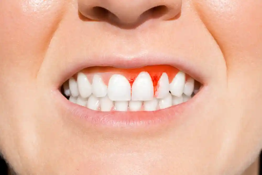
The removal of third molars that are impossible to save or produce pathology through surgical scalpel and separation steps is the most visionary preventive dentistry investment that saves the oral health ecosystem from destruction on a linear line. It does not just end periodic abscess oozing and bad breath; it definitively breaks the risk chain of cyst foci growing insidiously in the lower layers of the jawbone from completely melting the bone skeleton. When the chronic microbial load in the tissues is relieved, the life span of adjacent main chewing teeth is extended, and an unshakeable functional comfort is registered for the patient.
- Permanent Infection Elimination: Ending the necessity for frequent repeated antibiotic use, saving the body from systemic drug burdens.
- Temporomandibular Joint (TMJ) Protection: Resolving compression-induced muscle spasms (trismus) to reinforce the smooth opening and closing mechanics of the jaw joint.
- Prevention of Smile Crowding: Supporting facial aesthetics to remain permanent by protecting the alignment integrity of the front incisor teeth.
What Are the Frequently Asked Questions About Wisdom Teeth?
The most common practical concerns arising in the minds of patients planning this process to get rid of impacted wisdom tooth pain or to have a surgical extraction and their polyclinic medical counters are presented below:
Does it hurt a lot when the bone is being shaved during the impacted wisdom tooth operation?
No, absolutely not; you will not feel the slightest pain or ache. Prior to the procedure, strong clinical topical anesthetic gels are applied to the gingival mucosa, followed by local solutions delivered at a speed matching tissue resistance with computer-controlled digital anesthesia units to provide full insensitivity; since nerve transmission lines are completely shut down throughout the session, you only perceive the light touch of the devices and the water coolness, and the procedure is completed with high comfort.
Will my face swell a lot after the wisdom tooth is extracted, and when will that swelling and bruising go away?
In cases where intra-bone micro-shaving is performed in impacted tooth surgeries, the formation of a temporary edema (swelling) in the cheek area during the first 48 hours is a completely normal physiological reaction resulting from the repair reflexes of the extracellular matrix. Thanks to intermittent ice compresses (cold packs) applied from outside the cheek on the first day after the surgery and micro-edema reducing medications to be prescribed by the physician, this swelling quickly subsides starting from the 3rd day and completely disappears within 1 week without leaving any trace.
Is there any medical harm in constantly spitting out the blood that accumulates in my mouth after the extraction is finished?
Yes, this is a major medical misconception that is most frequently fallen into and sabotages healing in the post-extraction process. Making continuous powerful spitting motions to throw out blood or saliva from the mouth creates a high negative vacuum pressure inside the mouth; this pressure dislodges that vital blood clot freshly woven inside the socket and throws it out, preventing you from stopping the bleeding and leading to painful dry socket (alveolitis) inflammation; blood should be swallowed.
What is the risk of the lip remaining numb (paresthesia) in lower wisdom tooth surgeries, and is it permanent?
In anatomical variations where the lower wisdom tooth roots are very close or tangent to the lower jaw main nerve canal (inferior alveolar nerve), temporary micro-bruising may occur in the nerve fibers due to mechanical stretching during extraction, and the patient may feel a slight tingling in their lower lip after surgery. This condition is not a permanent paralysis; in our clinics, this risk is reduced to the minimum wavelength via 3D tomography planning, and temporary numbness that occurs completely subsides within a few weeks with B-vitamin courses.
Can I return to my normal professional and daily life immediately on the day the wisdom tooth extraction treatment is completed?
Yes, wisdom tooth operations are completely local and minimally invasive surgical steps that do not disrupt consciousness, do not affect motor skills, and do not involve the weight of general narcosis; therefore, you can get up from the chair the exact second the session ends and continue your normal daily, professional, social, or academic life rhythm uninterruptedly from where it left off; there is no clinical barrier to driving or participating in business meetings immediately after the procedure.
Who Are We? & Panorama Ankara Oral and Dental Health
Panorama Ankara is a licensed oral and dental health center offering institutional health assurance to its patients by combining an academic specialist staff and the latest global bio-technological digital infrastructure under a single roof in all specialty branches of dentistry, primarily oral, dental, and maxillofacial surgery, advanced implantology, microscopic endodontics, periodontology, and aesthetic dentistry. Instead of abandoning infected teeth that cannot be saved or impacted wisdom tooth nightmares to the simplicity of rough analog force that damages the jawbone, our center attaches vital importance to reaching permanent oral comfort by centering “bone-preserving atraumatic surgery” principles with evidence-based medical disciplines, and operates seamless 3D Digital Diagnostic Modeling and Atraumatic / Impacted Wisdom Tooth Surgery ecosystems with calendar discipline from the very beginning to the end of the clinical operation workflow.
Under the roof of Panorama Ankara, absolute sterilization, high patient safety, transparency, and honesty principles are operated with full compliance with medical codes in all diagnostic, radiological scanning, design, and micro-surgical interventions. In our clinical practices, while analyzing jawbone quality and root tunnel lines with 3D tomographies in milimetric sections, smart surgical cauteries that coagulate vascular integrity and ultrasonic piezo surgery units that do not damage surrounding bone are applied with milimetric precision under the supervision of our expert jaw surgeon staff. Our aim is not to impose old-fashioned, slow, bone-breaking, and distressing analog operation patterns on our patients that take weeks to heal, but to analyze each individual’s jaw skeletal quality, occlusion mechanics, and nerve pathways on the virtual screen in 3D before the operation, achieving maximum session comfort, seamless comfort standards without sutures, and smooth fixed implant infrastructures that you will use with an unshakeable confidence for a lifetime. If you are tired of your ruined teeth, persistent wisdom tooth pain, or bad breath when looking in the mirror, yet feel anxious about surgical risks or fear of pain and wish to start your smart digital treatment process from scratch in an expert corporate center environment with scientific guarantee; you can safely protect your health and smile integrity by contacting the expert staff of Panorama Ankara for detailed 3D scanning and digital planning analysis.