1. What is Periodontology?
Periodontology is a specialty branch of dentistry that examines the anatomical, physiological, and histological structures of the hard and soft tissues directly surrounding and supporting teeth and tooth-like clinical structures (such as dental implants), and diagnoses and treats the pathologies occurring in these tissues. Derived from the Latin words “peri” (around), “odons” (tooth), and “logos” (science), periodontology has the mission of protecting the fundamental building block of oral and dental health.
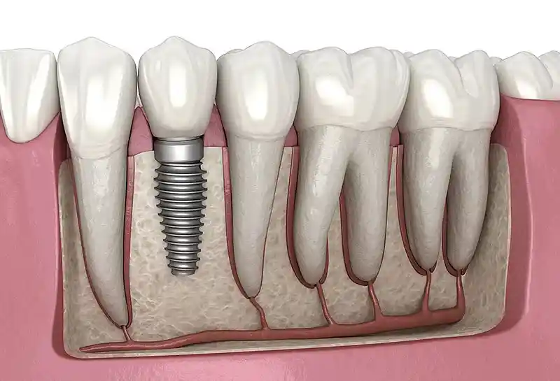
The ability of teeth to remain stable and function within the mouth does not solely depend on the sturdiness of the tooth’s own hard tissues. The supporting structures, termed the periodontium, consist of the gingiva (gum), alveolar bone (the part of the jawbone containing the tooth sockets), cementum (the layer covering the tooth root), and the periodontal ligament (fibers serving as a suspension system between the tooth root and bone). At Panorama Ankara, periodontology applications are based on preserving and improving the biological integrity of this complex system.

2. Which Diseases Does Periodontology Deal With?
The specialty field of periodontology covers a wide spectrum of diseases ranging from superficial inflammation of the gum tissue to the destruction of the jawbone and advanced infections that threaten systemic health. Periodontal pathologies are examined and treated under the following primary groups within international standards:
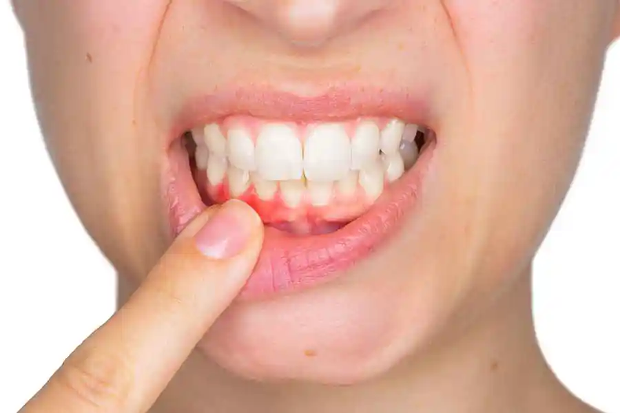
- Gingivitis (Superficial Gum Inflammation): The most common and reversible disease of the gums. Inflammation is limited only to the soft tissue; no loss has occurred in the bone support.
- Periodontitis (Advanced Gum and Bone Disease): A clinical picture in which inflammation spreads to deep tissues, leading to resorption in the periodontal ligament and alveolar bone. Chronic and aggressive forms are available.
- Necrotizing Periodontal Diseases: Acute conditions characterized by sudden tissue death (necrosis) in the gingival papillae and severe pain during sudden drops in systemic resistance, intense stress, or malnutrition.
- Peri-implant Diseases (Peri-implantitis and Peri-implant Mucositis): Inflammatory processes of the gum and bone tissue around dental implants applied instead of natural teeth. It leads to implant loss when not intervened early.
- Mucogingival Deformities: Conditions where the gum undergoes loss of function due to recession caused by genetic or acquired reasons, reduction in its thickness, or high frenulum (lip tie) attachments.
3. Why Do Periodontal Diseases Occur?
The primary and most important etiological factor of periodontal diseases is the microbial dental biofilm (bacterial plaque) layer that continuously accumulates on tooth surfaces and the gum line. The oral cavity is a complex ecosystem hosting hundreds of different bacterial species. When regular and effective mechanical cleaning is not performed, these bacteria organize to form a structural plaque layer.
When bacterial plaque is not cleaned, it hardens through the precipitation of calcium and phosphate ions in saliva, converting into dental calculus (tartar). The rough surfaces of dental calculus provide an ideal ground for pathogenic bacteria to colonize more rapidly. Endotoxins and enzymes secreted by bacteria stimulate the body’s immune system. In this process, the excessive or insufficient defense response (inflammatory reaction) given by the organism against infection unfortunately leads to the destruction of its own periodontal tissues and alveolar bone. In other words, a significant portion of tissue destruction stems from the host immune response spinning out of control, rather than directly from the bacteria themselves.
4. What Are the Symptoms of Gum Diseases?
Periodontal diseases mostly follow a chronic course and do not cause severe pain to the patient until they reach advanced stages. This insidious progression is the biggest factor in patients delaying treatment. However, with careful observation, the following clinical signs can be easily detected:
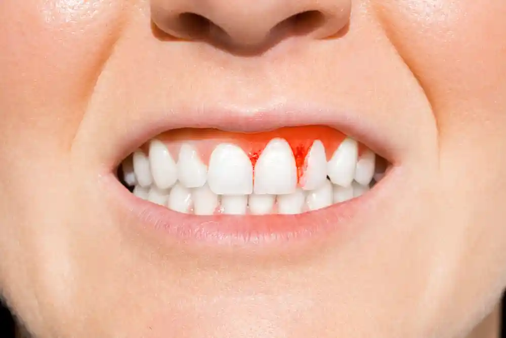
- Spontaneous Bleeding or Bleeding During Brushing: Healthy gums absolutely do not bleed. Bleeding is the primary evidence of active inflammation and the fragility of intra-tissue capillaries.
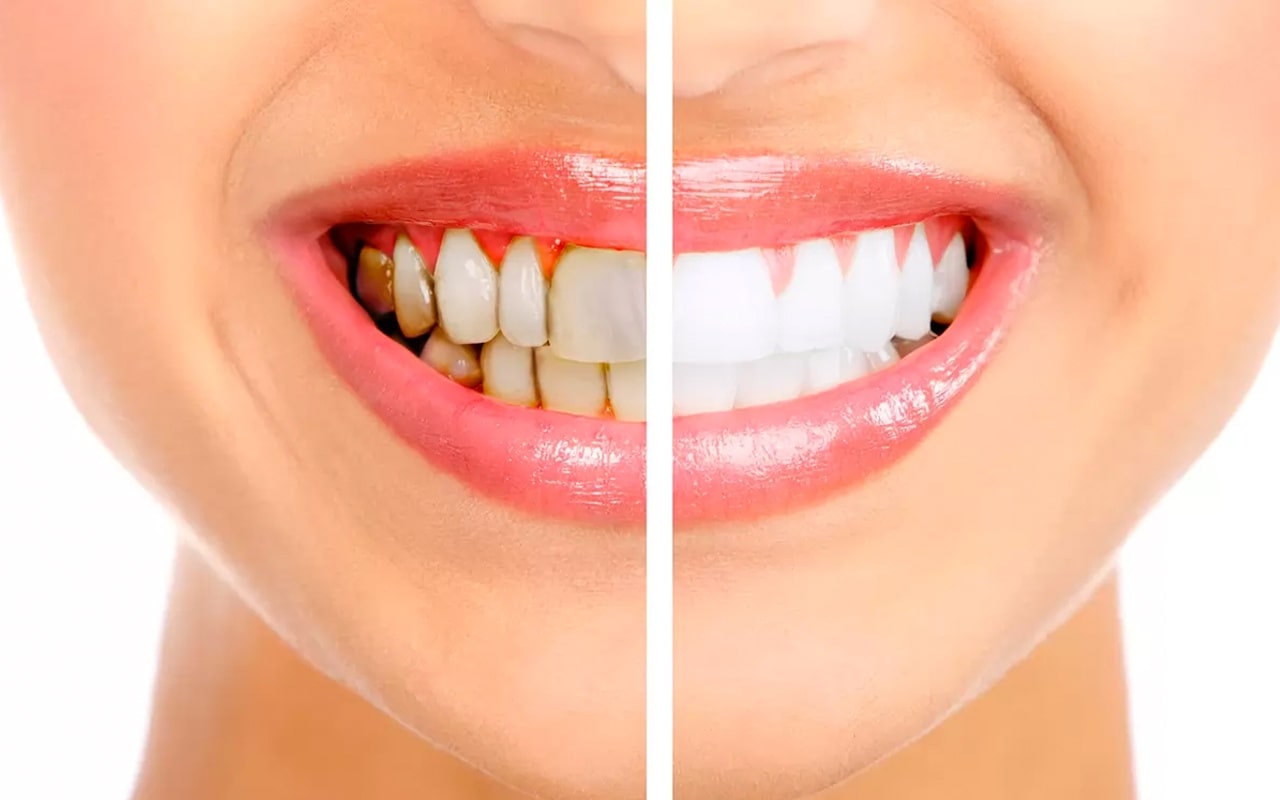
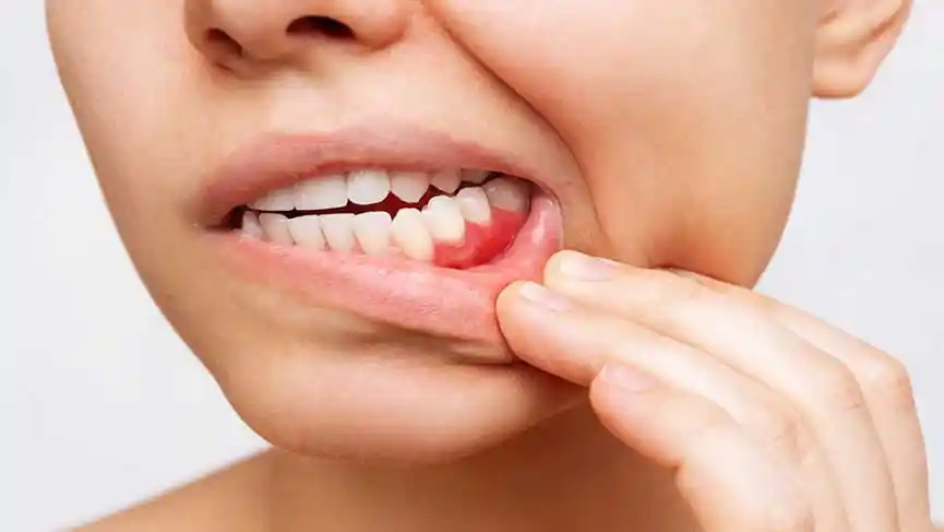
- Color and Shape Changes: The pale pink (coral pink) color and orange-peel appearance of healthy gums disappear; the tissue takes on a bright red or dark purple color, and its margins swell and round out (edematous structure).
- Persistent Halitosis (Bad Breath): Volatile sulfur compounds secreted by anaerobic bacteria multiplying in periodontal pockets lead to chronic bad breath that does not go away with brushing.
- Gum Recession and Sensitivity: Elongated appearance of tooth lengths as a result of the gum receding toward the root surface, and the initiation of hot-cold sensitivities due to the exposed cementum layer.
- Purulent Exudate (Pus Discharge From Gums): Active inflammatory discharge observed from inside the pocket spontaneously or when pressure is applied to the gum margin.
- Tooth Mobility (Shaking) and Displacement: Shaking of teeth, opening of gaps, or frontward fan-shaped angling as the supporting bone tissue falls below a critical level.
5. How is a Periodontology Examination Performed?
The periodontological examination process at Panorama Ankara involves a comprehensive clinical and radiological protocol to detect the extent of tissue destruction at a micro level. The examination is carried out by recording the following objective parameters:
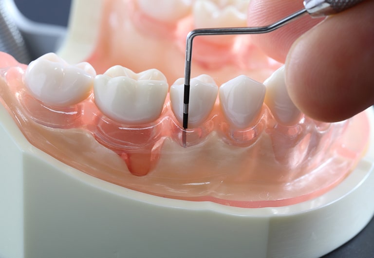
Periodontal Probing and Mapping: A special millimetric hand instrument called a “periodontal probe” is gently inserted into the pocket between the tooth and the gum. Measurements are taken from 6 different points around the tooth to identify the Probing Pocket Depth (PPD). While this depth is between 1-3 mm in healthy mouths, higher values are measured in pathological conditions.
Clinical Attachment Level (CAL) and Bleeding Index: The amount of attachment loss is calculated relative to the cemento-enamel junction of the tooth. Bleeding on Probing (BOP) is recorded by checking whether bleeding comes from the pocket 30 seconds after probing. This data provides information about the activity of the disease.
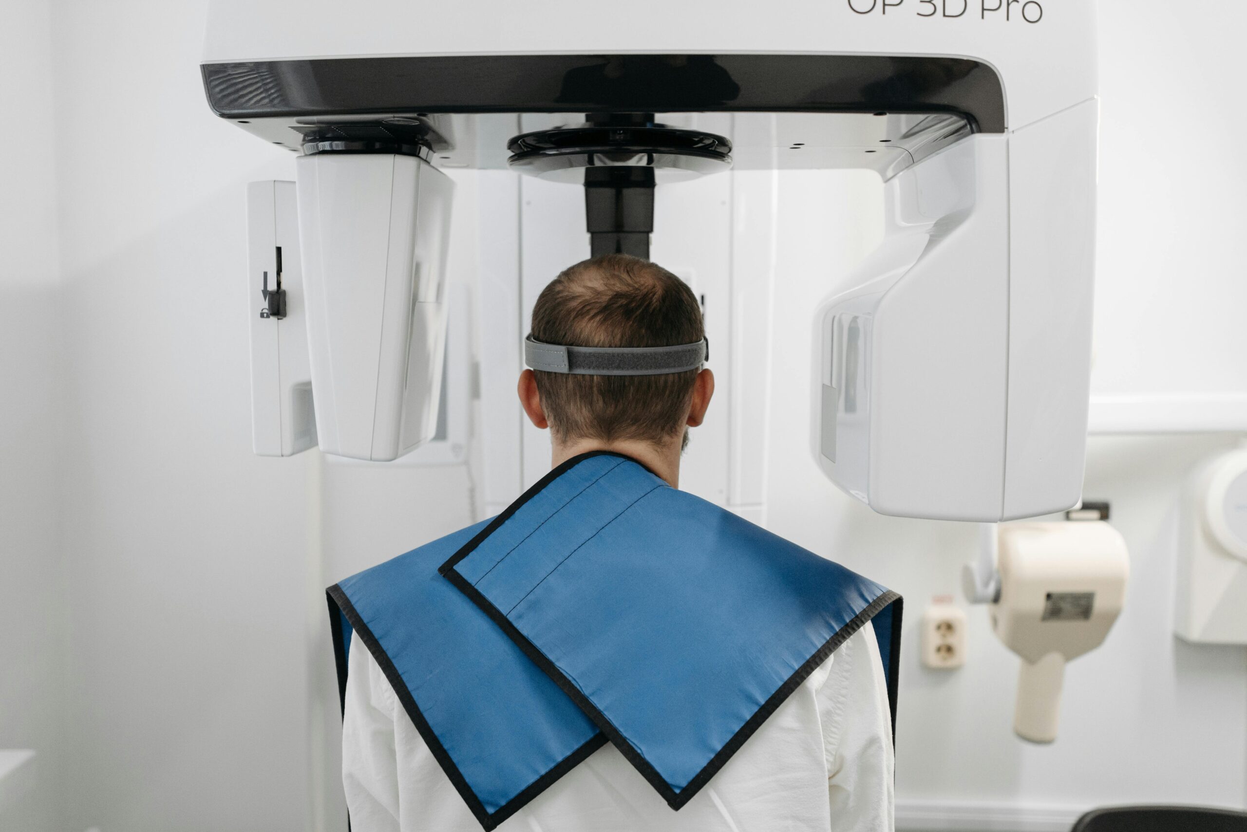
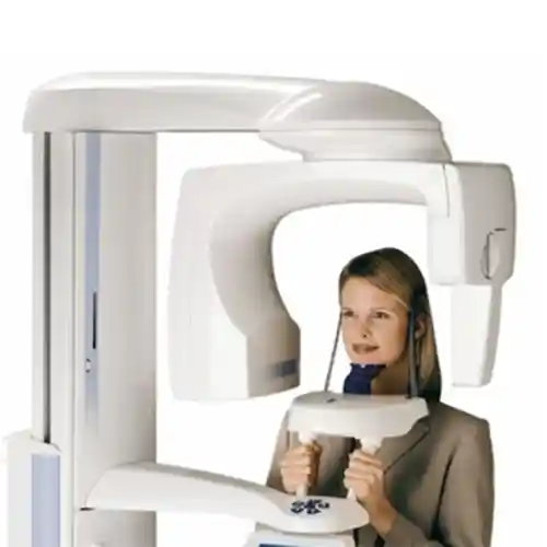
Radiological Evaluation: Clinical findings are supported by two-dimensional digital panoramic and periapical X-rays. The pattern of alveolar bone loss (horizontal or vertical bone resorption) and bone density are radiologially analyzed. 3D CBCT (Tomography) technology is utilized in advanced bone defects or pre-implant planning.
6. How is Gum Treatment Administered?
Gum treatment requires a phased approach according to the severity of the disease and the anatomical depth of the involved tissues. The fundamental philosophy of the treatment is to remove all pathogenic factors attached to root surfaces and to offer a sustainable oral environment to the patient.
The treatment begins with the initial non-surgical phase (Phase I periodontal therapy). Scaling and root planing procedures are applied in this stage. In advanced cases where inflammation has reached deep bone pockets, Phase II periodontal surgery (flap operations, grafting applications) steps in. Following all these active processes, the patient is taken into a periodic maintenance phase (Phase III) so that success can be permanent.
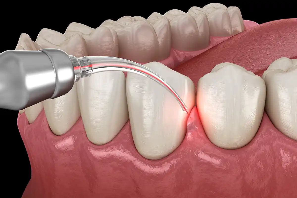
Panorama Ankara Approach: By preferring microscopic and laser-assisted methods that cause minimal damage to tissue at every stage of treatment, post-operative healing comfort is maximized.
7. What is Scaling (Dental Calculus Cleaning) and How is it Done?
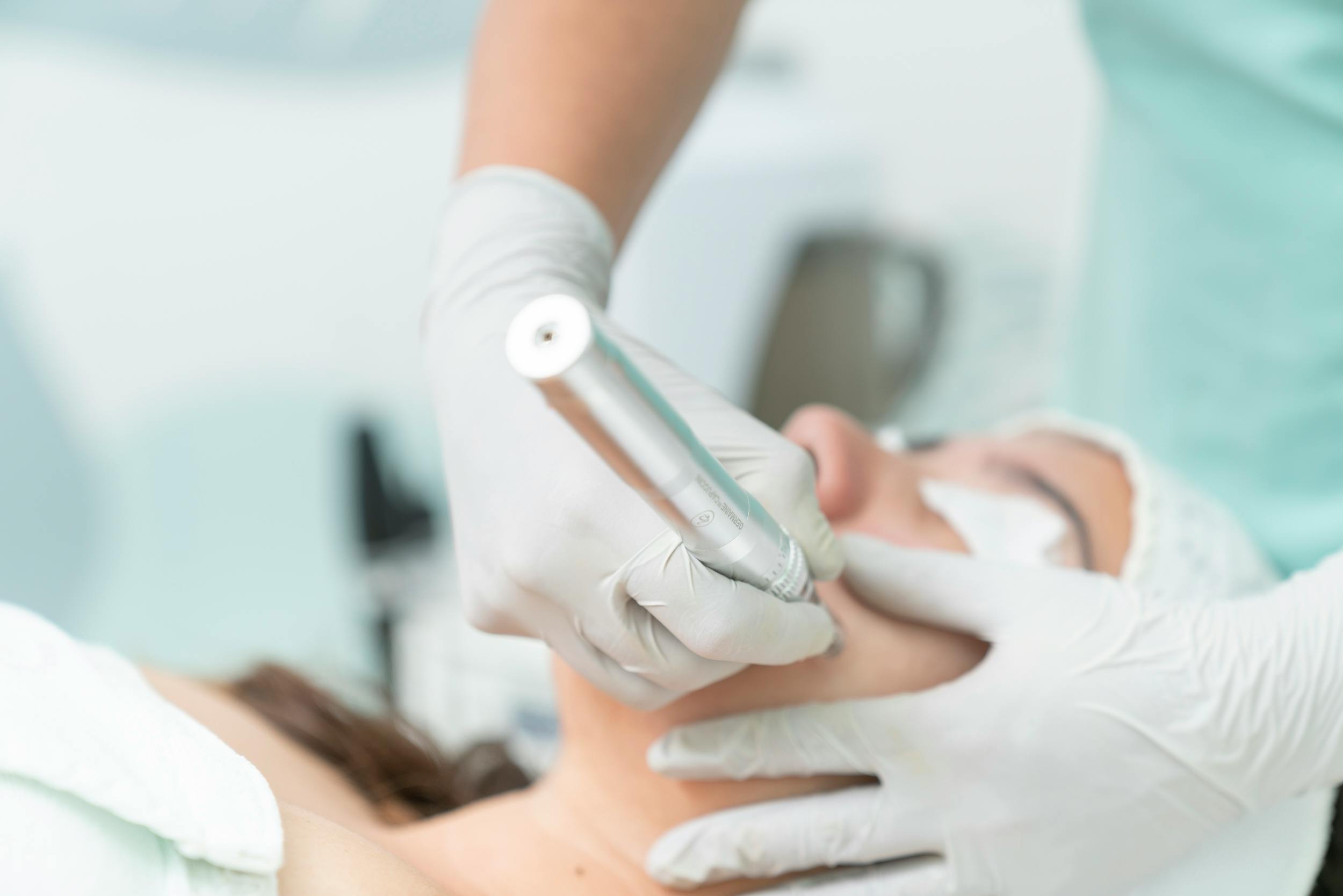
Scaling (Detertraj) is the procedure of removing hardened calcified deposits (tartar) and bacterial plaques that accumulate on tooth surfaces, interdental spaces, and immediately above the gum line (supragingival) using specialized clinical instruments. Scaling is the most fundamental and indispensable first step of periodontal treatment.
During the application, ultrasonic (piezoelectric) devices that produce high-frequency sound waves are used. The tips of these devices make micron-level vibrations, detaching and separating calculus masses without causing any harm to the tooth enamel. A water spray applied simultaneously washes away and removes debris from the area. Following ultrasonic cleaning, interdental surfaces that are difficult to reach are meticulously gone over with special hand instruments (scalers). In the final stage, polishing pastes are applied to tooth surfaces to smoothen the surface and delay new plaque attachment.
8. What is Root Planing (Curettage) Treatment?
In cases where inflammation has progressed below the gum line, namely toward the root surface (subgingival area), and formed a periodontal pocket, scaling alone remains insufficient. Subgingival Curettage / Root Planing treatment is applied at this stage.
The curettage procedure is performed under local anesthesia using precise hand instruments called Gracey Curettes, custom-designed to match the anatomical root structure and angle of each tooth. The purpose of the procedure is to scrape and remove subgingival calculus adhered to the root surface along with the necrotic cementum layer contaminated with bacterial toxins, and to obtain a completely smooth, hard, and clean root surface. Thanks to the cleaned root surface, loosened gum fibers can biologically reattach to the root (new attachment formation), thereby shrinking and eliminating pathological pockets.
9. How is Gum Recession Evaluated?
Gum recession (Recession) is the condition where the root surface becomes exposed to the oral environment as the gingival margin shifts below the cemento-enamel junction. Internationally recognized Miller or Cairo classification systems are used in evaluating recessions. In this evaluation, the depth and width of the recession, whether the interdental papillary bone is preserved, and the amount of attached gingiva band in the receded area are examined millimetrically.
The etiology of gum recession includes incorrect and aggressive tooth brushing, high and incorrectly localized frenulums, orthodontically moving teeth outside the bone boundaries, teeth clenching, and anatomically possessing a thin bone phenotype. Before a treatment plan is made, the primary factor causing the recession is identified and eliminated; then, surgical root coverage techniques are decided upon based on the aesthetic or sensitivity status.
10. What is a Gum Graft and In Which Situations is it Applied?
Gum graft applications (Plastic Periodontal Surgery) are micro-surgical operations performed to cover root surfaces in regions where recession has occurred or to increase the thickness and amount of weakened attached gum tissue in that region. It is examined under two main groups:
| Grafting Method | Tissue Source and Structure | Clinical Indication Area |
|---|---|---|
| Free Gingival Graft (FGG) | Taken as a block containing both epithelium and connective tissue from the patient’s palatal region. | Increasing the keratinized tissue band around the tooth, establishing a protective line before a prosthesis or implant. |
| Connective Tissue Graft (CTG) | The connective tissue in the inner layer of the palate is removed with a window method while protecting the upper epithelium. | Covering gum recessions in the anterior aesthetic zone, providing maximum color and tissue harmony. |
11. In Which Situations is Periodontal Surgery Preferred?
Periodontal surgery (Flap Operation) is preferred in the presence of active pockets whose depth remains over 5 mm despite non-surgical periodontal treatments (scaling and root planing), meaning when the disease cannot be brought under control. The internal portions of deep pockets are anatomically too distant for hand instruments to reach blindly, and surgical opening is mandatory.
In flap surgery, the gum tissue is slightly separated from the bone under local anesthesia (a flap is reflected), and root surfaces and intraosseous defects are completely cleaned under direct vision. Meanwhile, pathological granulation tissues causing the pocket are surgically enucleated. At the end of the operation, the gum is repositioned in accordance with the bone boundary and closed with stitches (sutures). This achieves a shallow and healthy gingival sulcus depth that the patient can clean by their own means.
12. Which Treatment Methods Can be Applied to Teeth with Bone Loss?
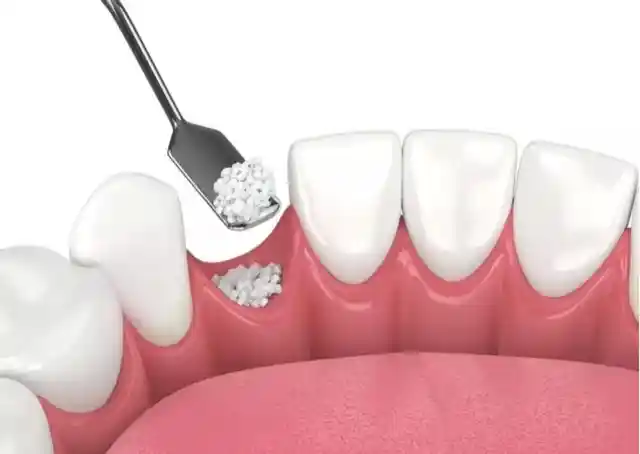
Resorptions occurring in the jawbone due to periodonititis are not always horizontal and uniform. Sometimes vertical, crater-like “infraosseous defects” form along the root of the tooth. In such vertical bone losses, Guided Tissue Regeneration (GTR) techniques are applied to biologically reproduce lost tissues.
In the reflected surgical field, biocompatible bone grafts (bone powders) are placed into the region with bone resorption. Collagen membranes (biological barriers) that serve as a barrier are laid over the grafts to prevent gum cells from infiltrating the region and to grant time to slow-growing bone cells. When necessary, enamel matrix derivatives (bio-active proteins) are injected into the site to enhance cell stimulation. This reduces the shaking of the tooth and functionally reconstructs the supporting bone structure.
13. Why is Gum Health Important Before Implants?
Although dental implants connect mechanically to the jawbone, the only barrier protecting them from bacteria in the oral environment is the surrounding gum tissue, just as it is with natural teeth. Placing an implant in a patient who has active gum inflammation or periodontitis directly endangers the biological future of the treatment.
Pathogenic bacteria in gum pockets rapidly migrate to the implant surface post-operation, leading to irreversible infections termed peri-implantitis that rapidly resorb the bone surrounding the implant. Periodontal ligament fibers present in the roots of natural teeth do not exist around implants; therefore, implants are far more vulnerable to infection compared to natural teeth. Eradicating infection completely from all intraoral tissues is an absolute prerequisite for a secure implant success.
14. What Are the Stages of Periodontal Treatment?
Periodontal treatment processes at Panorama Ankara are carried out in the following chronological order, adhering to the biological calendar of tissue healing:
- Hygiene Phase (Phase I): Scaling, curettage applications, correction of faulty and overhanging fillings, teaching correct brushing techniques to the patient.
- Re-evaluation Phase: The patient is recalled 4-6 weeks after the initial treatment, and all periodontal measurements are repeated. The healing response of tissues is examined, and the need for a surgical phase is decided upon in this stage.
- Surgical Phase (Phase II): Flap operations for deep pockets, grafting procedures for bone resorptions, or plastic surgery operations for aesthetic gum recessions.
- Supportive Periodontal Therapy (Phase III – Maintenance): Regular professional checks and maintenance cleanings performed at periods of 3, 6, or 12 months depending on the severity of the case to prevent recurrence of the disease.
15. What Should be Considered After Periodontal Treatment?
Critical elements our patients need to pay attention to for tissue stabilization and a comfortable recovery following gum treatments are as follows:
- First 24-Hour Care: It is normal to observe a slight pinkness in saliva or bleeding in the form of oozing after treatment. The mouth should not be continuously rinsed and spitting should be avoided. Excessively hot foods must be avoided on the day of the operation.
- Post-Operative Sensitivity: After calculus is cleaned and root surfaces are exposed, hot-cold sensitivities that can last for a few weeks may develop in teeth. This condition is temporary and is alleviated with sensitivity-relief toothpastes or fluoride varnishes applied in the clinic.
- Smoking Restriction: Smoking causes constriction (vasoconstriction) in peripheral vessels due to the chemicals it contains. This directly stops healing by reducing the blood flow and oxygen amount going to the wound site, leading to surgical failure.
- Mechanical Cleaning: Regions with surgical stitches should not be brushed normally during the first few days; they should be gently disinfected with antiseptic mouthwashes containing chlorhexidine prescribed by the dentist.
16. What Can be Done to Protect Against Gum Diseases?
Protecting and maintaining periodontal health depends on the daily care the patient shows at home rather than the treatments performed in the clinic. The basic prevention rules are as follows:
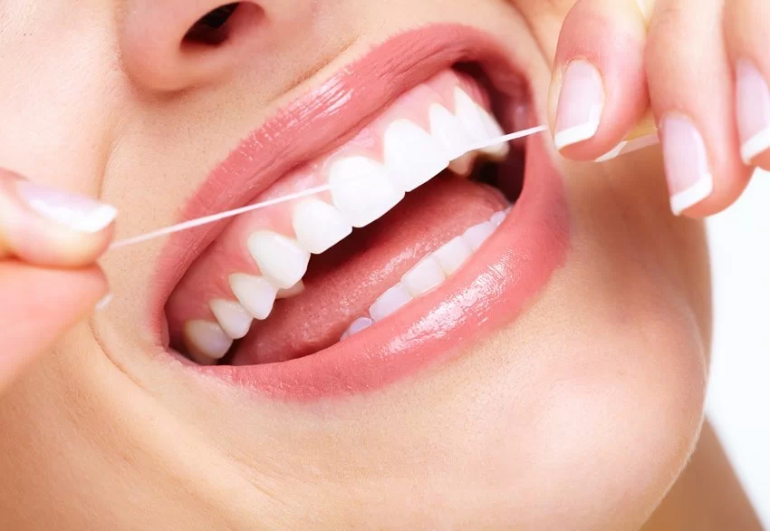
Brushing should be performed at least twice a day, with a soft-bristled toothbrush placed at a 45-degree angle to the gum line, using sweeping or circular motions from the gum toward the tooth (Bass Technique). The joining points of two teeth (interdental areas) where a toothbrush cannot reach are the locations where bacterial plaque accumulates most densely and diseases initiate. These areas must definitely be cleaned every day with the help of interdental brushes of appropriate thickness or dental floss. Oral irrigators, though they do not completely replace mechanical dental floss, provide great support in removing food debris by washing inside the pockets.
17. What Are the Factors Affecting Gum Health?
Gum health is in direct interaction not only with local oral hygiene but also with the general systemic balance of the body and lifestyle habits. These factors are:
- Diabetes Mellitus (Sugar Disease): There is a bidirectional relationship between diabetes and periodontal diseases. Tissue destruction progresses much faster in uncontrolled diabetic patients; simultaneously, infection in the mouth makes blood sugar regulation difficult.
- Hormonal Changes: Hormonal fluctuations during adolescence, pregnancy, and menopause increase the sensitivity of the gum against bacteria, paving the way for a “pregnancy tumor” (epulis) or severe gum enlargements.
- Genetic Predisposition: Even if some individuals pay attention to oral hygiene, they may be more prone to aggressive periodontitis forms due to immunological inheritance coming from the family.
- Medications Used: Certain blood pressure medications (calcium channel blockers), epilepsy medications (phenytoin), and immunosuppressive medications (cyclosporine) cause pathological overgrowths (hyperplasia) in the gums.
18. What Are the Factors Affecting the Success of Periodontal Treatment?
Clinical biological success rates are highly elevated in periodontal treatments; however, keeping this success permanent is possible by fulfilling certain conditions:
Patient Cooperation (Hygiene Continuity): Performing the treatment in the most perfect manner remains meaningless if the patient does not clean the plaque in the subsequent process. Periodontal success is a chain of responsibility carried out jointly by the dentist and the patient.
Status of Smoking: In patients who continue heavy smoking, the integration rates of surgical grafts with tissue and root coverage rates drop dramatically.
Regular Maintenance Appointments: Since periodontal diseases are of a chronic character, they tend to relapse (recur). Arriving without delay for the 3- or 6-month maintenance checks specified by the dentist ensures that new plaque accumulations at the initial level are cleaned immediately, protecting the main success for a lifetime.
19. What Are the Factors Affecting Periodontology Treatment Prices?
Periodontal treatment budgets are shaped individually according to the prevalence of the disease inside the mouth and the complexity of the surgical interventions needed. Elements affecting the price index are:
- Level of the Disease and Number of Teeth Involved: The session and workmanship cost of a localized curettage procedure involving only a few teeth versus a widespread advanced periodontitis treatment covering the entire lower and upper jaws (28-32 teeth) is different.
- Type of Surgical Intervention: The price dynamics change between standard non-surgical curettage prices versus flap operations where the gum is surgically reflected open or mucogingival plastic surgery operations.
- Use of Biological Materials: The amount of bone grafts (powders) placed into the defect in treating bone resorptions, the quality of the collagen membranes utilized, and the presence of reconstructive protein agents directly affect the material cost.
- Laser and Advanced Technology Integration: Utilizing dental laser systems included in operations for sterilization and tissue biostimulation purposes within the clinical workflow is among the factors shaping the price.
As Panorama Ankara Oral and Dental Health Polyclinic, we protect your gum and bone health, which is the main support of your teeth, with the most modern scientific protocols. Remember, even the healthiest teeth can only survive on a strong and inflammation-free foundation.