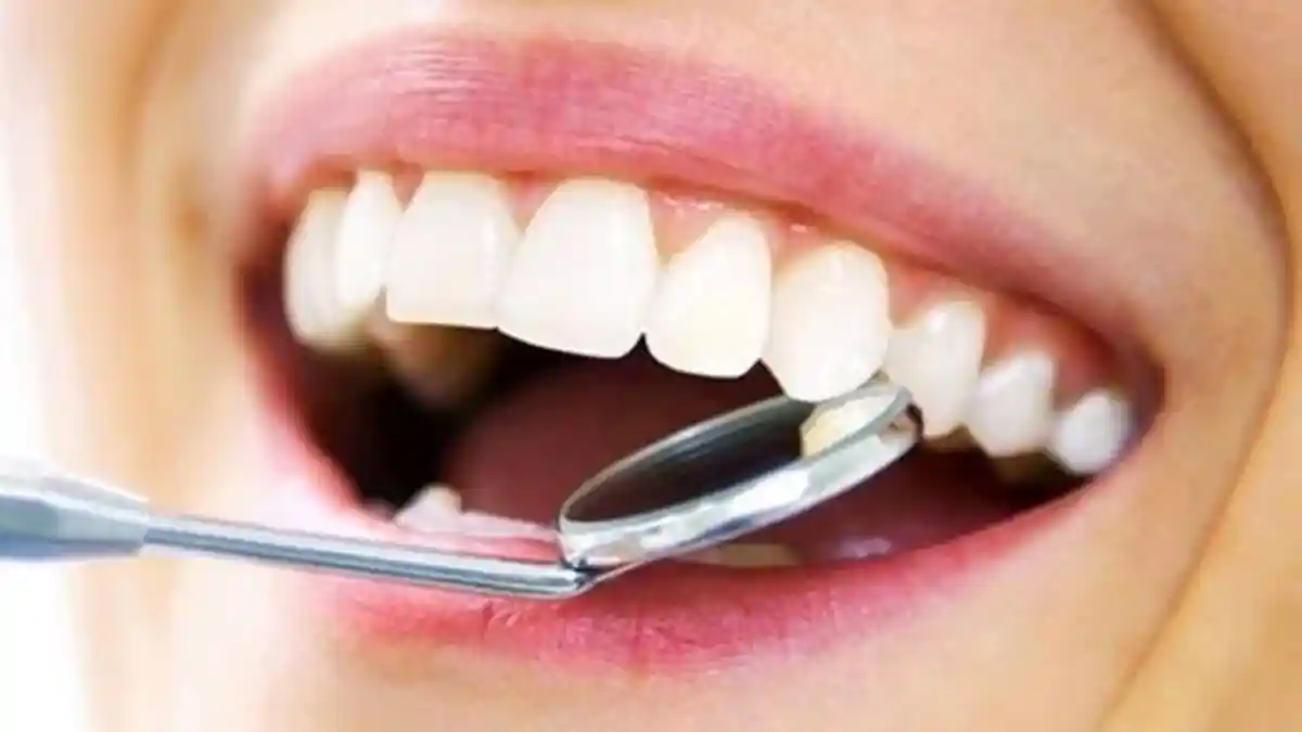
What is Laser Dentistry?
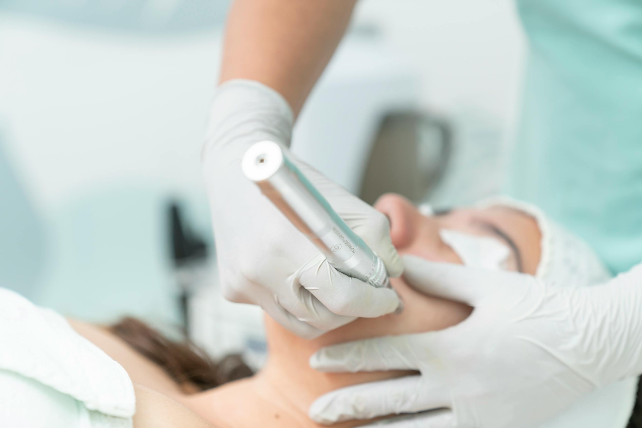
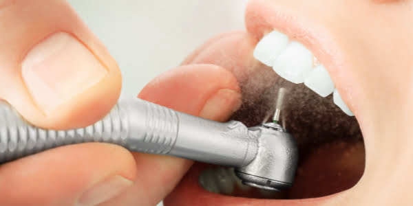
Laser dentistry is the utilization of monochromatic, linear, and concentrated light energy based on the principle of Light Amplification by Stimulated Emission of Radiation in intraoral hard and soft tissue treatments. The micro-cracks caused by rotating instruments (turbines and micromotors) on the enamel layer and the bleeding traumas created by the scalpel on soft tissues in traditional dentistry are completely eliminated thanks to the “target molecule” (chromophore) selectivity of laser energy. Hard tissue lasers target water and hydroxyapatite crystals in the tissue, while soft tissue lasers target hemoglobin and melanin pigments, vaporizing only the damaged area at the micron level without causing the slightest harm to surrounding healthy cells.
- Selective Photo-Ablation: Selectively vaporizing only the targeted infected cells or carious areas without touching healthy tissues.
- Mechanical Non-Contact: A wavelength technology that minimizes the stimulation of nerve conduction lines since it does not create frictional heat, vibration, or noise in the tooth.
- Bio-Stimulation Effect: Low-dose wavelengths stimulating cell mitochondria to activate local repair mechanisms.
In Which Treatments is Laser Dentistry Used?
Advanced technology laser systems constitute a versatile operational power with the widest boundaries in today’s clinical dental practices, capable of being integrated into almost all major specialty branches of dentistry. It possesses a wide range of indications, from cleaning dental caries to surgical bone contouring, and from sutureless gum surgeries to drying persistent root canal infections. Preventing arbitrary manual cuts and the necessity of needle numbing, these systems facilitate clinicians in achieving micron-level smooth and evidence-based results even in the most delicate dental restorations.
| Dental Clinical Branch Field | Integrated Laser Wavelength Type | Technological Contribution to the Treatment Process |
|---|---|---|
| Periodontology (Gum Treatment) | Diode, Nd:YAG, and Er:YAG Lasers | Eradicating pathogenic bacteria in deep pockets via photo-thermal radiation and bloodless gum contouring. |
| Endodontics (Root Canal Treatment) | Diode and Er,Cr:YSGG Laser Systems | Microscopic sterilization of main and capillary lateral canals inside root canals, ensuring sealing. |
| Restorative Dentistry | Erbium (Er:YAG) Hard Tissue Lasers | Vaporizing carious tissue in the tooth without the need for anesthesia and preparing the enamel surface for filling. |
| Oral and Maxillofacial Surgery | CO2, Er:YAG, and Nd:YAG Laser Infrastructure | Excision of soft tissue cysts, sutureless frenulum cutting (frenectomy), and bone shaving surgeries. |
Who is Suitable for Laser Dentistry?
Laser-assisted dental treatment approaches are medically suitable for all adult and pediatric patients with high levels of dental anxiety, who constantly postpone appointments due to the sound of needles and drills (dentophobia), and who have a low pain threshold. Since the system does not contain ionizing radiation and operates with tissue-friendly optical beam energy, it is applied as the primary safe option in sensitive patient groups whose general health profile presents local anesthesia risks (pregnant women, cardiac patients, those with high blood pressure, and individuals using blood thinners). However, the decision must be made by inspecting protective parameters in very specific cases with extreme sensitivity to light (photosensitivity) or where epileptic seizures can be triggered by light flashes.
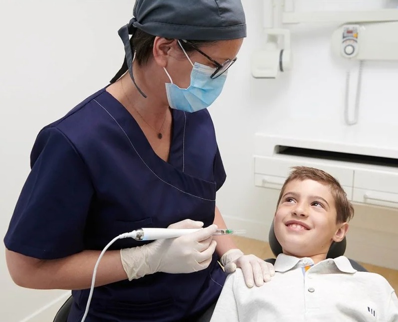
- Pediatric Age Group (Children): Children who do not develop dental trauma because they do not encounter drill vibrations and syringe appearances.
- Patients with a High Risk of Bleeding: Individuals with liver ailments or those using powerful anticoagulants (blood thinners) like aspirin.
- Individuals with Systemic Disorders: Chronic diabetes patients who exhibit a slow wound healing rate and an environment susceptible to infection.
Which Technologies are Used in Laser Dentistry?
The ability of laser dentistry to generate high resolution and tissue success in the clinical room depends on the existence of patented “Hard Tissue” and “Soft Tissue” laser device technologies that produce beams of different nanometer (nm) lengths across the electromagnetic spectrum. Each laser type is calibrated via software algorithms according to the absorption coefficient of the target component (water, melanin, hemoglobin, or hydroxyapatite) within the mouth. This technological diversity provides the physician with the opportunity to select the safest beam power for each patient’s specific tissue layer.
The primary laser technologies actively utilized in modern digital dental architecture are as follows:
- Diode Lasers (810 – 980 nm): Leading units in soft tissue cutting, hemostasis, and whitening, highly absorbed by hemoglobin and melanin.
- Erbium Lasers (Er:YAG 2940 nm / Er,Cr:YSGG 2780 nm): Hydro-kinetic systems showing maximum affinity for water molecules, operating both in enamel-bone shaving and soft tissue.
- Nd:YAG Lasers (1064 nm): Advanced surgical lasers that infiltrate into deep periodontal pockets to destroy pathogenic microbial colonies and provide tissue stimulation.
- Carbon Dioxide (CO2) Lasers (10600 nm): Dense units providing maximum coagulation in deep incisions and during the removal of large soft tissue tumors and cysts.
How are Laser Applications Used in Gum Treatments?
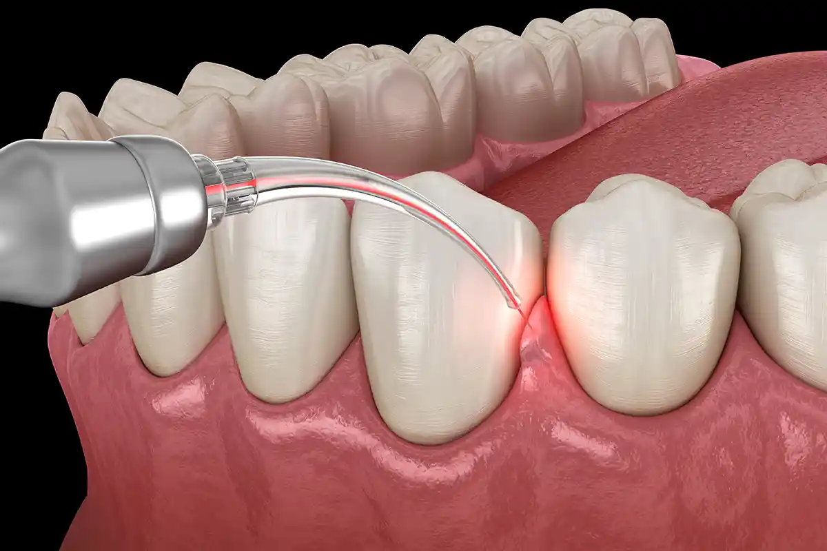
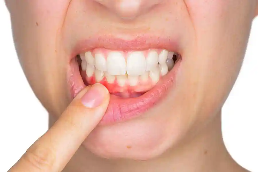
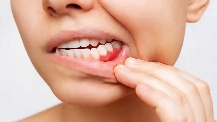
The application mechanism of laser technology in periodontal (gingivitis and periodontitis) treatments is based on destroying infection foci at microscopic depths with photo-thermal energy, which traditional scaling methods cannot reach. Micro laser fiber tips inserted into gingival pockets (sulcus) sterilize aggressive pathogenic bacteria (such as Porphyromonas gingivalis, etc.) that live without oxygen in that area and cause bone loss within seconds by bursting their cell membranes via radiation. While inflamed granulation tissues are vaporized during this procedure, healthy connective tissue fibers are treated passively; thus, tissue health is controlled without the need for deep periodontal surgeries.
- Laser-Assisted Curettage (LANAP): Cleaning the inflammation inside the pocket with beam wavelengths without utilizing scalpels and sutures.
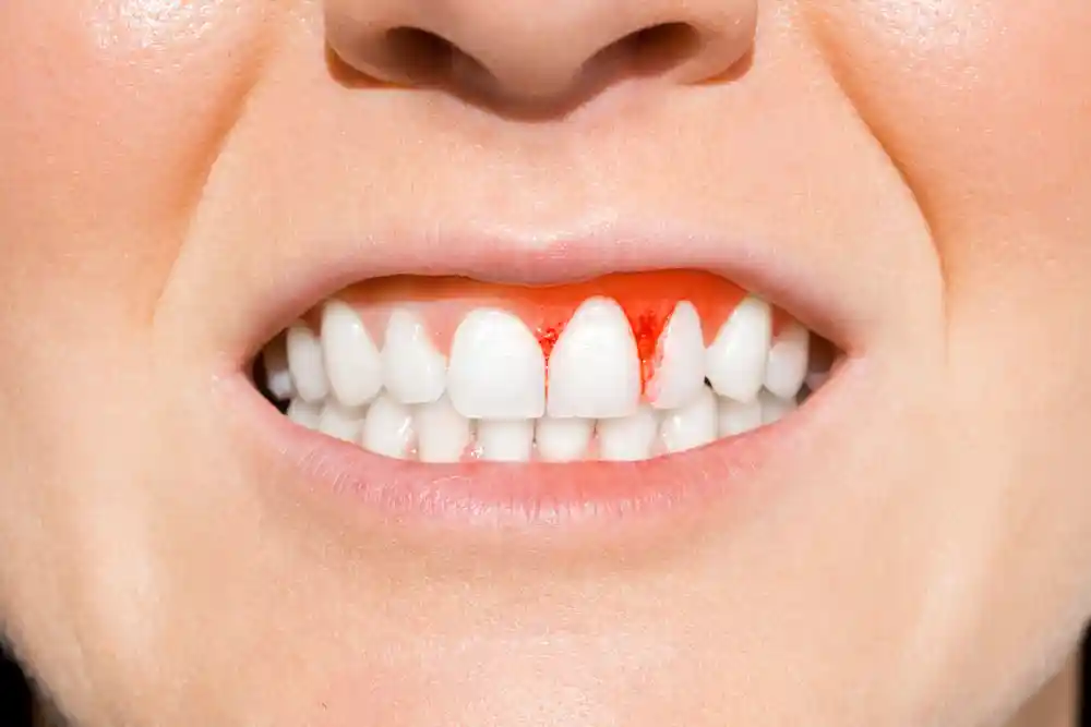
- Capillary Vessel Coagulation: Instantly halting brushing-induced bleeding by sealing edematous vessels in the gums.
- Tissue Attachment Triggering: The phase of biologically re-locking the tightening pink gingiva firmly back onto the cleaned root surface.
How Does Laser Dentistry Contribute to Aesthetic Dental Treatments?
In aesthetic dentistry and Smile Design practices, laser systems are preferred to establish a flawless visual “zenith harmony” by sharpening irregularities along the hard and soft tissue transition lines with an accuracy of one-tenth of a millimeter. Before porcelain veneers (laminates) or zirconium crown restorations are executed, the undulating symmetry lines of the gums are smoothly scanned and aligned with laser beams. This micron-level margin clarity provided by the laser allows restorations to seat onto the gingival boundary with zero margin of error, preventing artificial thicknesses and bringing the pink-white aesthetic harmony to its most natural form.
- Sharp Finish Line Calibration: Error-free transfer of the margin lines of teeth prepared for aesthetic restorations to computerized milling (CAD-CAM) units.
- Glaze Layer Protection: In removing old porcelain crowns, the power of laser energy to melt the adhesive resin, dropping the crown without harming the tooth.
How is Laser Gum Contouring Performed?
Laser gum contouring (gingivectomy/gingivoplasty) is an operation executed within seconds, mostly without even requiring local anesthesia, in cases where the gums appear excessively during smiling (gummy smile) or asymmetric mucosal curtains shorten tooth lengths. The physician smoothly follows the new gingival boundary line sketched on the screen according to facial golden ratio calipers on the mucosa using the micro tip of the laser handpiece. Because the laser beam vaporizes and removes excess tissue at the exact microsecond it touches it, while sealing vessel ends by welding them, the operation is completed with full micron resolution without a single drop of bleeding.
- Sutureless Pink Line Contouring: Elimination of tissue shrinkage and margin marks post-session since needles and suture threads are not utilized.
- Knife-Edge Morphology: Shaving thickened gingival margins with a laser to shape them into a thin form suitable for the natural sweeping mechanism of food.
How is Laser-Assisted Teeth Whitening Applied?
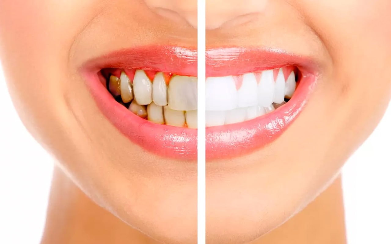
The laser-assisted in-office teeth whitening (bleaching) procedure is an advanced technology aesthetic cure that breaks down the chemical compounds of hereditary or chronic stains settled inside enamel pores into the fastest and most homogeneous phase. After specialized medical whitening gels containing peroxide are spread onto tooth surfaces, the optical head of the laser device with a specific wavelength (usually a diode laser) focuses on the teeth. Laser beams activate whitening crystals (chromophores) inside the gel photo-chemically within seconds, enabling the gel to infiltrate into the deepest tubules of the enamel layer; compared to traditional LED lamp whitening, this speed prevents overheating of the dental nerves (pulp) and eliminates permanent sensitivity risks, making the tooth 2-3 shades whiter.
- Photo-Chemical Activation: Accelerating the reaction speed of the gel through light power rather than heat, saving time.
- Minimum Sensitivity Coefficient: Absorption of stings during and after the procedure thanks to preventing thermal shock in the pulp chamber of the tooth.
How is Laser Dentistry Used in Root Canal Treatments?
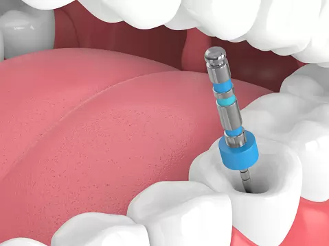
In endodontics (root canal treatment) practices, laser technology is preferred to establish a flawless disinfection barrier in the most micro-lateral tunnels (lateral canals) of root canals, where traditional medical liquids (sodium hypochlorite) cannot penetrate due to surface tension. After intra-canal mechanical instrumentation is finished, the “Laser-Assisted Activated Endodontic Disinfection” (LAFD) process is initiated by means of flexible laser fiber optic tips delivered into the canal. Beam energy bursts moisture molecules along the canal walls to generate shockwaves (acoustic streaming); these waves disrupt insidious bacterial colonies and biofilm layers on the canal walls at a cellular level, supporting the relapse-free drying of root-tip abscesses.
- Removal of the Smear Layer: Melting micro plaster debris (smear layer) left by rotating instruments on the canal wall with a laser to open up tubules.
- 3D Sealing Infrastructure: Completely pasive, gap-free locking of root tunnels completely purified of bacteria with bio-ceramic root canal fillings.
How are Laser Applications Performed in Intraoral Soft Tissue Procedures?
In the treatment of soft tissue lesions (fibromas, epulis, leukoplakias) developing in intraoral mucosa, cheek, and sublingual regions, laser systems offer a minimally invasive surgical comfort that saves the patient from heavy general narcosis processes in hospital environments. Pen-like micro-surgical laser tips separate the tissue from the lesion boundary lines by filtering them at a cellular level through the vaporization method. Since lymph and blood vessels are closed via thermal sealing at the exact moment the incision is made, there is absolutely no need for suturing; this safety is also actively applied in biologically drying up and instantly cutting the sting of painful aphthous ulcers and cold sores (herpes) that recur frequently in the mouth within the first 30 seconds.
- Aphthous Ulcer and Cold Sore Suppression: Instantly alleviating wound pain by deactivating early-stage herpes viruses with laser energy.
- Sutureless Excision Quality: Eliminating post-operative scar contraction risks thanks to removing mucosal tumors from the line without a suture burden.
How is the Pre-Laser Dentistry Evaluation Conducted?
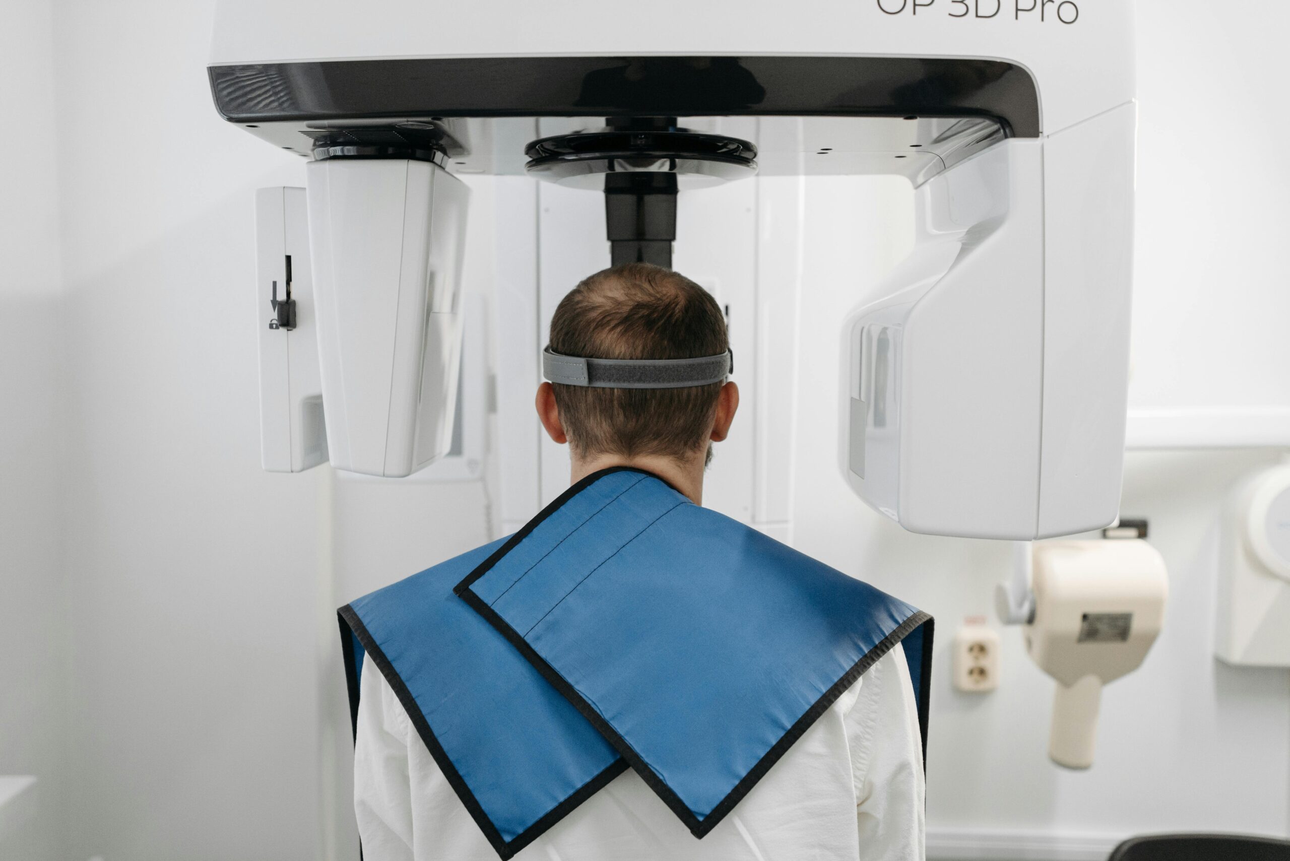
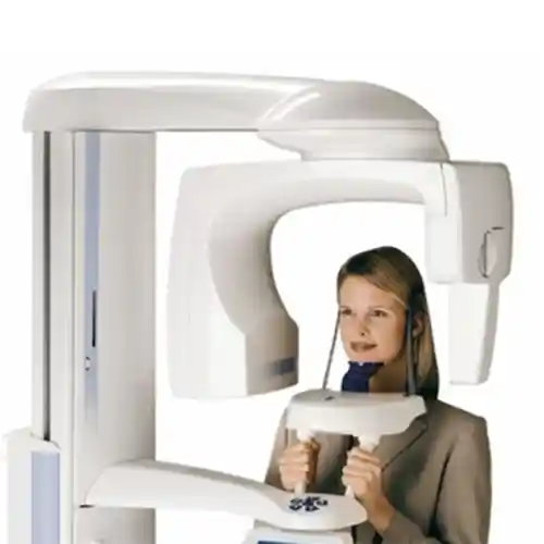
The clinical evaluation conducted before proceeding to the application session is a vital analysis stage where the patient’s hard and soft tissue quality is scanned to decide which specific laser wavelength and joule (energy) power will be selected for which indication. During the examination, the enamel calcification degree of the teeth and gingival pocket depths are mapped out millimetrically. The most critical digital support at this stage is the integration of CBCT dental tomography, which transcends the boundaries of classic two-dimensional radiographs to scan the jawbone and root tunnels with millimetric cross-sections in 3D; hard tissue density data obtained from the tomography finalizes the power calibration settings of the laser without error before the operation.
- Chromophore Density Analysis: Selecting the most accurate laser absorption mode on the computer based on water, pigment, and mineral ratios in the tissue.
- Systemic Contraindication Check: Scanning the patient’s history of medications causing photosensitivity and photospective immune responses through laboratory tests.
How Long Do Laser Dentistry Procedures Take?
Laser-assisted dental treatment procedures stand as one of the fastest, most dynamic, and practical processes that fit seamlessly into the busy pace of modern urban life, taking almost none of the patient’s time on the medical aesthetic and preventive dentistry calendar. Thanks to laser pulses operating at the speed of light (in millisecond pulses), clearing a cavity preparation of decay or contouring the mucosal boundary of a single tooth is usually completed within just 30 to 60 seconds; this speed cuts session durations in half compared to traditional analog methods.
- Sutureless Lip-Tie Cut (Frenectomy): Excising the tissue sutureless with a laser tip takes an average of 2 to 3 minutes.
- Full-Arch Laser Whitening Cures: Gel irradiation times are finished in approximately 15-20 minutes thanks to the speed of photo-chemical activation.
- Temporal Practicality: Chair time is minimal since stages like waiting for the anesthesia needle to numb and placing sutures at the end of the session are eliminated.
Are Laser Dentistry Applications Painful Procedures?
No, laser dentistry applications are medical procedures of a highly comfortable and non-invasive character that do not evoke pain, ache, or thermal stinging sensations within modern dental prophylaxis and surgical standards. The process can be managed smoothly without requiring traditional injection syringes or numbing local sprays in 80% of cases. The physiological reason for this is that laser beams are delivered in millisecond micro-pulses (shots) without physically contacting the tissue, and this duration is much shorter than the speed at which the human nervous system transmits pain signals to the brain (the axon conduction threshold); thus, the procedure ends comfortably before nerve endings can generate a sensation of pain.
- Neurological Gate Control: Preventing the stimulation of pain receptors (nociceptors) since pressure and frictional vibration do not form.
- Comfort Without Sedation: The luxury of even children and adults with the highest levels of dental phobia remaining calm in the chair without narcosis, purely through light comfort.
What Should Be Considered After Laser Dentistry?
To make the aesthetic and medical success of the session permanent, support the regenerative repair cycle initiated by laser beams in the underlying cell layers, and protect micro-cellular areas, the care discipline observed by the patient at home after the procedure is of vital importance. Since hard or soft tissue margins treated with a laser transition temporarily into a more delicate adaptation phase against external factors during the first hours, protecting the tissue from excessive irritant chemical and mechanical loads is a necessity for clinical safety.
The medical rules to be meticulously followed at home during the critical post-operative period are as follows:
- Excessively hot, ice-cold, or acidic foods must not be consumed for the first 24 hours following soft tissue contouring or whitening sessions.
- In cases undergoing laser peeling (depigmentation), the consumption of cigarettes, tea, and coffee must be ceased for at least 48 hours so that enamel pores do not rapidly re-absorb stains (white diet).
- Aggressive toothbrushing maneuvers should be avoided on the day of the procedure; starting from the next day, hygiene should be maintained with soft prophylaxis brushes recommended by the physician.
How Does the Healing Process Progress After Laser Dentistry?
The tissue recovery phase following the operation progresses rapidly, predictably, and comfortably with near-zero healing burden (no downtime) since it does not contain scalpel incisions and suture loads. The formation of a slight whitish/yellowish protective fibrin layer along the treatment boundary lines on the day of surgery is a completely natural and expected physiological repair reflex woven by epithelial cells to cover the wound surface; this layer must definitely not be scraped off by mistaking it for inflammation. The healing process flows along a linear line; since cell mitochondria divide rapidly via laser stimulation, the tissue normalizes completely within 3 to 5 days, regaining its fresh, firm form.
- The First 24 Hours: Spontaneous bleeding or leakage is not experienced since micro-vessels are sealed, and social life continues uninterrupted.
- 1st Week Milestone: Epithelial coverage completes its 100% integration with the extracellular matrix, verifying the pale pink healthy color harmony in the tissue.
What are the Advantages of Laser Dentistry?
The advantages brought to modern dentistry practices by advanced technology laser systems are not limited merely to the elimination of pain, but elevate the operational quality to a perfection beyond the limitations of the human hand and eye. Radically reducing material waste and the number of sessions in the clinic, this bio-technological flow transforms the dentistry chair from a room of fear into a comfortable medical spa experience.
- Sutureless and Bloodless Surgeries: Elimination of the necessity to place sutures thanks to executing surgical procedures with sealed vessels.
- High Sterilization Power: The heat of the beam instantly kills all bacterial flora in the operation field, reducing infection risks to zero.
- Maximum Tissue Conservation: Preserving the fracture resistance of the tooth by acting passively on healthy hard dentin and enamel tissue while cleaning decay.
- Scar-Free Healing: Elimination of post-operative tissue contraction aesthetic defects thanks to closing mucosal incisions with a smooth line.
How Does Laser Technology Support the Planning of Dental Treatments?
Laser technology supports the planning of evidence-based and entirely predictable treatment algorithms by completely removing clinical guesswork and arbitrary manual reductions. Because the physician models the patient’s intraoral digital data in 3D on the computer screen, the millimetric depth boundaries where laser beams will penetrate into the tissue and the joule energy powers can be simulated via software before even starting the operation; this digital convenience speeds up process management by ensuring the elimination of the margin of error.
- Micron-Level Calibration: The ability to custom-adjust the margin boundary and bone thickness belonging to each tooth from laser software panels.
- Multi-Disciplinary Synchronization: The ease of surgical, orthodontic, and aesthetic branch physicians making joint decisions on the same laser schedule.
With Which Procedures Can Laser Dentistry Be Applied Jointly?
Laser dental protocols can be planned within a combined synergy with other advanced technology dental applications to holistically elevate the aesthetic architecture of the face, jawbone rigidity, and smile integrity. While underlying mucosal and bone balances are sharpened with laser surgery, form defects in the upper layers can be treated simultaneously with different robotic units; the combination schedule is designed according to the result of the detailed facial analysis to be performed by the specialist physician.
- CAD/CAM Robotic Production: Following smooth gingival margins shaped with a laser, zirconium crowns emerging from digital milling devices seat into the mouth with perfect passive fit.
- Clear Aligner (Orthodontic) Treatments: While moving teeth with wires or aligners, loosening thick muscle attachments (frenulums) that obstruct movement with a laser within seconds (frenectomy).
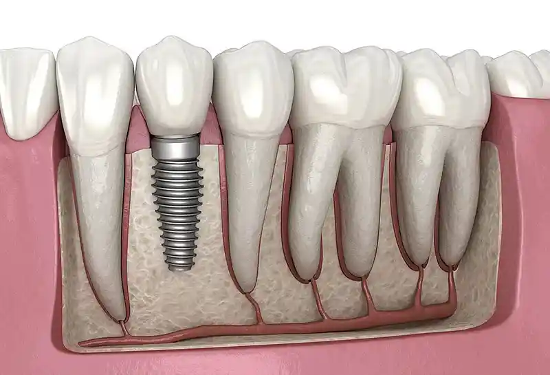
- Dental Implantology: Processes of opening the bone circularly and sutureless with a laser to place titanium screws and stimulating post-operative PRF membranes with a laser.
How Does Laser Dentistry Contribute to the Preservation of Oral Health?
The protective contribution of laser systems on the oral health ecosystem lies in removing microscopic bacterial fortresses (biofilm)—the main raw material supplier of insidious decay and gum diseases—by disrupting them at a cellular level, rather than merely cleaning tissues superficially. When bacteria cannot establish attachment matrices on enamel and root surfaces, they cannot produce acids; this biological purification balances the intraoral microflora, halts the jawbone resorption rate, and provides a complete protective medicine shield by eliminating persistent mouth odors (halitosis).
- Demineralizaton Barrier: Increasing resistance against acid erosions by hardening enamel pores with laser energy.
- Extending Tissue Lifespan: Preserving the attachment strength of periodontal ligaments, whose infection load is reset, to the jawbone.
What are the Frequently Asked Questions About Laser Dentistry?
Presented below are the most common practical concerns arising in the minds of patients planning to benefit from the high comfort standards offered by laser technologies in dental treatments and surgical processes, along with their uncensored medical answers:
Do laser beams cause radiation damage to my body while performing extractions or cutting inside the mouth?
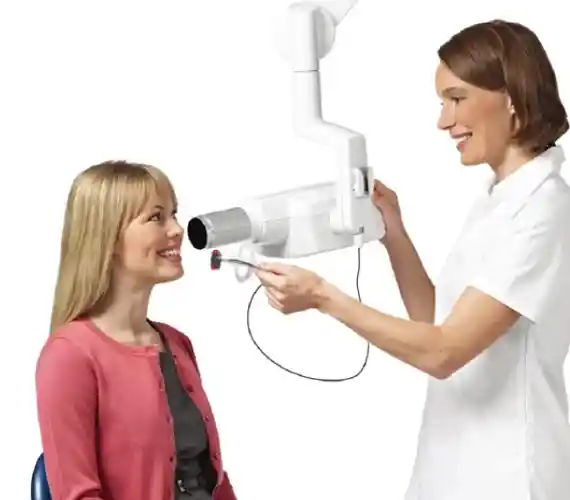
No, they strictly do not; this is a very common illusion of name similarity. Medical laser systems utilized in dentistry do not harbor ionizing “X-rays” that carry cancer risks like those in X-ray equipment. Lasers are optical systems utilizing completely safe visible light and wavelengths in the infrared spectrum; therefore, they have no side effects or radiation damage to general body biology or cell DNA structure, and are administered safely even in pregnant women.
Is cleaning decay with a laser safer compared to cleaning it with a normal drill (aerator)?
Yes, it is clinically a many times safer and enamel-friendly method. Traditional rotating diamond drills contact the tooth to emit high frictional heat and vibrations; these vibrations can form micro-cracks invisible to the naked eye, leading to the tooth breaking from the filling margin in the future. Hard tissue lasers, on the other hand, perform cleaning without physically touching the tooth at all, merely by bursting water molecules in the decay; it does not scratch or crack the enamel and provides maximum tissue conservation for the tooth.
Does the lack of suturing after gum surgeries performed with lasers lead to the opening up of that area?
No, this situation is the greatest microscopic safety advantage laser surgery presents to tissue. Because laser beams weld and seal (coagulate) capillary vessel ends at that incision boundary via photo-thermal effect while cutting soft tissue, bleeding or open leakage corridors do not remain at the wound margins. Since the tissue is left in a stabilized and sealed mode, cells lock onto each other smoothly on their own without needing to be wrinkled with suture threads.
Are session fees for laser dentistry applications very expensive compared to traditional analog treatments?
The technological infrastructure, periodic calibration certificates, and consumable costs of laser devices (Erbium, Nd:YAG systems) may appear high at first glance compared to analog syringes and scalpels; however, these smart systems reduce the patient’s number of sessions, eliminate suture/dressing appointments, and end anesthesia and medication waste. Most importantly, it is much more economical in the long run since it resets the budgets for repeating erroneous procedures with its micron-level precision power.
I have numerous implants and metal bridges in my mouth; do laser devices cause heating or dislodgement in my mouth?
No, there is absolutely no drawback or heating risk. Dental laser beams do not produce a powerful magnetic field or radio waves like MRI equipment in medical standards; they merely emit optical energy absorbed by specific cell pigments (melanin/hemoglobin) in the targeted tissue layer. Thanks to this biological safety, the beams cannot trigger a heating, loosening, or dislodgement reaction when they strike the titanium implant screws or porcelain zirconium metals in your mouth; it is completely safe.
Who We Are & Panorama Ankara Oral and Dental Health
Panorama Ankara is a licensed oral and dental health center that offers institutional health assurance to its patients by combining an academic expert staff and the latest global bio-technological digital infrastructure under a single roof, particularly across oral, dental, and maxillofacial surgery, advanced implantology, microscopic endodontics, periodontology, and aesthetic dentistry specialty branches. Attaching vital importance to completely eliminating arbitrary manual cuts, human error-induced marginal finish line inaccuracies, and those traumatic syringe needles and drill vibrations that cause chair anxiety in patients, our center operates uninterrupted Advanced Technology Laser Dentistry (Hard and Soft Tissue Laser) ecosystems from the very beginning to the end of the clinical operation workflow.
Under the roof of Panorama Ankara, absolute sterilization, high patient safety, transparency, and honesty principles are operated with uncompromising determination by regulation in all diagnosis, radiological scanning, design, and laser-assisted micro-surgical interventions. In our clinical practices, proprietary Erbium and Diode laser units that seal vessels instantly while focusing on tissue integrity with micron-level resolution are applied with millimetric precision under the supervision of our expert physician, surgeon, and periodontist staff. Our aim is not to impose old-type, slow, painful, and traumatic analog treatment molds that take weeks to heal upon our patients; but rather to analyze each individual’s jawbone quality, occlusion mechanics, and facial aesthetic golden ratio boundaries with the help of 3D tomographies on a digital screen prior to the operation, achieving maximum session comfort that is imperceptible from the outside without harming surrounding soft tissues and sensitive vascular pathways, a sutureless comfort standard, and healthy, smart smile designs to be used with smoothness for a lifetime. If you are tired of your missing teeth, old worn restorations, or gum bleedings when you look in the mirror, yet postpone your treatments due to fear of the dentist chair, needle phobia, and time limitations, and want to initiate your end-to-end digital smart treatment process with scientific assurance in an expert institutional center environment; you can safely protect your health and smile integrity by contacting the expert staff of Panorama Ankara for your detailed intraoral examination and laser comfort.