1. What is Orthognathic Surgery?
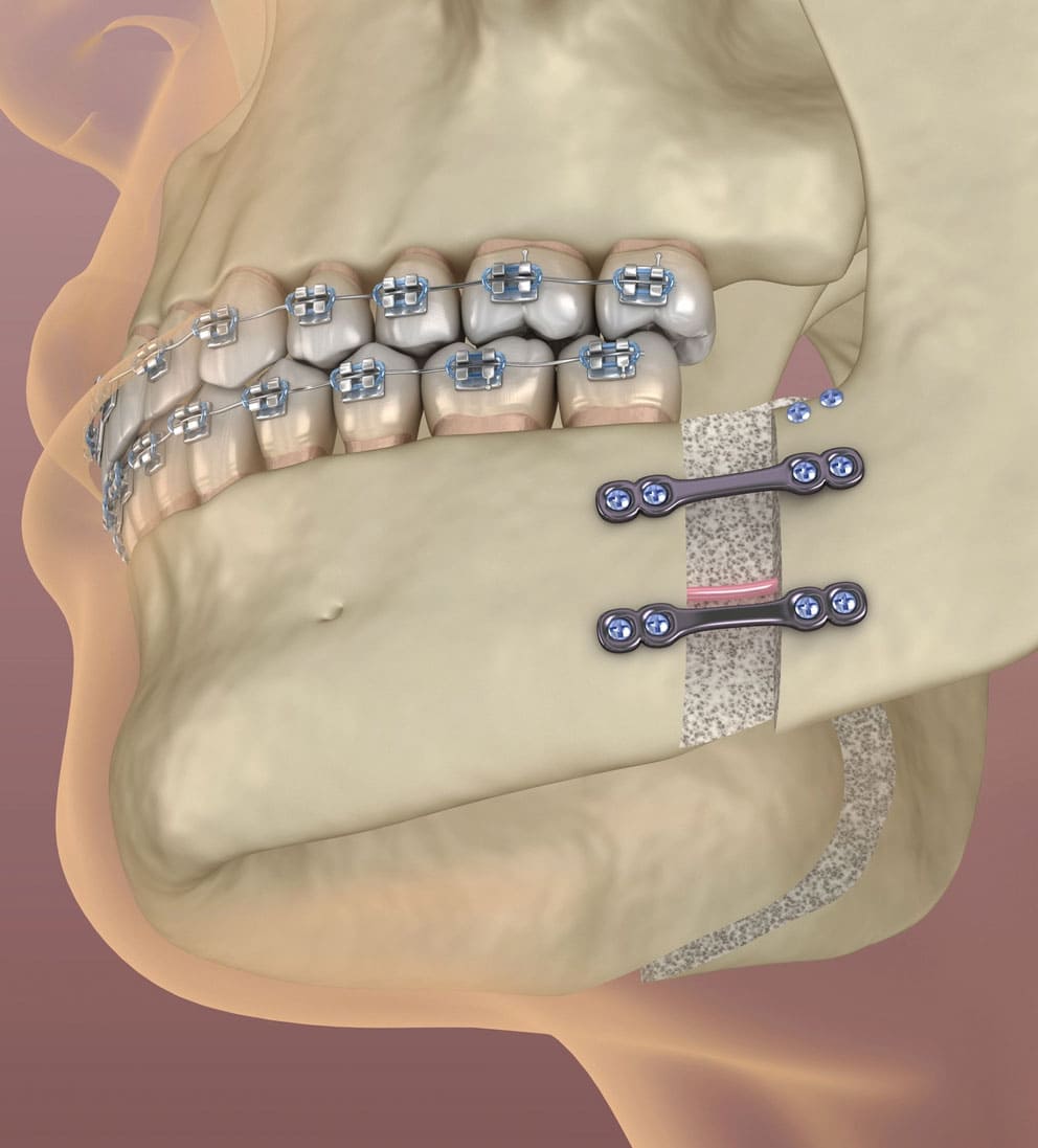
Orthognathic surgery is a major surgical procedure applied to correct flawed positional relationships of the lower jaw (mandible) and upper jaw (maxilla) bones, which constitute the facial skeleton, both among themselves and with the skull base. This method, which comes into play in skeletal anomalies too advanced to be corrected solely with dental braces (orthodontics), is based on the principle of surgically cutting the bones in a controlled manner (osteotomy), repositioning them into the correct anatomical location, and fixing them with titanium plates and screws. It is a multidisciplinary treatment type jointly conducted by plastic surgery and maxillofacial surgery specialists alongside orthodontists.
- Bone Repositioning: Moving the jawbones in a three-dimensional plane (forward, backward, upward, or downward).
- Rigid Fixation: Locking the cut bones to each other with titanium plates and screws to ensure they do not move during the healing process.
- Intraoral Approach: Since the operation is performed entirely from inside the mouth, no surgical scarring remains in the facial region.
| Component | Medical Definition | Role in Treatment |
|---|---|---|
| Osteotomy | Surgical cutting of bone | Allows mobilizing the jaw to move it. |
| Osteosynthesis Material | Titanium plates and micro-screws | Maintains stabilization until new bone heals. |
2. In Which Situations is Orthognathic Surgery Applied?
Orthognathic surgery is clinically indicated in situations where the jawbones have developed disproportionately due to genetic, syndromic, or acquired (trauma, etc.) factors during the growth and development process. Situations such as the lower jaw being excessively anterior or posterior relative to the upper jaw, upper jaw developmental deficiencies, and the front teeth not making any contact with each other during chewing (anterior open bite) are the primary application fields of this surgery. Furthermore, this operation is preferred in patients who have significant asymmetry between the right and left halves of the face, or in patients suffering from severe obstructive sleep apnea (cessation of breath during sleep) due to mandibular constriction, in order to widen the airway.
- Anterior Open Bite: The condition where a gap remains between front teeth while posterior teeth are in contact.
- Mandibular Prognathism / Retrognathism: The condition where the lower jaw is positioned excessively forward or backward.
- Maxillary Hypoplasia: The condition where the upper jaw remains developmentally posterior and flattened.
| Clinical Situation | Character of Pathology | Primary Goal of Surgery |
|---|---|---|
| Facial Asymmetry | Deviation of the chin to the right/left | Equalizing the facial midline with the chin point |
| Severe Sleep Apnea | Posteriorly retracted lower jaw obstructing the airway | Advancing the jaw to enlarge the respiratory passage |
3. Who is Suitable for Orthognathic Surgery?
The most fundamental medical criterion for this operation to be applied is that the individual’s skeletal growth and development process (bone development) must be completely finished. Since interventions performed while growth continues may disrupt surgical success due to the uncontrolled elongation of bones, it must be verified that bone age has matured via hand-wrist X-rays or cervical vertebra (neck vertebrae) analyses. Adult individuals who do not have any systemic illness that would pose a contraindication to general anesthesia, whose tooth and gum health is stable, and who possess the motivation to comply with the long orthodontic stages in the treatment process are suitable candidates for this surgery.
- Age Limit: Generally suitable for females aged 17-18 and males aged 18-20 and older.
- Periodontal Stability: It is mandatory that diseases such as gum recession or active bone resorption are absent.
- Systemic Suitability: Absence of contraindications such as uncontrolled diabetes, severe cardiovascular diseases.
| Patient Profile | Suitability Status | Clinical Rationale |
|---|---|---|
| Children During Growth | Not Suitable (With exceptions) | Bone growth can alter surgical planning. |
| Adults with Stopped Bone Development | Completely Suitable | The skeletal structure being fixed and predictable. |
4. How is the Evaluation Conducted Before Orthognathic Surgery?
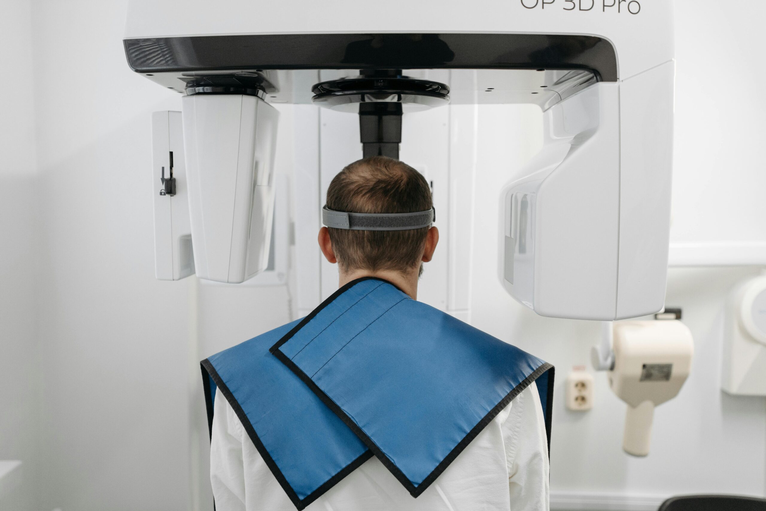
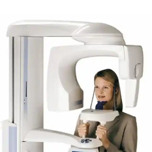
The clinical evaluation process before the operation is a vital analysis stage involving the use of extremely detailed diagnostic tools to leave no room for error. During the first examination, the orthodontist and jaw surgeon physically examine the patient’s facial profile, lip closure adequacy, smile line, and temporomandibular joint (jaw joint) functions. Subsequently, Cone Beam Computed Tomography (CBCT) scans are taken to millimetrically map bone thicknesses, the route of nerve channels (inferior alveolar nerve), and airway volume; digital impressions taken with intraoral scanners are integrated into these data.
- Craniofacial Analysis: Mathematical calculation of jaw angles via cephalometric X-rays.
- Soft Tissue Thickness Measurement: Estimation of how bone movement will reflect on the face skin.
- Physical Model Analysis: Examination of dental closure relationships in articulator devices.
| Evaluation Method | Criterion Measured | Clinical Importance |
|---|---|---|
| 3D Computed Tomography | Volumetric bone structure and anatomical nerve paths | Safely determining surgical incision lines |
| Digital Intraoral Scanning | Micron-level tooth closure relationship | Production of splints to be used during surgery |
5. How is Orthognathic Surgery Treatment Planned?
Orthognathic surgery planning is currently carried out through virtual surgical planning (Virtual Surgical Planning) softwares instead of plaster models and two-dimensional radiographs used in the past. After physicians create the patient’s 3D bone model in a computer environment, they cut the jaws with the help of virtual scalpels and slide them into their ideal positions. During this virtual sliding process, simulations are performed on how much the upper jaw will be recessed, how much the lower jaw will be advanced; surgical guides (splints) that will lock onto the teeth during surgery and guide the surgeon are manufactured in 3D printers in accordance with this plan.
- VSP (Virtual Surgical Planning): Simulating the surgery on a computer screen with zero margin of error.
- Splint Design: Production of intermediate and final plates keeping the lower and upper jaws locked during surgery.
- Facial Profile Simulation: Demonstrating to the patient what kind of facial appearance they will attain after surgery.
| Planning Phase | Technical Procedure Performed | Process Output |
|---|---|---|
| Data Fusion | Merging tomography data (DICOM) with scanner data (STL) | Realistic 3D Patient Model |
| Guide Production | Sending splints designed in CAD software to the 3D printer | Custom Surgical Splints |
6. Why is Orthodontic Treatment Applied Before Orthognathic Surgery?
In individuals with skeletal jaw disorders, teeth attempt to mask the existing anomaly by tilting or repositioning themselves over time to enable chewing (dental compensation). If the jaws are surgically corrected while these teeth are in their current crooked positions, the teeth will never meet on the operating table; for this reason, a pre-operative orthodontic treatment stage called “decompensation” must definitely be applied. In this stage, with the help of braces or clear aligners, teeth are brought to the ideal vertical angles where they should be relative to the jawbone they are attached to; although this temporarily makes the jaw disorder appear more belirgin from the outside, it prepares the ground for surgery.
- Decompensation: Correcting incorrect tooth angles and placing them vertically in their own bone base.
- Ensuring Arch Compatibility: Equalizing the width of the lower and upper dental arches.
- Preparing Closure: Ensuring teeth lock like puzzle pieces when the jaws are moved.
| Period | Tooth Position | Orthodontic Goal |
|---|---|---|
| Pre-treatment (Natural State) | Tilted/compensated teeth to save the bite | Tendency to hide existing bone constriction/crookedness |
| Pre-surgery (Post-orthodontics) | Independently aligned teeth at the correct angle in their bone | Capturing full contact when jaws move |
7. In Which Jaw Problems is Orthognathic Surgery Used?
Orthognathic surgery is a major solution for eliminating major craniofacial deformities that exceed traditional dentistry boundaries and are fixed entirely on a skeletal basis. Micrognathia cases where the lower jaw remains far behind, referred to as bird-face profile, and Class 3 malocclusions, popularly known as lower jaw prognathism, are within this scope. Furthermore, situations where one side of the face is longer than the other due to asymmetric growth, and excessive gingival display (gummy smile) occurring due to vertical overgrowth of the upper jaw bone which causes serious aesthetic discomfort, are radically solved with this surgery.
- Facial Length Incompatibilities: The middle or lower third of the face being excessively long or short.
- Laterognathism: The chin point being shifted to the right or left relative to the middle vertical line of the face.
- Restricted Airway Anomalies: Chronic snoring and respiratory tract constrictions stemming from jaw retraction.
| Jaw Problem | Anatomical Defect | Surgical Approach Applied |
|---|---|---|
| Class 2 Deformity | Lower jaw retrusion / Upper jaw protrusion | Advancing the lower jaw, positioning the upper jaw if necessary |
| Gummy Smile | Excessive vertical elongation of the upper jaw | Vertical bone block excision (impaction/recession) from the upper jaw |
8. How is Lower and Upper Jaw Surgery Performed?
Operations are conducted in a fully equipped hospital environment, in the operating theater, under general anesthesia, and under sterile conditions. In upper jaw surgery, the “Le Fort I” osteotomy technique is generally used; the upper jawbone is cut horizontally along the line above the tooth roots, mobilized, and shifted in the planned direction. In lower jaw surgery, the “Bilateral Sagittal Split Osteotomy” (BSSO) method is frequently applied; in this method, the lower jawbone is split vertically in two without harming nerve fibers, shifted, and subsequently, bone pieces are fixed to each other with titanium plates and screws, and the mucosa is closed with sutures.
- Le Fort I Osteotomy: Mobilizing the upper jawbone by cutting it from the sinus floor level.
- BSSO Technique: Splitting, elongating, or shortening the lower jaw ramus region in the sagittal plane.
- Genioplasty: Cutting the chin tip bone for aesthetic purposes to advance/recede or lengthen it.
| Surgical Region | Common Technique Used | Anatomical Risk Management |
|---|---|---|
| Upper Jaw (Maxilla) | Le Fort I Osteotomy | Protection of arterial bleedings and sinus mucosa |
| Lower Jaw (Mandibula) | BSSO (Bilateral Sagittal Split) | Protection of the integrity of the mental/alveolar lower nerve |
How is Digital Planning Technology Used in Orthognathic Surgery?
Digital planning technologies are the most important modern dental developments that increase the success rate of orthognathic surgery and minimize complication risks by shortening operation time. High-resolution tomography data obtained from the patient are combined with occlusion (bite) data in specialized softwares, and the rotation of the jaw joint (condyle) in its socket is simulated in three dimensions. Cutting guides that errorlessly show the surgeon exactly where and by how many millimeters to cut the bone during surgery, along with custom-bent titanium plates, are produced under computer control thanks to CAD/CAM systems.
- 3D Cutting Guides: Performing bone cutting not by hand, but with digital templates without error.
- Navigated Surgery: The surgeon being able to follow their position on the screen simultaneously during the operation.
- Custom Plates: Adaptability-high stabilizers pre-shaped according to the patient’s tomography.
| Technology Name | Clinical Function | Direct Benefit to Patient |
|---|---|---|
| CAD/CAM Intermediate Splint | Locking jaws in intermediate position during surgery | Reduces risk of incorrect positioning to zero. |
| IPS (Individual Patient Solutions) | Production of custom titanium systems for the patient | Shortens surgery duration, reduces edema. |
How Does Orthognathic Surgery Support Facial Aesthetics?
Although orthognathic surgery is a functional operation, it yields dramatic aesthetic results as it changes the fundamental structure of the facial skeleton. Bringing the bones into the correct position directly affects the tension and angle of the lip, cheek, chin tip, and neck skin covering them, imparting symmetry to the face in accordance with golden ratio rules. In a patient with lower jaw retrusion, the sagging in the dewlap region is resolved and the jawline (jawline) becomes pronounced post-surgery; in patients whose upper jaw retrusion is resolved, depressions alongside the cheekbones and nose wings are filled, achieving a younger and more dynamic facial profile.
- Nasolabial Angle Regulation: Bringing the angle of the upper lip with the nose base to aesthetic boundaries.
- Mentolabial Sulcus Aesthetics: Clarifying the natural curve between the lower lip and the chin tip.
- Facial Triangle Balance: Harmonious alignment of the forehead, nose, and chin tip in the profile plane.
| Deformity Type | Pre-Surgery Facial Aesthetics | Post-Surgery Aesthetic Change |
|---|---|---|
| Lower Jaw Protrusion | Hard, aggressive profile and sunken mid-face | Softened, balanced, and aesthetic profile lines |
| Short Face Syndrome | Aged, flattened appearance and inward-puckered lips | Younger and fresher facial lines with vertical dimension increase |
How Does Orthognathic Surgery Contribute to Chewing Function?
Patients with skeletal jaw disorders cannot mechanically grind foods sufficiently since their teeth do not fully contact each other, which can lead to chronic digestive system (gastrointestinal) problems. Orthognathic surgery brings the lower and upper dental arches into an ideal closure relationship (centric occlusion), ensuring chewing forces are distributed to all teeth equally and balancedly. The effective crushing function of posterior molars and the ability of anterior teeth to bite food healthily are restored; thus, excessive load on the chewing muscles (musculus masseter and temporalis) is lifted, preventing jaw joint degenerations.
- Increase in Occlusal Contact: Maximizing the surface area of teeth that contact each other.
- Chewing Force Distribution: Preventing excessive load on a single tooth to reduce fracture risk.
- TMJ Protective Effect: Eliminating asymmetrical pressures on the jaw joint disc.
| Closure Type | Chewing Efficiency | Long-Term Joint Health |
|---|---|---|
| Pathological Closure | 20% – 30% (Insufficient grinding) | High risk (Wear in joint, clicking sound, and pain) |
| Ideal Closure Post-Surgery | 90% – 100% (Full function) | Minimum risk (Stable and physiological joint load) |
How Can Orthognathic Surgery Affect Speech Function?
Speech is based on the shaping of air emerging from the lungs as it passes through narrow areas between the tongue, lips, palate, and teeth; therefore, the anatomical structure of the jawbones is directly influential on phonetics. For instance, a patient with an anterior open bite lisps because they jam their tongue into the space between teeth while pronouncing letters such as “S, Z, T,” while those whose lower jaw is excessively anterior experience difficulty in articulating (F, V, B, P) sounds. When the volume and boundaries of the oral cavity are restored to normal with orthognathic surgery, the tongue’s articulation movements are eased, and patients attain a much clearer and more intelligible diction following a short adaptation phase.
- Resonance Chamber Change: Clarification of voice tone through volumetric balancing of mouth and nasal passages.
- Standardization of Tongue Position: The tongue resting in the correct place on the palate during rest and speech.
- Elimination of Lisping: Anatomically closing air leaks between teeth.
| Existing Deformity | Affected Letter Groups | Post-Operation Improvement |
|---|---|---|
| Open Bite | S, Z, D, T (Fricative and plosive letters) | Air leakage ends, lisping is eliminated. |
| Protruded Lower Jaw (Class 3) | F, V, B, P (Lip-tooth contact letters) | Lower lip touches upper teeth comfortably, phonetics improve. |
How Long Does Orthognathic Surgery Take?
The duration of the orthognathic surgery operation varies from clinic to clinic and case to case depending on the number of jawbones to be intervened in and the complexity of the deformity. Single jaw operations involving only the lower or upper jaw are completed on average between 1.5 to 2.5 hours, while double jaw (bimaxillary) surgeries where both jaws are moved simultaneously usually take 3 to 4.5 hours. If extra procedures such as chin tip aesthetics (genioplasty) or cheekbone modifications are included in the operation, this duration can extend for an additional 30-45 minutes.
- Single Jaw Operation Duration: Approximately 90 – 150 minutes.
- Double Jaw (Bimaxillary) Duration: Approximately 180 – 270 minutes.
- Anesthesia Duration: In addition to the surgery time, there is an approximately 30-minute awakening/preparation phase.
| Surgery Scope | Estimated Surgical Duration | Hospital Stay Duration |
|---|---|---|
| Only Lower or Upper Jaw | 2 Hours | 1 Night |
| Double Jaw + Chin Tip (Genioplasty) | 4 Hours | 2 Nights |
How Does the Healing Process Progress After Orthognathic Surgery?
Recovery following the operation is a patience-requiring process progressing in stages according to bone biology and soft tissue repair rules. The first 48-72 hours following the operation is the phase where edema (swelling) and possible bruising in the facial region reach their peak; once this phase is surpassed, swelling decreases rapidly within the first week, and residual minimal edema fades away completely within 3-6 months. Priming bone healing (the microscopic fusion of bones to each other) occurs in the first 6 weeks; during this period, specialized medical elastics attached between the patient’s jaws restrict jaw movements and support stabilization.
- First 3 Days: Edema control via ice application (cryotherapy) and the rest period in the hospital.
- 10th Day: Check-up of intraoral sutures and the subsidence of significant swelling on the face.
- 6th Week: Completion of bone fusion and slow initiation of normal jaw movements.
| Time Frame | Clinical Condition / Symptom | Expected Healing Rate |
|---|---|---|
| First 1 Week | Significant edema, temporary numbness in lips, congestion | Soft tissue healing begins (40% edema subsides) |
| 3rd Month | Bones are fully fused, sensation begins to return | Facial lines become defined (90% healing is complete) |
What Should Be Considered After Orthognathic Surgery?
Success of the operation depends as much on the patient’s meticulousness in following the maintenance rules and physician instructions post-operation as it does on the surgery itself. Patients must strictly avoid all physical traumas, impacts, and sudden jaw movements for the first two months that could disrupt bone stabilization. Perfect oral hygiene must be ensured after every feeding using antiseptic oral rinses prescribed by the physician and specialized soft surgical brushes so that surgical incision lines inside the mouth do not get infected; furthermore, the use of tobacco products, which delay healing and disrupt microcirculation, is strictly prohibited.
- Elastic Use: Fitting the guide elastics between jaws in the order shown by the physician without omission.
- Sleeping Position: The double-pillow position where the head is erect/elevated for the first two weeks to reduce edema.
- Activation Restriction: Avoiding pressing activities such as heavy sports, straining, and lifting heavy loads for the first 4 weeks.
| Application | Correct Conduct | Incorrect Conduct to Avoid |
|---|---|---|
| Oral Care | Rinsing with gargle after every liquid food | Forcing suture lines with hard brushes |
| Physical Activity | Light-paced short walks | Contact sports with high risk of impact (Football, etc.) |
How Should Nutrition Be After Orthognathic Surgery?
Nutrition management is one of the clinical phases of orthognathic surgery requiring the most discipline, because since bones have not yet fused, even the slightest chewing force can lead to breaking of the plates or bone displacement. For the first 3 to 4 weeks, the patient must strictly apply a “liquid diet,” consuming foods in the form of soups passed through a strainer, protein powders, fruit purees, and medical nutrition solutions with the help of a straw or specialized syringes. After the 4th week, with the radiological approval of the physician, transition is made to soft foods (yogurt, puree, rice, soft omelet) that can be crushed with a fork in baby-food consistency; hard foods requiring chewing are permitted only after the 8th week.
- Clear Liquid Period (Days 1-15): Bone broth soups, compotes without pulp, freshly squeezed strained fruit juices.
- Soft Puree Period (Days 15-30): Blended vegetable meals, custard, pureed meats.
- Calorie Tracking: High-protein and high-calorie nutritional regimen to prevent weight loss and muscle wasting.
| Post-Op Period | Food Consistency | Forbidden Foods |
|---|---|---|
| First 1 Month | Completely pulpless Liquid / Fluid | Bread, meat, any solid food requiring chewing |
| 2nd Month | Soft Solids (Crushable with a fork) | Nuts, chips, hard-shelled or fibrous foods |
Does Orthodontic Treatment Continue After Orthognathic Surgery?
Orthognathic surgery is the major intermediate stage where the skeletal relationship is corrected, not the final end of treatment; therefore, orthodontic treatment definitely continues after the surgery. Approximately 4-6 weeks after the jaws are brought to the ideal anatomical position with surgery, the orthodontist re-activates the wires or clear aligner mechanisms on the teeth. In this final phase, termed “post-operative orthodontics,” millimetric closure details of teeth, closing of microscopic gaps, and full locking of chewing lines are ensured; this process generally takes between 4 to 9 months.
- Fine-Tuning Phase: Providing micro-movements ensuring teeth interlock like a jigsaw puzzle piece.
- Occlusal Stabilization: Consolidating the attained new closure so it is permanent and non-reverting.
- Debonding: Removing the wires after the ideal closure is attained and transitioning to the retention (protection) period.
| Orthodontics Phase | Average Duration | Primary Clinical Goal |
|---|---|---|
| Pre-Surgery Orthodontics | 12 – 18 Months | Resolving dental compensation, preparing teeth for surgery |
| Post-Surgery Orthodontics | 4 – 9 Months | Performing closure detailing and terminating treatment |
What are the Advantages of Orthognathic Surgery?
Clinical and aesthetic advantages provided by orthognathic surgery are of a nature to radically and positively change the entire life quality of patients. Treatment anatomically eliminates a structural flaw that has caused the individual to smile while hiding or suffer chewing difficulty throughout their life. While increasing chewing quality functionally protects stomach health, enlarging the airway increases sleep quality, reducing daytime fatigue and cardiovascular risks; aesthetically, the symmetry and golden ratio attained provide maximum self-confidence in the individual’s social life.
- Permanent Skeletal Solution: Provides a permanent facial skeleton bone structure that will not degrade even in old age.
- Respiratory Improvement: Definitive treatment of snoring and sleep apnea, especially in patients whose lower jaw is advanced.
- Psychosocial Rehabilitation: Cessation of social anxiety thanks to the dramatic and positive change in facial aesthetics.
| Advantage Category | Clinical Health Impact | Daily Life Outcome |
|---|---|---|
| Functional Advantages | Full chewing, comfortable breathing, flawless phonetics | Healthier digestion and uninterrupted night sleep |
| Aesthetic Advantages | Balanced profile, defined jawline, symmetrical face | Confident posture in photos, high self-esteem |
Why is Long-Term Follow-up Important After Orthognathic Surgery?
Long-term clinical follow-up after orthognathic surgery is of vital importance in preserving ideal results attained via surgery and preventing “relapse” tendencies in a timely manner. Muscles and connective tissues surrounding the jawbones (masticatory muscles, tongue, etc.) may continue to apply micro-forces to the bones to return to their former pathological positions they have been accustomed to for years. For this reason, bone densities must be checked periodically with radiological X-rays for the first few years after surgery, and the condition of night protective aligners or stabilizing retainer wires must be regularly monitored by the orthodontist.
- Relapse Control: Detecting whether bones or teeth are shifting slightly toward their old positions.
- TMJ Tracking: Monitoring the long-term health of the joint disc adapting to the new jaw position.
- Plate/Screw Control: Tracking whether titanium materials trigger any reaction under the mucosa.
| Follow-up Period | Clinical Check Performed | Objective |
|---|---|---|
| 1st Year Post-Surgery | Control tomography and occlusion analysis | Approval of the full hardness (consolidation) of bone fusion |
| Subsequent Years (Once a year) | Physical examination of retention appliances and joint | Preservation of attained aesthetic and functional success for a lifetime |
What are the Frequently Asked Questions About Orthognathic Surgery?
Is there severe pain during or after the surgery?
Because bone cuts are made during orthognathic surgery operations, sensory nerve fibers (nervus alveolaris inferior) are temporarily suppressed or numbed. Due to this natural numbness, patients feel almost no acute pain in the first days after surgery; they only feel pressure linked to facial tension, and this situation is completely controlled with standard painkillers administered in the hospital.
Do the titanium plates and screws fitted during surgery need to be removed in the future?
Titanium plates and screws utilized in orthognathic surgery are 100% biocompatible materials with the human body and can safely remain there for a lifetime after the bone heals. Unless rare conditions such as infection, mucosal opening, or the patient being psychologically uncomfortable develop, there is no medical requirement for these plates to be removed with a second surgery.
Is numbness in the lips permanent after surgery?
Seeing temporary loss of sensation or tingling in the lower lip and chin tip after lower jaw surgeries (BSSO) is a normal clinical presentation expected to occur. This situation, stemming from the stretching of nerve fibers during the operation, gradually resolves within 3 to 6 months on average depending on the regeneration speed of nerve cells; very rare cases may exhibit permanent local sensation reduction at a microscopic level, but this situation does not affect motor functions (lip movement).
Does a visible scar remain on my face after the surgery?
All incision lines and surgical approaches utilized in orthognathic surgery are performed entirely from the gingival mucosa inside the mouth. Since no incision is made from the outer surface of the skin (outside the cheek or chin), no surgical scars, suture marks, or scar tissue visible from the outside remain in the facial region post-surgery.
| FAQ Focus Area | Clinically Verified Fact |
|---|---|
| Pain Level | Due to nerve numbness, pain is minimal in the first week, there is a feeling of pressure. |
| Risk of Visible Scarring | Zero scarring remains outside since all surgery is done from inside the mouth. |
| Titanium Plates | They are biocompatible, do not cause allergy, and can remain in the mouth for a lifetime. |
Who We Are? Panorama Ankara Oral and Dental Health Polyclinic
As Panorama Ankara, we offer high-standard healthcare services in craniofacial surgery and advanced orthodontics by bringing together innovative surgical technologies and an expert academic physician staff. In our modern and fully equipped clinic in Ankara, we meticulously manage all diagnostic, three-dimensional digital planning, and post-operative follow-up processes of our patients requiring orthognathic surgery under a single roof. We plan the pre-operative decompensation treatments of our patients with our artificial intelligence-supported virtual surgical softwares (VSP) and 3D intraoral scanners, and by ensuring full coordination with maxillofacial surgery teams, we perform the production of custom-made splints that reduce the margin of error to zero during the operation; we have made it our mission to attain not just a functional closure, but an aesthetic, balanced, and confident future for our patients.
- Technological Infrastructure: Three-dimensional computed tomography-supported digital virtual surgery planning systems.
- Expert Coordination: Synchronized case management of experienced orthodontists and jaw surgeons in their fields.
- Holistic Follow-up: Complete patient tracking from pre-operative orthodontics to the moment of operation and the post-operative detailing (finishing) phase.