1. What is Endodontics?
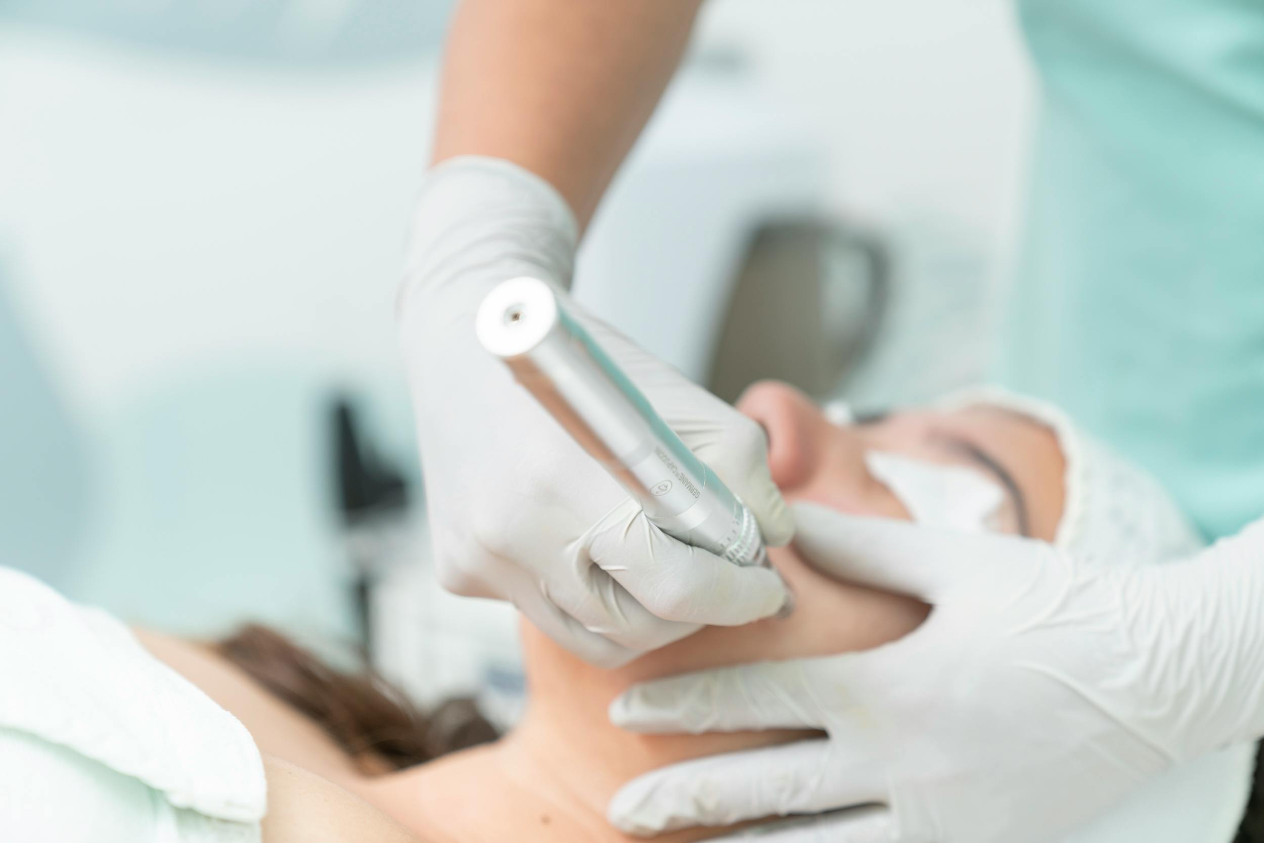
Endodontics is a specialty branch of dentistry that examines, diagnoses, and microscopically treats diseases of the pulp (the tooth nerve and vascular bundle) located inside the tooth, as well as the hard and soft tissues surrounding the tooth root. Etymologically derived from the combination of the Greek words “endo” (inside) and “odons” (tooth), this branch serves as the most critical line of defense for retaining natural teeth in the mouth by adapting microscopic bioengineering principles to dentistry.
Endodontic applications carried out within Panorama Ankara do not consist of a mechanical canal shaping procedure alone. Under advanced dental microscopes, the side canals and micro-architecture of root canals, which cannot be noticed by the naked eye, are mapped out. Our fundamental clinical philosophy is not to fill the tooth like an inanimate object, but to functionally retain even highly infected teeth in the mouth by re-establishing the biological balance with the periapical (around the root apex) bone tissue.
2. Which Tooth Problems Does Endodontics Deal With?
The endodontic specialty undertakes the treatment of bacterial, traumatic, or structural destructions that bypass the integrity of the dental hard tissues (enamel and dentin) and infiltrate into the pulp chamber. Accordingly, the clinical problems dealt with are listed as follows:
- Irreversible Pulpitis: Acute or chronic inflammations caused by bacterial toxins where the tooth nerves exceed their boundary of self-healing.
- Pulp Necrosis and Gangrene: The death of the vital tissue inside the tooth as a result of the blood supply being cut off, and its putrefaction due to the invasion of microorganisms.
- Acute and Chronic Periapical Abscesses: The clinical picture where the infection in the root canal leaks out of the root apex, leading to localized inflammatory accumulations and swellings in the jawbone.
- Dentin Hypersensitivity: Deep hard tissue erosions where the pulpal tissue is neurologically overstimulated.
- Root Resorptions: The initiation of the tooth root melting from the inside (internal) or outside (external) due to cellular anomalies or chronic traumas.
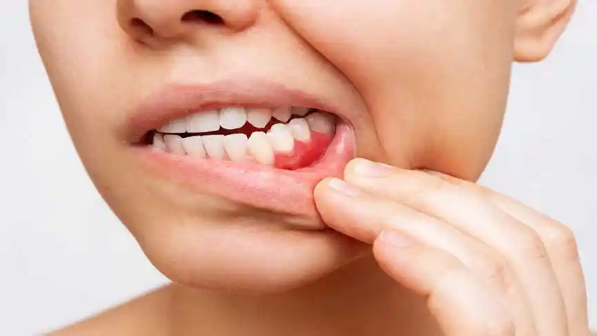
- Combined Periodontal-Endodontic Lesions: Complex tissue destructions where gum disease and tooth nerve infection merge on the root surface.
3. In Which Situations is Root Canal Treatment Applied?
Root canal treatment (Endodontic treatment) is applied to prevent tooth loss in all situations where the pulp tissue of a tooth cannot biologically maintain its vitality or is colonized by microorganisms. The clinical scenarios where this surgical intervention is mandatory are as follows:
- Deep Interdental and Surface Cavities: In cases where the decay layer completely dissolves the dentin thickness and opens directly into the pulp chamber.
- Prosthetic Preparations and Excessive Reductions: In cases where the pulp chamber becomes exposed or is subjected to excessive thermal stress during tooth reductions performed for prosthetic purposes (crowns, etc.) or aesthetic corrections.
- Structural Crack and Fracture Lines: In deep vertical or horizontal micro-cracks that initiate from the dental crown and extend toward the root surface, disrupting the pulp integrity.
- Failed Old Restorations: In cases where secondary cavities developing over time underneath deep fillings infect the pulp.
- Intraosseous Chronic Lesions: In cases where, even if there is no distinct decay in the tooth, the tooth nerve dies silently due to a past trauma and initiates bone resorption (a lesion) at the root apex.
4. How is an Endodontic Examination Performed?
Since the internal structure of root canals cannot be viewed directly, the endodontic examination process applied at Panorama Ankara clinics is managed with a scientific workflow that combines digital and clinical diagnostic tests:
Evaluation of Subjective Symptoms: The patient’s description of pain (spontaneous pain, throbbing, factors initiating the pain) is listened to chronologically.
Clinical Vitality Tests: Special cold sprays (Endo-Ice, -50°C) or hot stimuli are applied to measure the tooth’s response to thermal changes. When necessary, Electric Pulp Test (EPT) devices, which numerically measure the electrical excitability of the nerve fibers inside the tooth, are utilized.
Physical Touch Tests (Percussion and Palpation): Gentle pressure is applied to the tooth and the gum region corresponding to the root apex to test whether the infection has spread to the jawbone and periodontal fibers.
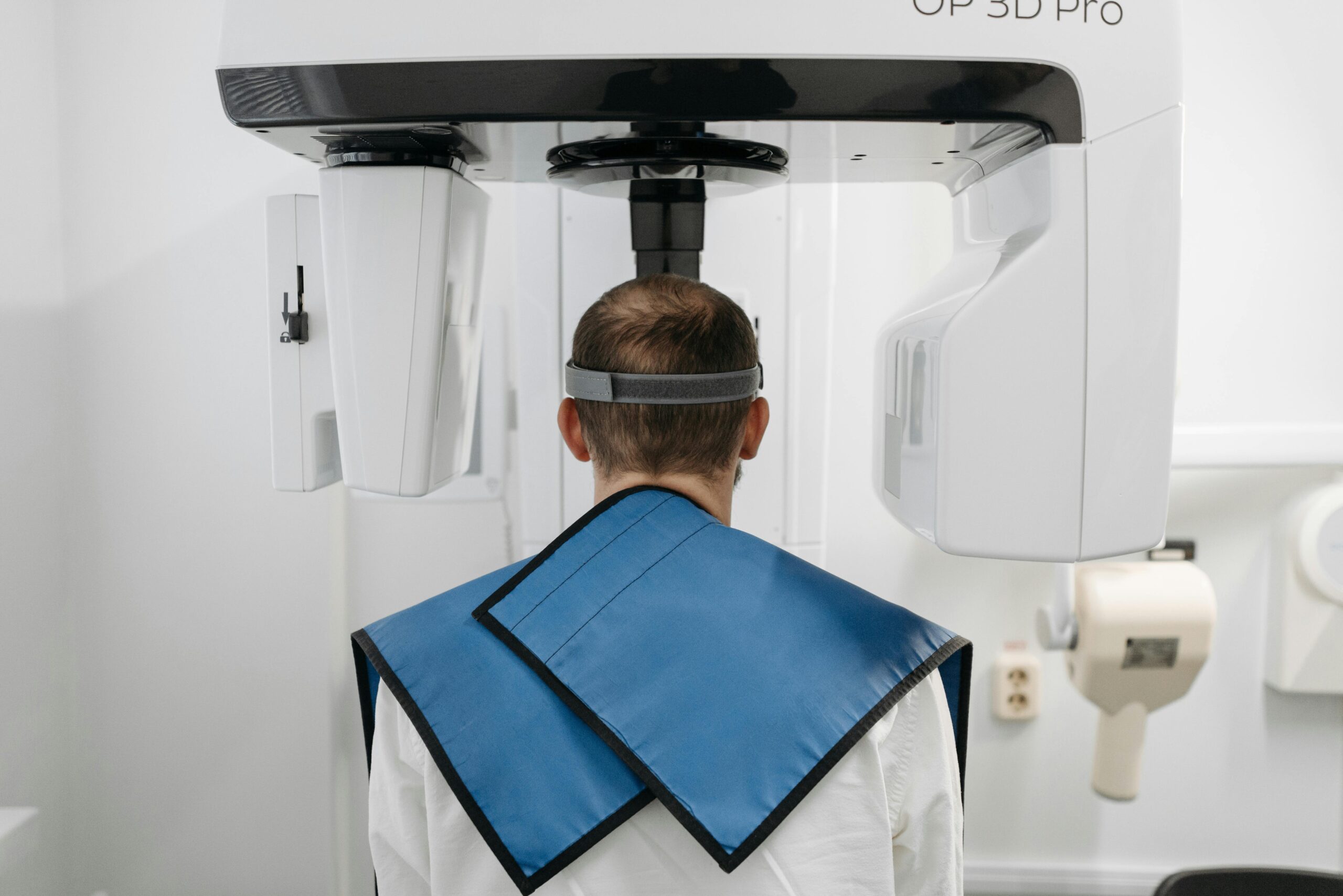
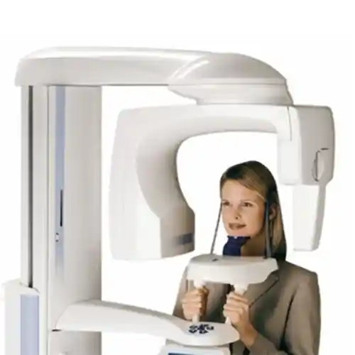
3D Digital Radiology: In addition to traditional two-dimensional periapical X-rays, high-resolution CBCT (Computed Tomography) scans providing millimetric cross-sections are performed, particularly to identify anatomical variations (C-shaped canals, fourth canal/MB2, etc.) and root fractures in multi-canalled molar teeth.
5. How is Root Canal Treatment Performed?
Modern endodontics is a microscopic engineering process where mechanical cleaning and chemical disinfection are carried out simultaneously. At Panorama Ankara, root canal treatment is brought to life under microscopic vision with the following technical steps:
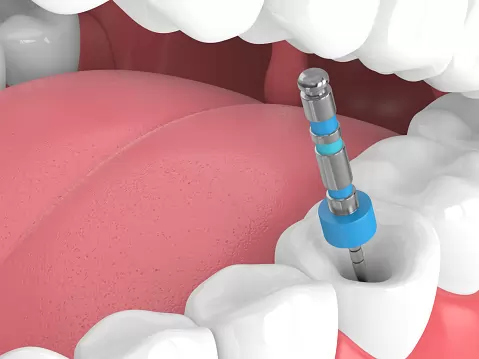
First, the tooth and surrounding tissues are completely desensitized with local anesthesia. Subsequently, absolute isolation from the oral environment is ensured by fitting a rubber dam over the tooth. After the access cavity is opened, the entry orifices of the root canals are identified under a dental microscope. With the help of electronic apex locator devices, the anatomical length (working length) of the root canals is measured millimetrically.
With new-generation nickel-titanium (NiTi) rotary instrument systems, the root canals are shaped into a conical form, remaining loyal to their anatomical outlines. During shaping, special chemical disinfectant solutions (Sodium Hypochlorite, EDTA) are introduced into the canals, and these solutions are activated with ultrasonic or laser waves to eradicate all bacteria in the micro-canals. Finally, the cleaned canals are three-dimensionally and hermetically filled with biocompatible sealers and a natural thermoplastic material called gutta-percha.
6. What Are the Symptoms Indicating the Need for Root Canal Treatment?
The clinical symptoms indicating that a tooth needs a root canal treatment differ according to the level of the pathology. The most distinct indicators are as follows:
- Unprovoked Night Pains: Severe, throbbing pains that initiate suddenly without any stimulus, triggered by the increase in head blood pressure when shifting to a lying position in the evening, and demonstrate resistance to painkillers.
- Prolonged Thermal Sensitivity: The lingering of an aching sensation in the tooth for minutes (sometimes hours) after consuming a cold or hot food, even if the food is removed from the mouth.
- Acute Sensitivity During Chewing: A sudden pain sensation similar to an electric shock radiating toward the root apex when pressure is applied to the tooth or when encountering a hard food.
- Fistula (Abscess Pimple) Formation: The inflammation at the root apex penetrating the bone to emerge at the gum margin in the form of a small pimple, introducing a periodic bad taste into the mouth.
- Crown Discoloration: The crown color becoming matte, turning gray, or shifting to dark brown as the tooth loses its vitality as a result of trauma or internal infection.
7. What is Tooth Pulp and Why is it Treated?
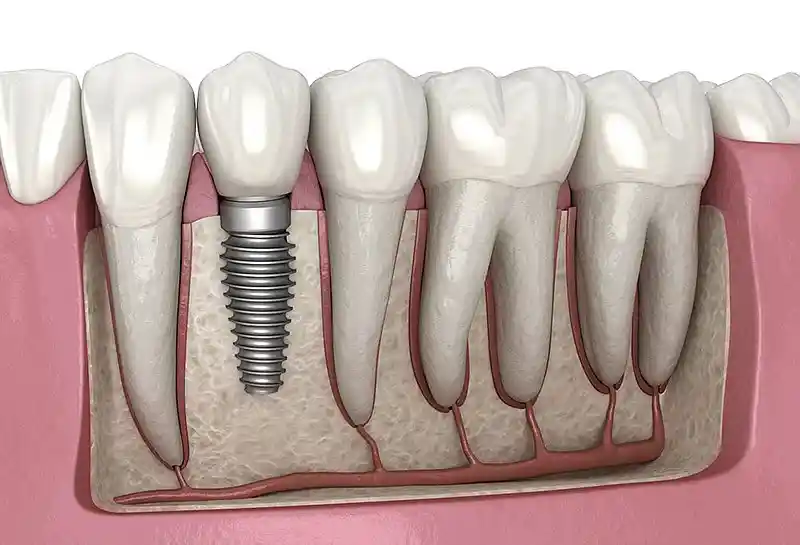
The dental pulp is the specialized connective tissue that gives vitality to the tooth, located beneath the enamel and dentin layers of the tooth within the space that extends from the pulp chamber in the crown portion down to the root apices. The pulp consists of a rich network of blood vessels, lymph vessels, nerve fibers (Raschkow plexus), and odontoblast cells capable of granting new dentin tissue to the tooth. Undertaking vital tasks during the developmental period of the tooth, the pulp primarily serves as a sensory warning and defense mechanism after development is completed.
When bacterial infections occurring in the pulp are left untreated, the inflammatory edema inside this closed box increases the pressure within the tissue. Because small vessels are crushed under high pressure, the nutrition of the pulp is impaired and the tissue is rapidly driven into necrosis (death). The necrotic pulp becomes an uncontrolled breeding ground for pathogenic microorganisms. If this space is not cleaned endodontically, the infection leaks into the jawbone through the openings at the root apices (foramen apicale), causing bone resorption, cyst formations, and even focal infection sites that can spread through the bloodstream to the entire body.
8. In How Many Sessions is Root Canal Treatment Completed?
The number of sessions for a root canal treatment varies depending on the biological condition of the tooth at the moment of the operation, the chronicity degree of the infection in the canals, and the health of the periapical tissues:
Treatments Finishing in a Single Session: In situations where the tooth nerves are vital (acute inflammatory states), no bone destruction (a lesion) is present at the root apex, and the infection has not yet spread widely to the root canals, the treatment can be successfully completed within a single session (in approximately 45-60 minutes).
Treatments Finishing in Multiple Sessions: In chronic cases where the tooth has lost its vitality, active inflammatory or oozing discharge comes from the canals, or large cystic structures or abscesses are present at the root apex, the treatment requires multiple sessions. In the first session, the canals are cleaned, and antiseptic medications based on Calcium Hydroxide or new-generation Bio-Ceramic materials are placed inside the canal to wait for 7-14 days. After ensuring that the infection is completely dried up, the permanent filling stage is initiated in the second or third session.
9. What is Retreatment (Repeat Root Canal Treatment)?
In cases where a root canal treatment performed in the past fails due to various technical or biological reasons, the procedure of removing the old canal filling materials, recleaning, reshaping, and refilling the root canals is called Retreatment (Canal Renewal).
The reasons for a root canal treatment failure include the presence of additional canals overlooked during the initial treatment, resistant bacterial species such as “Enterococcus faecalis” remaining in the canal due to insufficient irrigation, or oral fluids leaking back into the root canal (coronal micro-leakage) as a result of the upper restoration (the crown) leaking over time. In the retreatment procedure, old hardened fillings are completely removed from the root under microscopic view with the help of special solvent agents and ultrasonic tips. The canals are redisinfected at a micro-level, filled with new biomaterials, and the tooth is saved.
10. How is a Tooth Protected After Root Canal Treatment?
Because a tooth undergoing root canal treatment loses its vital vascular structure, it loses its elasticity modulus over time, its water content decreases, and it takes on a relatively more brittle (fragile) structure. Furthermore, the removal of extensive tooth structure along with the decay to perform the treatment reduces the structural resistance of the tooth. Therefore, protecting the tooth mechanically is highly critical:
In extensive material losses, performing standard composite fillings alone can lead to vertical cracks forming in the tooth walls during chewing. When a tooth fractures vertically, it unfortunately must face extraction. In order to protect the tooth from this risk, support is drawn from glass fiber-reinforced post (Fiber-Post) systems that are placed into the root canal and distribute the force evenly along the root. The superstructure of the tooth, on the other hand, must be armored with monolithic zirconium crowns resistant to vertical forces or computer-manufactured porcelain overlay restorations that protect the sturdy walls of the tooth.
11. What is Root End Resection (Apicoectomy)?
Apicoectomy (Endodontic Surgery) is the procedure of surgically cutting the root end and completely scraping the inflammatory tissue, cyst, or granuloma structure out of the bone in that region, in cases where the chronic infection at the root apex does not heal despite normal or repeated (retreatment) root canal treatments.
The procedure is carried out microscopically under local anesthesia. A small window is opened in the gum to reach the bone tissue at the root apex. Measurements are taken, and the root surface is reduced. The infected root tip is removed, and a retrograde preparation is shaped at the root end. The apex is filled with a bio-compatible material, sealing the root canal against bacteria. The reflected gum is closed with stitches. Healing takes place rapidly, and the tooth is preserved in the mouth.
12. Which Treatment Methods Can be Applied to Teeth with Bone Loss?
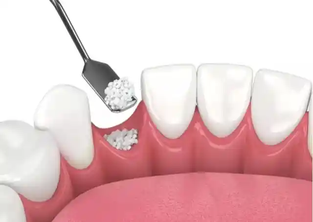
Resorptions occurring in the jawbone due to periodontitis are not always horizontal and uniform. Sometimes vertical, crater-like “infraosseous defects” form along the root of the tooth. In such vertical bone losses, Guided Tissue Regeneration (GTR) techniques are applied to biologically reproduce lost tissues.
In the reflected surgical field, biocompatible bone grafts (bone powders) are placed into the region with bone resorption. Collagen membranes (biological barriers) that serve as a barrier are laid over the grafts to prevent gum cells from infiltrating the region and to grant time to slow-growing bone cells. When necessary, enamel matrix derivatives (bio-active proteins) are injected into the site to enhance cell stimulation. This reduces the shaking of the tooth and functionally reconstructs the supporting bone structure.
13. Why is Gum Health Important Before Implants?
Although dental implants connect mechanically to the jawbone, the only barrier protecting them from bacteria in the oral environment is the surrounding gum tissue, just as it is with natural teeth. Placing an implant in a patient who has active gum inflammation or periodontitis directly endangers the biological future of the treatment.
Pathogenic bacteria in gum pockets rapidly migrate to the implant surface post-operation, leading to irreversible infections termed peri-implantitis that rapidly resorb the bone surrounding the implant. Periodontal ligament fibers present in the roots of natural teeth do not exist around implants; therefore, implants are far more vulnerable to infection compared to natural teeth. Eradicating infection completely from all intraoral tissues is an absolute prerequisite for a secure implant success.
14. What Are the Stages of Periodontal Treatment?
Periodontal treatment processes at Panorama Ankara are carried out in the following chronological order, adhering to the biological calendar of tissue healing:
- Hygiene Phase (Phase I): Scaling, curettage applications, correction of faulty and overhanging fillings, teaching correct brushing techniques to the patient.
- Re-evaluation Phase: The patient is recalled 4-6 weeks after the initial treatment, and all periodontal measurements are repeated. The healing response of tissues is examined, and the need for a surgical phase is decided upon in this stage.
- Surgical Phase (Phase II): Flap operations for deep pockets, grafting procedures for bone resorptions, or plastic surgery operations for aesthetic gum recessions.
- Supportive Periodontal Therapy (Phase III – Maintenance): Regular professional checks and maintenance cleanings performed at periods of 3, 6, or 12 months depending on the severity of the case to prevent recurrence of the disease.
15. What Should be Considered After Periodontal Treatment?
Critical elements our patients need to pay attention to for tissue stabilization and a comfortable recovery following gum treatments are as follows:
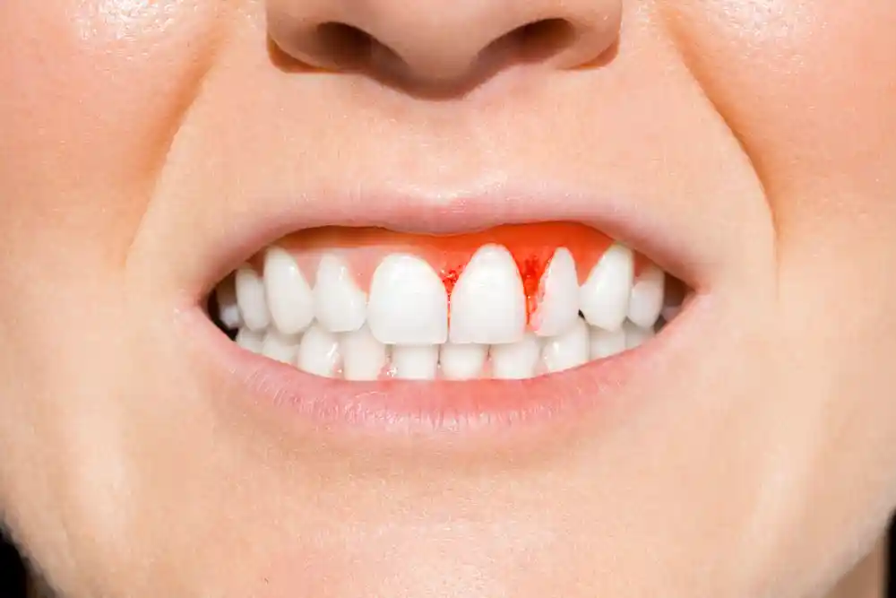
- First 24-Hour Care: It is normal to observe a slight pinkness in saliva or bleeding in the form of oozing after treatment. The mouth should not be continuously rinsed and spitting should be avoided. Excessively hot foods must be avoided on the day of the operation.
- Post-Operative Sensitivity: After calculus is cleaned and root surfaces are exposed, hot-cold sensitivities that can last for a few weeks may develop in teeth. This condition is temporary and is alleviated with sensitivity-relief toothpastes or fluoride varnishes applied in the clinic.
- Smoking Restriction: Smoking causes constriction (vasoconstriction) in peripheral vessels due to the chemicals it contains. This directly stops healing by reducing the blood flow and oxygen amount going to the wound site, leading to surgical failure.
- Mechanical Cleaning: Regions with surgical stitches should not be brushed normally during the first few days; they should be gently disinfected with antiseptic mouthwashes containing chlorhexidine prescribed by the dentist.
16. What Can be Done to Protect Against Gum Diseases?
Protecting and maintaining periodontal health depends on the daily care the patient shows at home rather than the treatments performed in the clinic. The basic prevention rules are as follows:
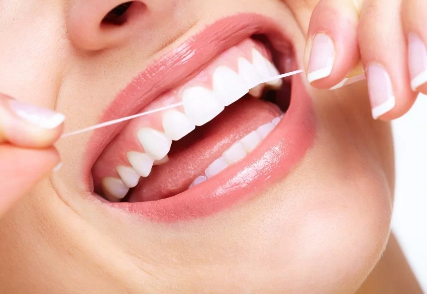
Brushing should be performed at least twice a day, with a soft-bristled toothbrush placed at a 45-degree angle to the gum line, using sweeping or circular motions from the gum toward the tooth (Bass Technique). The joining points of two teeth (interdental areas) where a toothbrush cannot reach are the locations where bacterial plaque accumulates most densely and diseases initiate. These areas must definitely be cleaned every day with the help of interdental brushes of appropriate thickness or dental floss. Oral irrigators, though they do not completely replace mechanical dental floss, provide great support in removing food debris by washing inside the pockets.
17. What Are the Factors Affecting Gum Health?
Gum health is in direct interaction not only with local oral hygiene but also with the general systemic balance of the body and lifestyle habits. These factors are:
- Diabetes Mellitus (Sugar Disease): There is a bidirectional relationship between diabetes and periodontal diseases. Tissue destruction progresses much faster in uncontrolled diabetic patients; simultaneously, infection in the mouth makes blood sugar regulation difficult.
- Hormonal Changes: Hormonal fluctuations during adolescence, pregnancy, and menopause increase the sensitivity of the gum against bacteria, paving the way for a “pregnancy tumor” (epulis) or severe gum enlargements.
- Genetic Predisposition: Even if some individuals pay attention to oral hygiene, they may be more prone to aggressive periodontitis forms due to immunological inheritance coming from the family.
- Medications Used: Certain blood pressure medications (calcium channel blockers), epilepsy medications (phenytoin), and immunosuppressive medications (cyclosporine) cause pathological overgrowths (hyperplasia) in the gums.
18. What Are the Factors Affecting the Success of Periodontal Treatment?
Clinical biological success rates are highly elevated in periodontal treatments; however, keeping this success permanent is possible by fulfilling certain conditions:
Patient Cooperation (Hijyen Continuity): Performing the treatment in the most perfect manner remains meaningless if the patient does not clean the plaque in the subsequent process. Periodontal success is a chain of responsibility carried out jointly by the dentist and the patient.
Status of Smoking: In patients who continue heavy smoking, the integration rates of surgical grafts with tissue and root coverage rates drop dramatically.
Regular Maintenance Appointments: Since periodontal diseases are of a chronic character, they tend to relapse (recur). Arriving without delay for the 3- or 6-month maintenance checks specified by the dentist ensures that new plaque accumulations at the initial level are cleaned immediately, protecting the main success for a lifetime.
19. What Are the Factors Affecting Periodontology Treatment Prices?
Periodontal treatment budgets are shaped individually according to the prevalence of the disease inside the mouth and the complexity of the surgical interventions needed. Elements affecting the price index are:
- Level of the Disease and Number of Teeth Involved: The session and workmanship cost of a localized curettage procedure involving only a few teeth versus a widespread advanced periodontitis treatment covering the entire lower and upper jaws (28-32 teeth) is different.
- Type of Surgical Intervention: The price dynamics change between standard non-surgical curettage prices versus flap operations where the gum is surgically reflected open or mucogingival plastic surgery operations.
- Use of Biological Materials: The amount of bone grafts (powders) placed into the defect in treating bone resorptions, the quality of the collagen membranes utilized, and the presence of reconstructive protein agents directly affect the material cost.
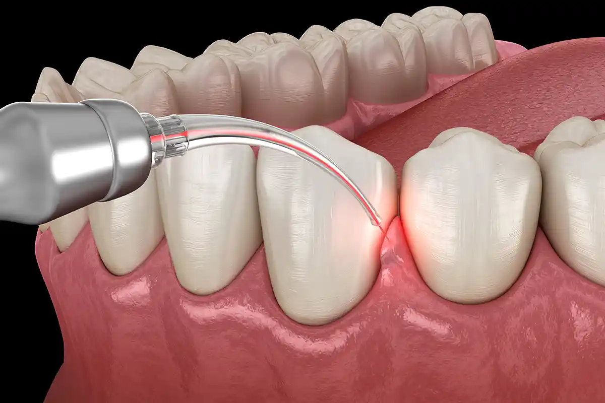
- Laser and Advanced Technology Integration: Utilizing dental laser systems included in operations for sterilization and tissue biostimulation purposes within the clinical workflow is among the factors shaping the price.
As Panorama Ankara Oral and Dental Health Polyclinic, we protect your gum and bone health, which is the main support of your teeth, with the most modern scientific protocols. Remember, even the healthiest teeth can only survive on a strong and inflammation-free foundation.