What Are Graft, Membrane, and PRF Applications?
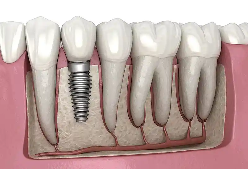
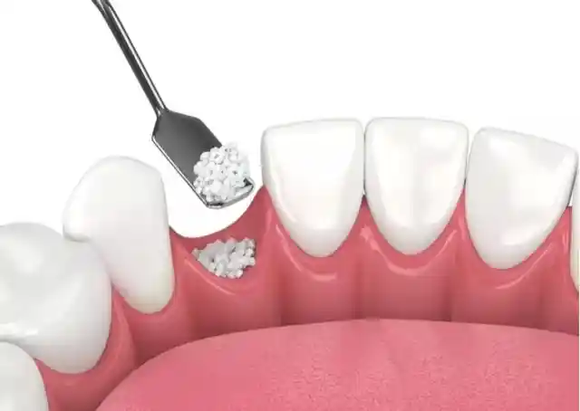
Graft, membrane, and PRF applications represent a triple biomaterial combination protocol in modern oral and maxillofacial surgery that mimics and accelerates the self-repair mechanisms (regenerative capacity) of tissues. Severe resorption (atrophy) occurs in the jawbone and surrounding mucosal bed as a consequence of tooth extractions, chronic cysts, or heavy periodontal diseases. Graft (bone powder) acts as a three-dimensional structural scaffold for new bone cells to adhere to within these resorbed zones. Membrane serves as a protective barrier preventing soft tissue cells from infiltrating into the bone grid. PRF (Platelet Rich Fibrin) functions as a biological engine pumping millions of living cells and growth factors that initiate healing into the surgical field. When these three elements unite, hard and soft tissues in the jaw region are smoothly reconstructed at a cellular level.
- Foundation of Tissue Engineering: Triggering the body’s own osteogenic (bone-forming) potential with the support of biological raw materials.
- Triad Dependence: Sinergy and coordination between bone graft, barrier membrane, and the patient’s own blood components.
- Implantological Infrastructure: A technique to bring inadequate and thin jawbones to a volume capable of safely supporting titanium implant screws.
In Which Situations is Graft Application Preferred?
Jawbone grafting (bone graft injection) is preferred when the bone hard tissue volume remains anatomically and millimetrically inadequate for implant placement, correcting jaw asymmetries, or halting gum recession. Following a tooth loss, the bone tissue in that region undergoes rapid resorption because it loses its function, triggered by gravity and facial pressures. In scenarios where bone thickness falls below 4-5 mm, the implant screw might breach the bone boundaries, inducing paresthesia risks; grafting is deployed to build safe surgical corridors along these risky margin lines.
The primary clinical situations where graft applications are conclusively selected in surgical practices include:
- For socket preservation purposes to prevent the collapse of the extraction space (alveolar socket) and maintain its volume,
- In sinus lifting operations executed to increase vertical bone height as a result of maxillary sinus cavities sagging downward in the upper jaw molar region,
- For filling wide anatomical spaces and cavities developing within the jawbone post-chronic cysts, tumors, or trauma,
- During horizontal ridge split procedures where the jawbone becomes excessively thin transversely due to long-term edentulism.
What is Membrane Application and How is it Used?
Membrane application, within the framework of “Guided Bone Regeneration” (GBR) guidelines, is the process of utilizing sterile and bio-compatible medical membranes that protect and cover the surgical area packed with bone grafts like a cap. In human anatomy, soft tissue cells (epithelial cells) divide and migrate approximately 10 times faster than bone-forming osteoblast cells. If the area packed with grafts is not covered with a barrier, gum cells rapidly infiltrate that space to pack between the bone powder, completely obstructing the ossification process. While mechanically blocking gum cells from moving downward, the membrane grants time and a sheltered chamber for the bone graft in the sub-layer to smoothly evolve into actual bone tissue (preserving homeostatic balance).
- Resorbable Membranes: Typically manufactured from bovine or porcine collagen; they dissolve on their own post-operatively without creating a secondary surgical removal burden.
- Non-Resorbable Membranes: Titanium-reinforced or reinforced artificial membranes; they protect the bone like a mold during vertical ridge reconstructions and are cleared from the line months later.
- Barrier Locking: The phase of stabilizing the membrane onto the jawbone with the aid of micro-surgical pins (screws) to prevent graft micro-leakage.
What is PRF Application?
PRF (Platelet Rich Fibrin) application is a 100% autologous (natural) cell enrichment technology in modern regenerative dental surgery that utilizes the patient’s own biological healing power at a maximum coefficient. At the moment of operation, a small volume of blood is drawn from the patient’s arm vein into sterile tubes and spun in high-speed centrifuge devices utilizing proprietary revolution formulas, without adding any chemical additives. Once the rotation cycle concludes, red blood cells precipitate to the base of the tube while an intense “living yellow fibrin clot gel” (PRF) separates at the upper tier. This gel is exceptionally rich in connective tissue fibers, growth factors (VEGF, TGF-beta), and leukocyte cells; when placed onto the wound site, it accelerates tissue repair velocities at a microscopic level.
“PRF is a miraculous biological blanket manufactured from the patient’s own blood; the exact second it is draped over the wound site, it recruits stem cells there to compress the healing timeline by half.”
Who Are Graft, Membrane, and PRF Applications Suitable For?
These advanced technological regenerative surgical protocols are medically suitable for any adult patient who wishes to undergo implant treatment but encounters bone inadequacy barriers, exhibits cyst-induced damages in the jawbone, and aims to support surgical steps completely with natural healing velocities. Because the system positions the patient’s own blood components (PRF) at the center, its bio-compatibility profile resides at a maximum level. However, individuals managing uncontrolled severe diabetes, those who have recently received radiation therapy (radiotherapy) to the head and neck region, or patients utilizing potent bisphosphonate group medications that block bone metabolism are not suitable candidates for these surgical bone construction processes.
| Patient Profile / Bone Status | Surgical Suitability Status | Expected Bone and Cellular Response |
|---|---|---|
| Those with Bone Resorption Due to Chronic Edentulism | Fully Suitable / Primary Indication | Provides total osseointegration for implant screws by scaling up the width and height of the bone. |
| Sensitive Patients Requesting Fast Healing and Minimal Swelling | Fully Suitable / PRF Doping Mandatory | Lowers post-operative pain and micro-edema risks by 60% thanks to PRF membranes. |
| Heavy Smokers Continuing Intense Tobacco Consumption | Risky / Controlled Suitability | Cigarette smoke constricts capillaries; it radically downscales graft adherence success and ossification lifespans. |
| Patients with Active Jawbone Inflammation (Osteomyelitis) | Not Suitable (Infection Phase) | Grafting steps are postponed until active intraosseous microbial infection is completely dry. |
What Are the Materials Used in Graft Applications?
Materials utilized in bone grafting operations are classified into four primary groups according to the biological raw material sources they are derived from and their histological mechanisms of action within the jawbone. Which material will be selected is calibrated by the specialist clinician according to the dimension of the defect, the patient’s budget parameters, and the targeted ossification velocity. All materials are proprietary products that have passed medical-grade sterilization audits.
- Autogenous Grafts (Gold Standard): Living bone blocks harvested by scraping from the patient’s own body (symphysis or ramus region); it stands as the fastest ossifying type because it houses vital cells.
- Allografts (Human-Derived): Freeze-dried sterile human bone grafts obtained from medical bone banks.
- Xenografts (Animal-Derived): Safe bone grafts harvested from bovine or porcine sources, cleared of organic proteins leaving strictly a sterile calcium mineral skeleton; it stands as the most frequently used type.
- Alloplastic Grafts (Synthetic): Artificially manufactured calcium phosphate or hydroxyapatite-based bio-ceramic crystals in laboratory environments.
How Do Membrane Applications Support Bone Healing?
The supporting power of barrier membranes over bone cellular architecture relies on the principles of “selective cell filtration” and “surgical field stabilization.” When draped over the bone graft, the membrane isolates that region from the pressure exerted by intraoral mobile expression muscles, preventing bone particles from shifting (migration). Thanks to its microscopically porous structure, it intercepts harmful cell infiltration while allowing blood flow, nourishing fluids, and oxygen molecules critical for ossification to seep smoothly into the field; this safeguards sub-tissue homeostatic balances to verify smooth bone formation.
- Particle Isolation: Preventing bone grafts from scattering via mucosal movements and escaping into facial spaces.
- Angiogenesis Guidance: Facilitating micro-capillaries advancing beneath the membrane to nourish the newly forming bone mass.
How Does PRF Application Contribute to the Healing Process?
The revolutionary contribution of Platelet Rich Fibrin (PRF) gel over the healing schedule stems from the capacity of its constituent cytokines and Growth Factors to execute linear and cumulative release into the tissue bed. The PRF network positioned across the surgical field continues to project tissue repair signals uninterruptedly for an average of 7 to 14 days. These signals keep macrophage cells in the region active to accelerate wound debridement, stimulate fibroblasts to weave new gingival attachments, and escalate bone cell division velocities to elevate post-operative healing comfort to the peak.
- Rapid Epithelialization: Closing the gum incision lines with a sutureless seal via new epithelial cells during the initial 24-48 hours.
- Edema and Pain Absorption: Maintaining the post-operative swelling coefficient at a minimum boundary thanks to its anti-inflammatory cytokine configuration.
Why Are Graft and Membrane Applications Critical in Implant Treatment?
Long-term clinical success in dental implant surgeries directly depends on maintaining the bone tissue enveloping the titanium screw as a cortical shield at least 1.5 – 2 mm in thickness; graft and membrane stand as the most uncompromising guarantee of this reinforcement. If an implant is forcefully driven into a thin or resorbed jawbone, the implant threads uncover in a short turnaround, the gums turn purple, and an insidious implant inflammation termed “peri-implantitis” is triggered, causing the screw to loosen and shed. Graft and membrane thicken thin bone walls to ensure titanium surfaces lock 100% with bone cells (osseointegration), laying the foundation for the implant to remain in the mouth for a lifetime.
- Horizontal and Vertical Bone Gain: Ensuring the implant remains fully embedded and safe within the bony matrix.
- Aesthetic Pink Line Support: Establishing flawless pink aesthetic harmony at the crown neck region by preventing recession of the bone-nourished gums.
How is Graft Application Executed in Bone Inadequacy?
Grafting in bone inadequacy cases is a precise oral surgery procedure executed under local anesthesia blocks in polyclinic operating rooms subject to absolute sterilization guidelines. First, the gum tissue in the resorbed region is relaxed along a smooth line using a scalpel or cautery and lifted from the bony membrane (periosteum) to reflect a flap. Following micro-perforations drilled onto the exposed flat bone surface to stimulate bleeding, paste or granular graft materials are millimetrically positioned across the defect area; the operation is finalized by draping a membrane over the grafts, locking it with PRF sheets, and closing the gum tissue tension-free with primary sutures.
- Cortical Perforation: The phase of micro-drilling the bone to enable stem cell infiltration from the underlying vital spongy bone into the graft field.
- Block / Granule Selection: The calibration of selecting screw-retained bone blocks (onlay grafts) in extreme resorptions, versus granular powders in superficial resorptions.
How is PRF Application Used in Gum and Soft Tissue Treatments?
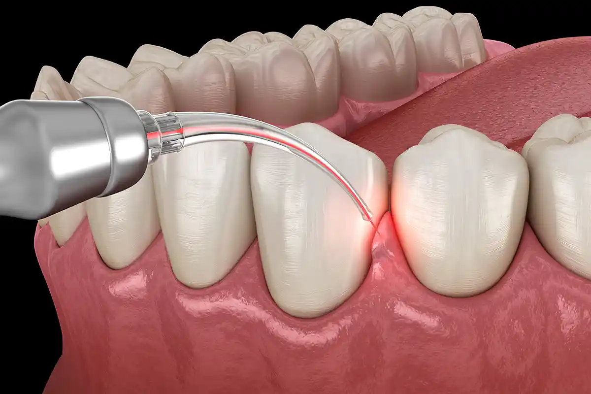
The application scope of PRF gels in soft tissue and periodontics surgeries extends priceless comfort for patients who do not wish to have an extra tissue piece (graft) harvested from their palate. In gum recession treatments or black triangle camouflage, thick PRF membranes derived from the patient’s blood are stretched and sutured over the receded root surfaces. Thanks to the high collagen and leukocyte power it incorporates, PRF directly stimulates the weaving of a new and vital gingival margin in that region; because it terminates the obligation to harvest tissue from the palate, it eliminates the patient’s home post-operative wound-site sting burden.
- Exposed Root Coverage Power: Isolating the root cementum layer with a vital fibrin shield in gum recessions.
- Zeroing Palatal Trauma: The relationship of maximizing the post-operative comfort coefficient since no donor site is opened.
Which Evaluations Are Executed Prior to Graft, Membrane, and PRF Applications?
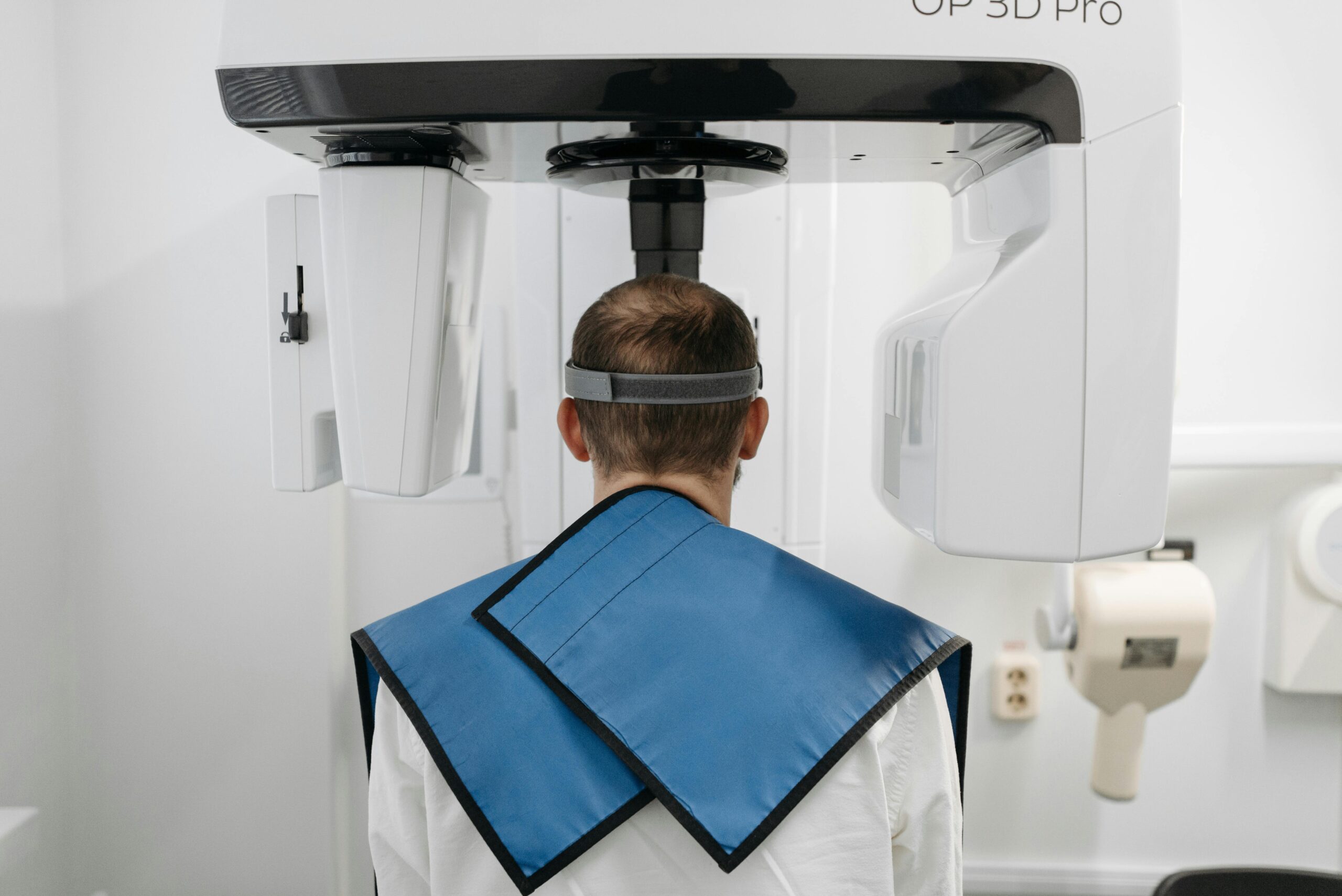
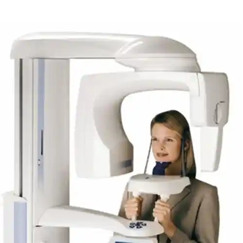
The clinical evaluation conducted before proceeding to the regenerative operation phase is the most vital analysis step millimetrically charting the surgical route and verifying that aesthetic success remains stable without recurrence. To transcend the limitations of two-dimensional classic X-rays, the clinician requests a CBCT 3D dental tomography scan from the patient first. Measuring the width, height, density, and distance of the bone to anatomical nerve canals at a 1:1 scale within the tomography interface, the exact required graft volume (injektör dosage in cc) is rationally determined before the surgery.
- Bone Defect Morphology: Selecting the most accurate material blend according to the layout of the resorption (one-walled, multi-walled defect classification).
- Systemic Analysis Audit: Screening the patient’s HbA1c (blood sugar) values and bone metabolism velocity parameters through laboratory tests.
- Oral Hygiene Scoring: Auditing preliminary prophylaxis checks since grafting cannot be executed in oral cavities housing dental calculus and inflammation.
How Are Graft, Membrane, and PRF Applications Planned?
Planning this triple regenerative protocol is executed across a staged timeline based on the chronicity of bone loss, the patient’s general phototype biology, and the criteria of whether simultaneous implant placement (immediate placement) can be achieved. Hasty steps are avoided during workflow planning; because the evolution of bone grafts into the patient’s own vital bone biologically demands an ossification timeline spanning months. In planning, the maturation phases of bone gain are meticulously woven into the patient’s social calendar.
- Stage 1 (Preparation Phase): The process of cleaning dental calculus, filling cavities, and verifying a sterile operational foundation.
- Stage 2 (Surgical Phase): Centrifuging blood, separating PRF, and millimetrically transferring the graft and membrane combination to the jaw.
- Stage 3 (Ossification Waiting Period): The phase of leaving the tissue at rest untouched for 4 to 6 months for the graft to evolve into genuine bone.
- Stage 4 (Implantation / Prosthesis): Placing titanium screws into the newly generated hard bone bed or mounting the superstructure crown restoration.
How Long Do Graft, Membrane, and PRF Applications Take?
Because they incorporate millimetric suturing and stabilization steps demanding a deep level of concentration from the surgeon, graft, membrane, and PRF combination operations require more meticulous time management regarding active chairside session duration compared to a standard tooth extraction. The active surgical phase of the process shapes smoothly between 30 to 60 minutes per session, depending on the volume of the bone defect to be intervened with and the width of the field; the blood spinning (centrifuging) stage executed during the session is advanced concurrently, imposing no additional time burden onto the schedule.
- Single-Tooth Site Grafting: A comfortable session lasting an average of 30 – 40 minutes, encompassing cleaning, graft placement, and PRF draping.
- Extensive Sinus Lifting / Block Bone Surgeries: An operative duration of approximately 60 minutes because it incorporates skeletal reshaping and pin stabilization steps.
Are Graft, Membrane, and PRF Applications Painful Procedures?
No, because these regenerative operation components are executed under potent local anesthetic solutions or computer-controlled digital anesthesia units engineered within modern medical aesthetics and jaw surgery standards, they conclusively do not cause even the slightest pain, ache, or sting to the patient during the session. Since nerve conduction gates are entirely locked starting from the moment of injection, you perceive strictly the gentle touch of instruments and water coolness throughout the operation; it is verified that you step out of the operatory room with a complete session comfort and psychological relief.
- Deep Anesthetic Blockade: The security of pre-operatively numbing the main nerve trunks nourishing the jawbone.
- Sedation Alternative: The luxury of managing the workflow with zero stress in a sedation (semi-sleep) mode for patients presenting with extreme chairside anxiety or panic attacks.
How Does the Healing Process Advance Following Graft, Membrane, and PRF Applications?
Although it incorporates surgical flap sutures, the post-operative tissue healing phase advances through steps exponentially faster, more painless, and predictable compared to conventional bone surgeries, thanks to the intense growth factor shield extended by PRF membranes. Experiencing a temporary micro-edema (swelling) in the cheek region during the initial 48 hours following surgery due to intraosseous stimulation and vascular stabilization steps is a completely natural physiological reaction exhibited by the extracellular matrix. The healing process flows along a linear path; at the conclusion of the first week, mucosal sutures reach a smoothness ready to be removed, and the ossification cycle in the sub-layer enters its maturation phase in insidious silence.
- First 48 Hours (Peak Edema Phase): The phase where swelling is most visible; this process is easily absorbed via intermittent cold pack compresses applied from outside the cheek.
- Days 7 to 10: Epithelial coverage concludes, sutures are cleared from the line without stinging during clinical check-ups, and cheek swelling subsides completely.
- Month 4 and Beyond (Ossification): Graft particles making way for vital, vascularized actual human bone to transition into a phase fully prepared for implantation.
What Should Be Considered Following Graft, Membrane, and PRF Applications?
To make the aesthetic and medical success of the regenerative bone operation permanent, prevent the displacement of graft particles packed underneath the tissue, and protect suture lines, the care discipline observed by the patient at home post-procedure is of vital importance. Because newly transferred bone grafts have not yet completely ossified during the initial days and are susceptible to leakage, full dedication to pressure and hygiene rules is a clinical requirement.
The medical rules that must be meticulously followed during the critical period post-procedure include:
- Intraoral fluids should be swallowed continuously and must not be vacuumed and spit out for the first 24 hours to preserve the clot balance (spitting triggers bleeding).
- Rigid, crusty, or granular foods must be cut for the first week to protect suture lines from mechanical tearing; a puree-like soft diet at room temperature should be maintained.
- In cases undergoing sinus lifting, nasal cleaning must not be executed by forceful blowing for at least 2 weeks to avoid scaling up upper jaw bone pressure and blowing out the grafts; the mouth should be kept open while sneezing.
- Sauna, hammam, solarium, and heavy sports activities that will escalate capillary pressure in the tissue to increase leakage and bruising risks must be postponed for at least 1 month.
What Are the Advantages of Graft, Membrane, and PRF Applications?
Among holistic maxillofacial restorations, this triple biomaterial combination stands as the most visionary surgical solution bringing patients who have lost hope in dental treatments due to bone resorption to permanent fixed teeth comfort. Thickening jawbones that have thinned down to a razor’s edge with a strength beyond non-surgical options shapes this methodology into one of the most popular and evidence-based tracks of modern implantology.
- Renders the Impossible Possible: It transports borderline cases, historically told implants cannot be placed due to bone absence, safely into a treatable phase.
- 100% Natural Cellular Acceleration: Ensures complete adaptation by zeroing out risks of the body exhibiting reactions against foreign materials, thanks to the deployment of PRF.
- Rapid and Comfortable Wound Closure: Drags post-operative infection, pain, and abscess complication coefficients down to a minimum boundary via the power of growth factors.
- Permanent Bone Infrastructure: The newly generated bone architecture functions as a permanent protective shield matrix surrounding the titanium screws for a lifetime.
Which Treatments Can Graft, Membrane, and PRF Applications Be Combined With?
Graft, membrane, and PRF technologies can be planned in an integrated combination alongside other medical surgical and restorative dental protocols to holistically elevate the face’s aesthetic architecture, bone rigidity, and masticatory function quality. While sub-tier bone foundations are reinforced via surgery, form losses in the upper tiers can be simultaneously treated with distinct technologies.
- Dental Implant Treatment: The chain of placing titanium screws either in the very same session alongside bone grafting or after the bone matures.
- Sinus Lifting (Sinus Elevation): Operations where the sagging sinus membrane is elevated and the cavity underneath filled with graft and PRF.
- Surgical Extractions of Impacted Teeth: The process of packing massive cavities remaining in the jawbone post-heavy wisdom teeth extractions with PRF to ensure rapid ossification.
What Are the Frequently Asked Questions About Graft, Membrane, and PRF Applications?
The most common practical anxieties arising in the minds of patients planning to recourse to these regenerative applications due to jawbone inadequacy, along with their uncensored medical responses, are presented below:
Will these bone grafts utilized cause tissue rejection or an allergic reaction in my body?
No, they absolutely will not. Xenograft (animal-derived) or allograft (human-derived) bone grafts utilized in modern clinical practices undergo thermal and chemical processing at exceptionally high temperatures in international laboratories; all organic proteins, cells, and DNA remnants within their configuration are completely purged, leaving strictly a sterile calcium mineral skeleton. Therefore, the risk of the body identifying this powder as a foreign enemy and initiating tissue rejection or an allergy reaction is medically zero; the bone integrates with full compliance.
Is fasting or special preparation required prior to surgery for the PRF gel derived from my blood?
No, there is no extra medical preparation involved requiring the patient to fast or follow a diet before giving blood for a PRF application. The exact second you enter the operating room following your normal routine examination, your blood is drawn into tubes from your arm vein by our nurse within seconds right before surgery initiates, and it is spun and prepared in the device while you are in the chair; the workflow is a comfortable clinical step that does not consume time.
Following the surgery, I feel bone graft particles leaking out into my mouth or meeting my tongue; is this normal?
Yes, experiencing small, chalk-powder-like granular particles leaking into your mouth when you spit or touch your tongue against the suture lines during the initial few days following surgery is completely normal and expected. A few surplus granules out of the thousands of micro bone grafts packed into the surgical field may seep through the suture margins into the oral cavity; this condition conclusively does not compromise the success or volume of the core bone mass inside, and there is no cause for concern.
How much time does it take for permanent teeth (implant-supported prostheses) to be fitted into the zone where graft and membrane were placed?
The evolution of placed bone grafts into the patient’s vital, vascularized actual bone tissue capable of bearing masticatory loads (ossification biology) requires a fixed waiting timeline averaging 4 to 6 months, depending on the patient’s age and the dimension of the defect. The grafts mature throughout this waiting timeline; once the duration concludes, a new 3D tomography is captured to verify bone quality, safely advancing to the assembly phase of titanium screws or porcelain crowns.
Can I return immediately to my normal social and professional life, to my work, on the day the operation is completed?
Yes, graft, membrane, and PRF applications are entirely local micro-surgical procedures that do not involve general narcosis and do not disturb consciousness or motor skills; therefore, you can comfortably step up from the chair the exact second the session concludes to resume your normal daily, academic, or professional life rhythm uninterrupted. Adhering to ice compress rules and avoiding hot foods strictly to manage the mild swelling that may form in the cheek on the first day is a comfortable detail that will not affect your practical life.
Who Are We? & Panorama Ankara Oral and Dental Health
Panorama Ankara is a licensed oral and dental health center extending institutional health assurances to its patients by combining an academic expert staff and the latest global bio-technological digital equipment under a single roof, particularly in oral, dental, and maxillofacial surgery, advanced implantology, orthodontics, periodontology, and aesthetic dentistry specialties. Attaching vital importance to keeping the bone within the mouth by reviving it at a cellular level through evidence-based medical disciplines—rather than condemning advanced jawbone resorptions, cyst defects, and sinus sagging barriers to the convenience of surgical extractions or removable dentures—our center operates original Graft, Membrane, and PRF-Supported Jawbone Restoration ecosystems, which deliver high success curves across clinical operational workflows, without compromise.
Across all radiological, scanning, design, and micro-surgical interventions executed under the roof of Panorama Ankara, absolute sterilization, high patient safety, transparency, and honesty principles are operated with uncompromising determination as per our charter. In our clinical practices, while analyzing the patient’s jawbone quality with 3D tomographies in millimetric sections, intelligent surgical electrocauteries that coagulate vascular integrity and proprietary centrifuge units that separate vital fibrin membranes from your own blood are meticulously applied under the supervision of our expert jaw surgeon cohort. Our goal is not to subject our patients to failure risks by forcing implants into inadequate bone tissues utilizing arbitrary frameworks; it is to analyze every individual’s facial golden ratio boundaries and skeletal bone density on a digital screen prior to the operation, achieving maximum session comfort, a sutureless comfort standard, and smooth fixed tooth restorations that you will utilize with unshakable confidence for a lifetime as if it were your own native bone. If you are tired of your missing teeth when looking in the mirror, yet postpone your treatments due to fear of bone resorption or pain, and wish to initiate your smart regenerative treatment workflow from start to finish with a scientific guarantee in an expert institutional center environment, you can safely protect your health and smile integrity by contacting the expert staff of Panorama Ankara for your detailed 3D tomographic analyses.