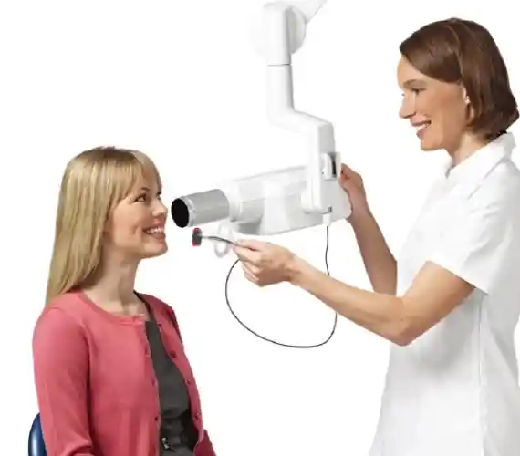
What is a Periapical X-Ray Device?
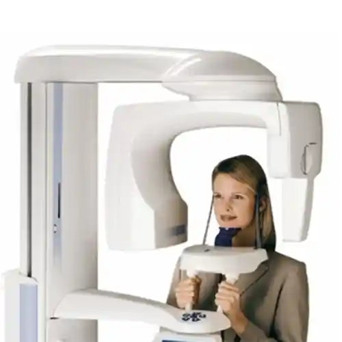
A periapical X-ray device is a precise X-ray generator falling into the category of intraoral radiography, developed in dentistry to perform localized and high-resolution scans. Unlike panoramic systems that scan by rotating around the entire jaw, these devices feature a small tube head (X-ray tube) that focuses strictly on the targeted micro-region. Linear X-rays emitted from the device strike a small digital sensor or analog film placed inside the mouth, ensuring uninterrupted data transmission. Digital periapical units utilized in modern dental clinical practices possess the technological infrastructure to project even micron-level structural changes within the deep layers of the tooth onto the screen, thanks to their high contrast capability.
- Localized Focusing: Sharpens the hard tissue layers of only the 2-3 teeth subject to the clinical complaint.
- Flexible Positioning: Allowing the horizontal and vertical angles of the device head (tube) to be freely calibrated according to the anatomical inclination of the tooth.
- Digital Radiovisiography (RVG) Integration: An intelligent sensor infrastructure that transfers the image to the computer within milliseconds, eliminating the wait for darkroom film development.
How is a Periapical X-Ray Taken?
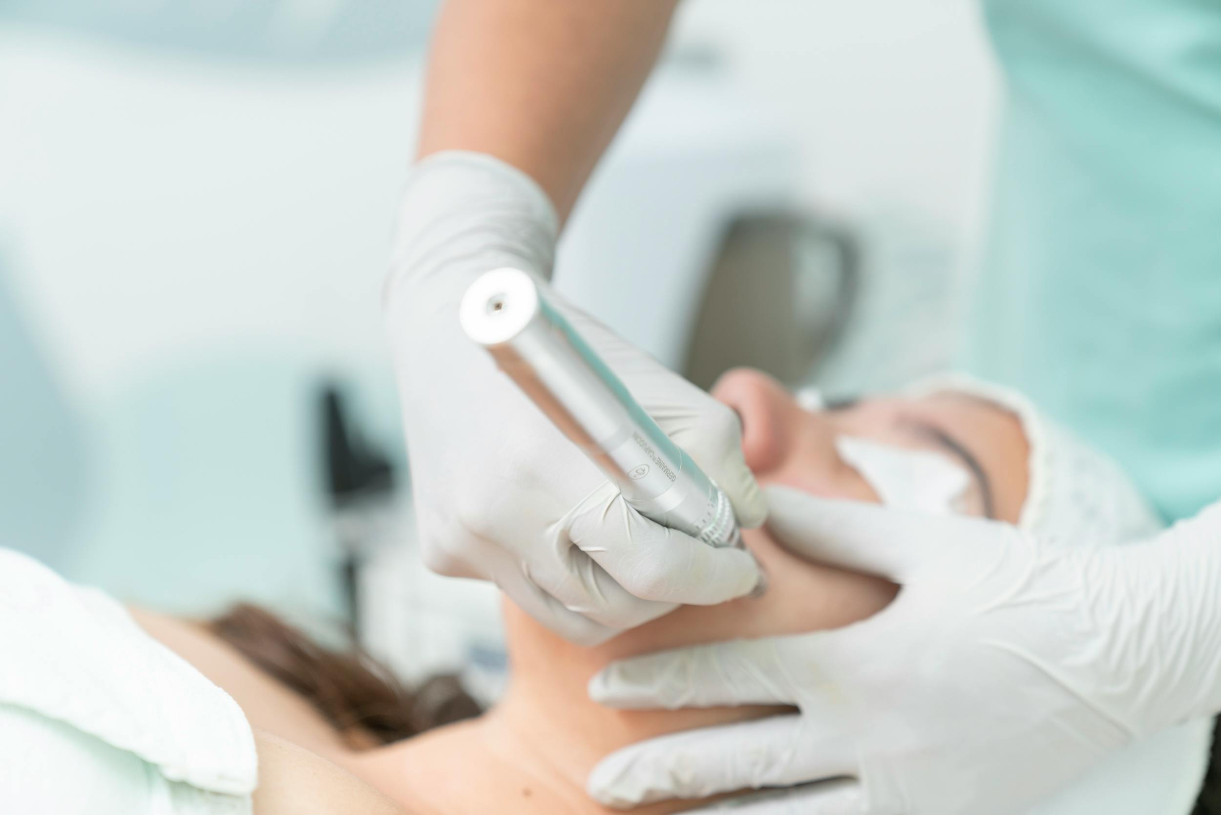
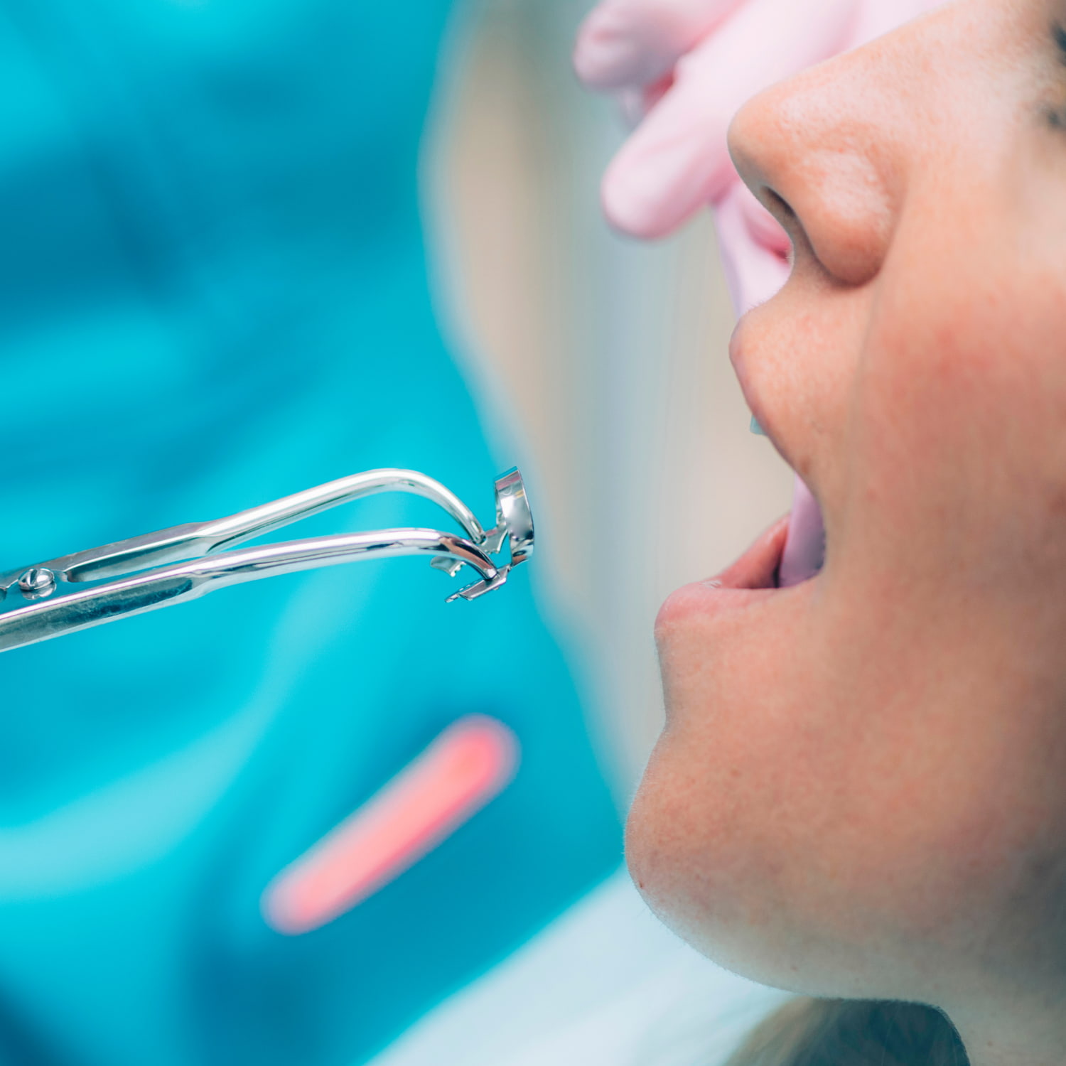
Taking a periapical X-ray is a non-invasive and highly practical radiological procedure completed within seconds in dental clinical settings without the patient even needing to stand up from the chair. At the beginning of the procedure, safety measures are implemented to protect the patient’s chest and thyroid regions; subsequently, intraoral preparation begins. A small digital sensor (or phosphor plate) is positioned parallel to the posterior (lingual or palatal) side of the target teeth without requiring numbing or incisions. The patient is instructed to gently bite a specialized plastic biting apparatus (positioner) with their teeth to keep the sensor stationary. The radiology technician positions the device tube from the outer part of the cheek at the correct angle and presses the button to complete the scan.
- Sensor Positioning: Placing the digital receptor into the mucosal bed so that it stands perfectly parallel to the long axis of the tooth.
- Parallel Technique Application: Delivering the beams perpendicular (at a 90-degree angle) to the sensor to prevent elongation or foreshortening (distortion) in the image.
- Instant Exposure Blockade: An irradiation phase lasting only milliseconds, initiated by the technician pressing the button from outside the room or from a safe distance.
In Which Situations is a Periapical X-Ray Used?
Small intraoral films are utilized in situations where microscopic dental pathologies and root apex details—which might be overlooked by the wide-angle structure of panoramic X-rays—need to be clarified. This high-resolution vision is required to diagnose insidious decay hidden beneath the gums or behind fillings during mirror examinations, root fractures, and localized resorption in the alveolar bone. The dentist introduces this local scan as a clinical necessity to mechanically verify the accuracy of the steps taken before starting treatment or during the session.
The primary situations where periapical X-ray technology is actively deployed in dentistry include:
- Detecting insidious decay originating in the interproximal spaces where teeth touch one another,
- Mapping the boundaries of root-tip abscesses (periapical lesions) within the bone, which are often the source of persistent toothaches,
- Urgent diagnosis of whether horizontal or vertical cracks/fractures have formed in the tooth roots following an impact or trauma,
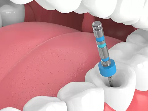
- Millimetrically verifying the length of root canals (working length) during active root canal treatment sessions.
Who is a Periapical X-Ray Suitable For?
The periapical X-ray method is suitable for any adult individual who presents with a localized dental complaint, is scheduled for a filling, root canal treatment, or extraction, and possesses the ability to cooperate. Because the system involves no systemic narcosis or injection of medicated contrast fluids, it can be utilized with high safety regardless of the patient’s general health profile. However, in certain sensitive cases involving a severe gag reflex or a severely limited mouth opening, parameters can be flexed as introducing a rigid sensor into the mouth might prove challenging. For pregnant patients or those with a suspected pregnancy, this screening is postponed until post-delivery unless an urgent/vital dental infection focus is present.
- Those with Localized Pain: Patients experiencing a persistent ache strictly in a specific tooth rather than throughout the entire mouth.
- Patients in Active Treatment Phasing: Individuals whose root canal treatments or filling workflows are actively ongoing in the clinic.
- Pediatric Age Group: Children who can cooperate, scanned to evaluate the resorption status of primary tooth roots or permanent tooth germs.
Which Structures Can Be Visualized with a Periapical X-Ray?
Despite possessing a narrow field of view (FOV), a periapical film lays out all anatomical tissues within its focused micro-region with a clarity close to cellular smoothness. Because overlapping bone shadows common in wide-angle films are eradicated in this method, a topographic reading extending from the internal chambers of the tooth to the bone cells can be executed. This facilitates the early capture of anomaly lines within the internal architecture of the tissue.
The primary structures that can be visualized at a micron level with this radiological method include:
- Tooth Crown and Enamel: The enamel and dentin hard tissues in the visible outer layer of the tooth, along with hidden decay spaces underneath existing fillings,
- Pulp Chamber and Root Canals: The volume of the nerve chamber that carries the vital sensation of the tooth and the morphological trajectory of the canals within the root,
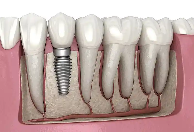
- Periodontal Ligament (PDL) Space: The microscopic thickness line of the fiber network that cushions the tooth, located between the tooth root and the bone,
- Lamina Dura and Alveolar Bone: The integrity of the dense bone socket (socket wall) surrounding the root and the trabecular bone lattice,
- Root Apex Morphology: The anatomy of the absolute tip of the root and the neurovascular entry portal opening from this point into the bone.
What Are the Advantages of a Periapical X-Ray?
The most revolutionary advantage periapical X-ray technology brings to modern dentistry is that its diagnostic resolution quality is exponentially higher compared to panoramic films. While a panoramic X-ray presents the entire mouth as a general map, the periapical system narrows its focus to provide magnified details within the millimetric layers of the tooth. This prevents major tooth loss by ensuring insidious pathologies are caught at an incipient stage. The technical differences between the two radiological systems are presented in the comparison table below:
| Radiological Parameter | Digital Periapical X-Ray (Intraoral) | Panoramic Dental Radiography (Extraoral) |
|---|---|---|
| Image Resolution and Detail | Extremely high; clearly displays fine cracks and canal orifices. | Moderate; provides general outlines, can cause shadowing in micro-details. |
| Regional Magnification (Distorsiyoni) | Virtually non-existent; tooth dimensions project onto the screen at a 1:1 ratio. | Harbors a magnification or anatomical curve deformation rate of 15% to 25%. |
| Soft Tissue/Air Space Obstruction | The sensor stands directly adjacent to the tooth inside the mouth, immune to shadows. | Shadows from the spine, sinus cavities, and tongue can obscure the image. |
| Irradiation Field and Dosage Power | An ultra-low targeted dose is dispatched exclusively to a narrow 2-3 cm field. | Utilizes a wider radiation corridor covering the entire jaw and facial region. |
Why is a Periapical X-Ray Critical in Root Canal Treatment Planning?
Clinical success in endodontic (root canal treatment) practices relies on the principle of millimetrically cleaning infected nerve tissues down to the root apex and sealing them in a leak-proof manner; periapical radiography is the most uncompromising guide for this surgical craftsmanship. Prior to treatment, the curvature of root canals (degree of calcification) is screened with this film. The most critical phase is the “working length determination” stage; the clinician introduces a medical guide file into the canal to capture a periapical film and measures on-screen how many millimeters remain to the anatomical boundary of the root tip (apical foramen), conclusively preventing under-cleaning or excessive trauma to vital tissues.
- Canal Anatomy Discovery: Pre-operative identification of hidden lateral canals or unusual anatomical curvatures within the root.
- Sealing Quality Control: The phase of verifying that the canal sealer and gutta-percha materials fill the root canal down to the apex without voids.
- Post-Treatment Monitoring: Chronological tracking of the success and tissue compatibility of the root canal filling in subsequent months.
How is a Periapical X-Ray Used in the Evaluation of Periapical Lesions?
Periapical lesions are localized inflammatory foci (abscesses) formed when bacteria leaking from the root tip into the jawbone due to a loss of tooth vitality resorb the bone tissue in that region. Because they absorb fewer X-rays, these lesions manifest on periapical radiographs as dark, blackish circular shadows (radiolucent areas). The high quality of the film empowers the clinician to clearly outline the boundaries of the lesion, establishing a differential diagnosis between a root-end cyst and a chronic abscess. Following root canal treatment, the visual shrinking of these black shadows over time to make way for new white bone tissue is the clearest radiological indicator that the treatment has proven its biological success.
- Measuring Destruction Dimension: Rational tracking of the damage inflicted by inflammation in the surrounding alveolar bone in mm².
- Adjacent Root Protection: Auditing whether the infection focus has infiltrated the root cells of neighboring healthy teeth.
- Surgical Decision: Serving as a criterion to determine when a root canal treatment alone will not suffice for extensive lesions, indicating the necessity of an apicoectomy (root-end resection).
How Does a Periapical X-Ray Enable the Inspection of the Tooth Root and Surrounding Tissues?
The cellular resolution of intraoral small films enables tracking the microscopic integrity of the periodontal ligament attachments anchoring the tooth root and the socket bone (lamina dura) with sharp lines. In a healthy oral architecture, this black fiber line surrounding the root is homogeneous and thin; however, when excessive load is imposed on the tooth (occlusal trauma or bruxism) or when an infection initiates, this line widens and thickens. The periapical system conveys these micro-stretches and bone density reductions by generating light-dark contrast on the screen, rendering pathological destruction processes underneath the tissue visible before clinical symptoms manifest.
- Detection of Root Resorption: Early capture of tendencies where a tooth root undergoes internal or external self-resorption.
- Analysis of Bone Pockets: Screening vertical bone resorption defects developing along the tooth root due to periodontal (gum) diseases.
- Scar and Fissure Management: Establishing accurate diagnoses by identifying shadows generated by microscopic cracks on the root surface.
Can a Periapical X-Ray Be Used Prior to Implant Treatment?
Yes, a periapical X-ray can be safely utilized prior to implant treatment and particularly during the healing phases following implant placement as a complementary tracking tool. Although 3D tomography or panoramic radiographs are primary necessities at the very inception of implant planning to measure the transverse width of the bone, a periapical film extends extra security to the surgeon by evaluating the root angles of vital teeth adjacent to the edentulous span scheduled for implant placement at a micron level. Post-operatively, these local films verify the bone integration (osseointegration) around the implant with the highest clarity.
- Root Angle Mapping: Detecting whether adjacent tooth roots exhibit tilt or curvature toward the intended implant socket.
- Marginal Bone Tracking: Faultless cataloging of the bone level status at the implant neck region over the years.
Is Preparation Required Prior to a Periapical X-Ray?
A periapical X-ray scan is a non-invasive diagnostic step executed with zero prior preparation and high practicality, requiring no fasting or ingestion of chemical solutions by the patient beforehand. The sole preparation prior to entering the scanning room is the removal of all removable metal objects located in the head and neck regions that could mechanically block the passage of X-rays and generate flares (artifacts) on the image. Because these flares degrade film quality and lower diagnostic accuracy, strict compliance with this rule is a clinical requirement.
The primary objects that must be removed prior to the scan include:
- Removable partial dentures with metal clasps present within the oral cavity,
- Earrings, piercings in the ears, and metal necklaces around the neck,
- Glasses, hearing aids, and large metal hair clips or pins.
How Long Does a Periapical X-Ray Scan Take?
The intraoral small film application is the fastest radiological procedure that fits perfectly into the time constraints of active urban life, concluding within seconds without even requiring the patient to step out of the chair. The net duration during which the device focuses on the target tooth and performs the scan by emitting X-rays consists of a microscopic window of only a fraction of a second (averaging 0.1 to 0.2 seconds). Including positioning the sensor inside the mouth and bringing the device tube close to the cheek, the total process is completed in 1-2 minutes at most; in digital configurations, images project onto the clinician’s screen instantaneously.
- Active Irradiation Time: 0.1 – 0.3 seconds on average (A millisecond-level micro-beam pulse period).
- Clarity Speed on Screen: It takes approximately 3 seconds for the image to land on the screen as sensor data streams into the software.
- Time Convenience: Darkroom chemical-bath processing phases have been completely eliminated thanks to digital sensors (RVG).
What Should Be Considered During a Periapical X-Ray Scan?
Strict rules that the patient and the technician must follow during that millisecond active irradiation window when the device trigger is pressed directly dictate the resolution quality of the film. The most vital medical rule is that despite the absence of injections or incisions, the patient must stand completely stationary at the moment of exposure, avoid moving their head, and refrain from swallowing. A minor movement at the micron level generates “motion blur,” rendering the film unreadable and forcing a repeat scan, causing the patient to receive unnecessary radiation.
- Stable Position: Standing motionless like a statue once the device tube is positioned against the cheek until the exposure concludes.
- Bite Apparatus Balance: Maintaining homogeneous pressure with the teeth on the intraoral sensor holder, avoiding any slippage.
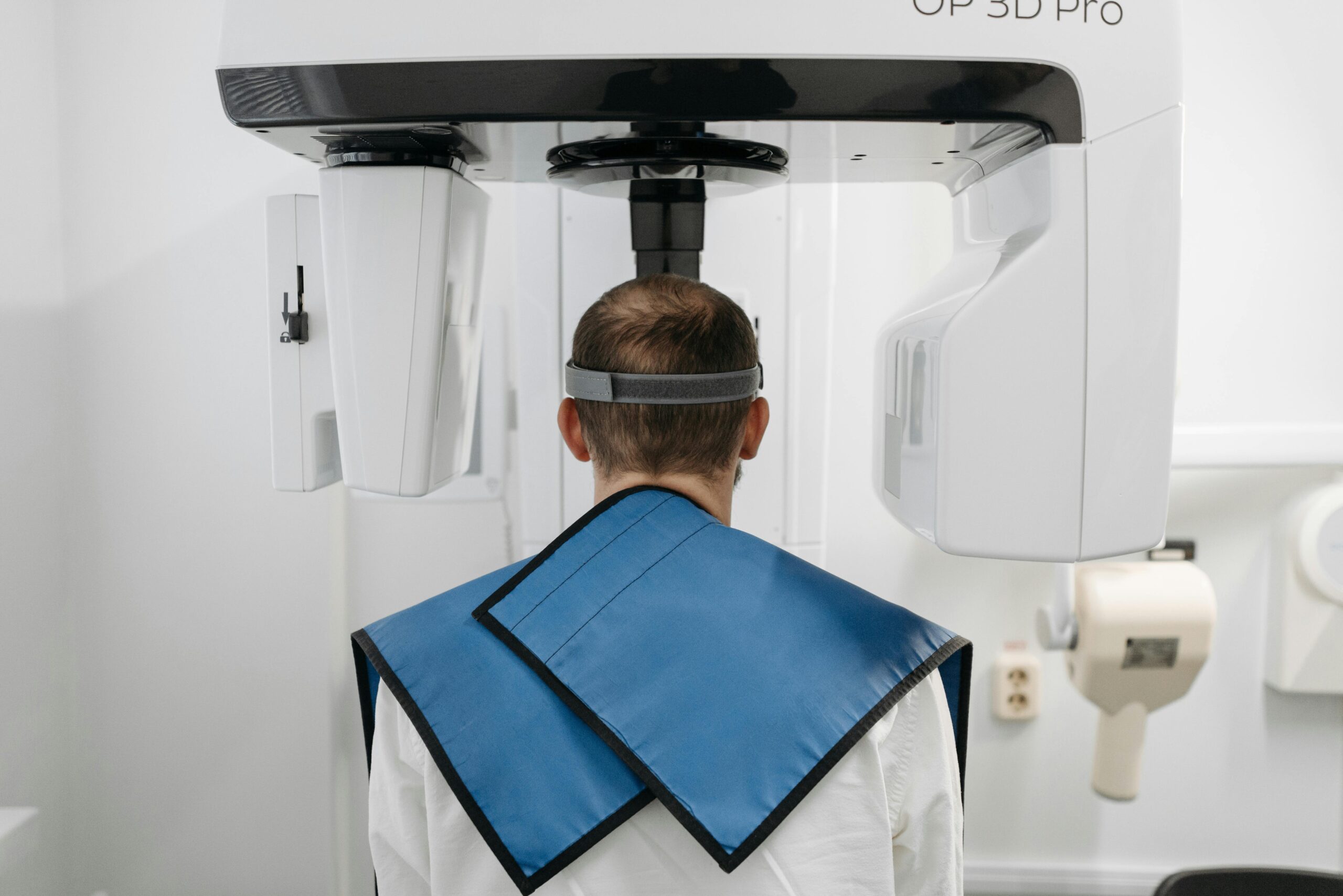
- Lead Protection: The strict obligation to fully drape a lead apron equipped with a thyroid shield over the patient’s torso during the procedure.
Is a Periapical X-Ray a Safe Imaging Method?
Yes, although it involves ionizing radiation, digital periapical radiography stands at the forefront of the safest imaging methods worldwide, engineered within the framework of modern radiology standards and “ALARA” principles. Because the light-capture sensitivity of digital RVG sensors is 80% higher compared to older films, a microscopically low X-ray dose is sufficient to capture a sharp image. When compared to the background radiation amounts we naturally absorb from nature or cosmic rays in daily life, the risk coefficient emitted by a single periapical radiograph resides within entirely negligible safety boundaries.
- Narrowly Targeted Corridor: Beams are dispatched strictly to a localized 2-3 cm tissue field, demonstrating no propagation to the rest of the body.
- High Safety Protocol: The assurance of auditing the devices within the polyclinic structure against dosage leaks through periodic calibrations.
Can a Periapical X-Ray Be Used on Children?
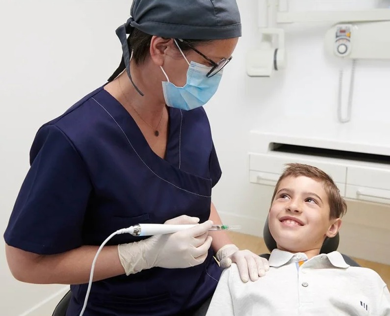
Yes, periapical radiography can be used with high safety and frequency on pediatric patients in pediatric dentistry (pedodontics) and preventive orthodontics practices. During children’s mixed dentition periods, the development levels of permanent tooth germs underneath un-shed primary teeth and root resorption morphologies are screened most clearly with these local films. During scans of pediatric patients, the “pediatric dose” mode is selected from the device panel; this mode radically decreases the irradiation time compared to adults, placing the child’s sensitive tissues under full protection.
- Pediatric Protection: Utilizing specialized thyroid collars to safeguard the developmental thyroid glands of children.
- Early Diagnosis Convenience: Detecting deep decay in primary teeth before it compromises the architecture of the underlying permanent tooth.
- Rapid Adaptation: The comfort of completing the scan within a second, preventing children from growing restless in the chair.
Which Dental Treatments Does a Periapical X-Ray Facilitate Planning For?
Local intraoral small films facilitate the faultless and evidence-based planning of operational decisions at the moment of treatment across nearly all specialties of dentistry. While wide-angle films establish the initial diagnosis, the periapical system functions as a tactical tool providing simultaneous mechanical verification to the clinician during active treatment. The table below summarizes the primary dental treatments whose planning and execution are directly supported by periapical radiography:
| Supported Dental Treatment | Intra-Session Convenience Provided by the X-Ray | Achieved Clinical Success Outcome |
|---|---|---|
| Composite / Aesthetic Fillings | Micron-level detection of how close decay has approached the vital nerve chamber (pulp) of the tooth. | Preserves tooth vitality by preventing unnecessary root canal treatments. |
| Surgical Tooth Extractions | Identifying root fracturing, curvature (dilaceration), or hypercementosis statuses within the bone. | Presents a minimally traumatic surgical map by reducing root fracture risks during extraction. |
| Tissue Grafting and Perio Surgeries | Assessing the depth boundaries of vertical bone loss channels (periodontal defects) between two teeth. | Intra-operative planning of the exact millimetric margins up to which bone powder will be packed. |
How Does a Periapical X-Ray Contribute to the Diagnostic Process?
The greatest contribution of a periapical X-ray to the diagnostic process is making “micro hidden foci”—which can be missed by the eye or two-dimensional wide-angle films—visible with high resolution power. Interproximal micro-decay or vertical root fractures that cannot be discerned on panoramic films due to the overlapping of cranial bone architectures become clear with this method. Magnifying the image within a digital interface, the clinician can capture even the smallest thickening along the tooth’s ligament line; this enables pinpointing the root cause underlying insidious toothaches of unidentified origin, granting an opportunity for early intervention.
- Micro-Detail Visibility: Screening millimetric fracture lines and micro-leakage areas across the root surface.
- Eradication of Illusions: Completely zeroing out superposition (shadow overlapping) errors generated by adjacent tissues.
How Are Periapical X-Ray Results Evaluated?
Evaluating the acquired digital small film is an analytical reading workflow where the dentist operates endodontic anatomy and radiology expertise in tandem. Opening the film on a computer screen, the clinician first evaluates color variations along the enamel-dentin boundary; subsequently, distances to the pulp are measured. Descending along the root, the periodontal space line is monitored, and the bone density map at the root apex (apical zone) is screened. Dark radiolucent shadows report infection and bone resorption, while bright radiopaque fields represent healthy bone architecture or filling materials.
- Contrast Adjustment Management: Modifying brightness configurations within digital software to evaluate the blockage (calcification) degrees of root canals.
- Bone Margin Registration: Inspecting the continuity of the socket wall line (lamina dura) anchoring the root directly from the screen.
Can One Return to Daily Life Immediately After a Periapical X-Ray?
Yes, a periapical X-ray scan is a completely non-invasive diagnostic step that induces no fatigue, restrictions, or numbness on the patient’s motor skills, consciousness, nervous system, or general functionality. No needles are pricked into the skin, no injections are performed, and no medicated contrast fluids are used during the procedure; therefore, the exact minute the session concludes, the patient can resume their normal daily, social, professional, or academic life rhythm uninterrupted. There are no clinical barriers to driving or attending business meetings immediately after the procedure.
- Zero Social Interruption: The freedom to resume normal life comfort right where it was left off the moment one steps out of the room.
- Practical Process Management: The ease of capturing the scan within seconds without even standing up from the chair during a dental treatment session to continue with the procedure.
What Are the Frequently Asked Questions About Periapical X-Rays?
The most common practical anxieties arising in the minds of patients advised to undergo an intraoral small film scan during dental treatments or diagnostic pathways, along with their clear, uncensored medical answers, are presented below:
Will I feel pain during the periapical X-ray scan, or will the radiation hurt me?
No, you will absolutely feel no pain, ache, or soreness. The procedure is entirely non-invasive; a small plastic sensor is simply placed inside your mouth, and the device is brought close to your cheek from the outside. As for radiation waves, they possess a physical nature imperceptible to human senses, so you will feel no heat, sting, or electricity during the scan.
I already had a wide panoramic X-ray, why is this small film additionally required?
A panoramic X-ray delivers the entire jaw as a general map, but due to its wide angle, it may not display microscopic capillary canals inside tooth roots or starting millimetric cavities with perfect clarity. A periapical film, conversely, narrows its focus exclusively to that suspicious tooth to provide micron-level sharpness and resolution; this detail is mandatory to avoid errors, especially in procedures requiring millimetric precision like root canal treatment.
Are implants or metal fillings in my mouth a barrier to taking this small film?
No, fixed bridges, metal fillings, or titanium implants within your mouth pose absolutely no medical barrier to undergoing a periapical X-ray; since these structures harbor no magnetic field, they bear no risk of flying out or heating tissue like in MRI devices—they are completely safe. Only the metal portions form bright white zones on the film, and the clinician possesses the clinical experience to read past these boundaries.
Can breastfeeding mothers undergo a periapical small film scan, and is it necessary to discard milk afterward?
Yes, breastfeeding mothers can safely undergo a periapical X-ray scan if required. The X-rays emitted by the device are energy waves moving at the speed of light; the moment they strike tissue, they pass through and dissipate, harboring absolutely no radiation character that accumulates, gets stored in the body, or compromises the biological structure of breast milk. Therefore, there is no need to pump breast milk and throw it away after the scan; the mother can nurse her baby as soon as she leaves the room.
How long do I need to wait to receive the results after a periapical film is captured?
You absolutely do not need to wait. In modern digital RVG-sensor systems, data streams into the computer software via cable the exact second the exposure trigger is pressed, and the film becomes ready in high resolution on the computer screen within an average of 2-3 seconds. Because the image hits your physician’s screen instantly, you can transition directly to the treatment or canal measurement phase that very minute without losing time.
Who Are We? & Panorama Ankara Oral and Dental Health
Panorama Ankara is an institutional oral and dental health center providing healthcare services at international standards to its patients by combining an academic expert staff and the latest technological digital infrastructure under a single roof, particularly in oral, dental, and maxillofacial surgery, implantology, endodontics, and aesthetic dentistry. Attaching vital importance to millimetric root craftsmanship during treatments, screening insidious decay at its incipient stage, and executing surgical steps in a completely evidence-based, faultless manner, our center welcomes its clients without making them stand up from the chair, using high-resolution and ultra-low radiation dose premium Digital Periapical Radiography (RVG) systems within its clinical infrastructure.
Across all radiological screenings and clinical interventions performed under the roof of Panorama Ankara, absolute sterilization, high patient safety, transparency, and honesty principles are operated with uncompromising determination as per our charter. In our clinical practices, smart sensor technologies that calibrate the ionizing beam duration at a millisecond level based on the patient’s age and tissue sensitivity are applied under the supervision of our expert medical staff. Our goal is not to conduct risky and incomplete workflows with arbitrary patterns; it is to analyze the quality of every tooth root, ligament integrity, and the millimetric boundaries of root-tip abscesses on a computer screen at the exact moment of treatment, achieving maximum session comfort, a faultless root canal treatment standard, and the highest success rates without damaging surrounding tissues and vascular pathways. If you are tired of your unremitting, insidious toothaches or old worn-out fillings when looking in the mirror, yet harbor concerns regarding radiation risks and wish to initiate your treatment process with a scientific guarantee in an expert center environment, you can safely protect your health and smile integrity by contacting the expert staff of Panorama Ankara for detailed periapical analyses.