What is a Panoramic X-Ray Device?
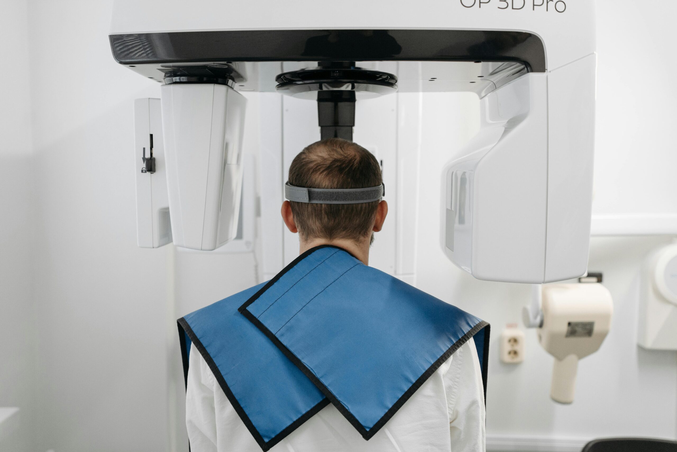
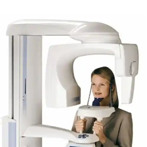
A panoramic X-ray device (orthopantomograph) is an extraoral imaging system operating on tomographic motion principles, designed in dentistry to generate a comprehensive map of the entire oral structure. The device features an X-ray source and a digital sensor panel that rotate synchronously around the patient’s head. During this scanning motion, a narrow beam of radiation is delivered in perfect alignment with the curved anatomy of the jaw arch. The acquired data is processed within seconds via digital software to produce a wide-angle panoramic visual where all teeth are aligned in a single plane without overlapping.
- Extraoral Scanning: Managing the entire imaging workflow from outside the mouth, eliminating the need to place a film intraorally,
- Linear Tomography Mechanism: Establishing a clear focal layer (focal trough) to sharpen only the structures located within the dental arch,
- Digital Sensor Technology: Housing sensitive receptors that instantly transfer the image to the computer screen instead of old-school chemical-bath films.
How is a Panoramic X-Ray Taken?
Taking a panoramic X-ray is a practical radiological procedure that is highly comfortable for the patient, completely painless, and completed within seconds. When the patient is escorted into the X-ray room, radiation protection protocols are completed first; then, the patient is positioned in the center of the device. Guided by the technician, the patient’s chin is placed onto the support apparatus of the device, and they are instructed to bite a sterile, disposable bite block with their front teeth. Temporal supports are stabilized to prevent the head from tilting to the right or left. Once the device is activated, the mechanical arm executes a circular rotation around the patient’s head to complete the scan.
- Positioning: Aligning the patient’s head on a perfectly symmetrical axis with the aid of laser guidance lines,
- Bite Block Activation: Ensuring the front teeth meet edge-to-edge to prevent the jawbones from overlapping on one another,
- Active Scanning Phase: The device head sweeping around the jaw within seconds by tracing a 360-degree semi-arc.
In Which Situations is a Panoramic X-Ray Requested?
A panoramic X-ray is routinely requested in dentistry from every new patient coming for a general examination to screen the overall status of intraoral hard tissues. Cysts, root-tip abscesses, jawbone resorption, or impacted tooth positions hidden beneath the skin and gums cannot be discerned with the naked eye during an intraoral mirror and explorer examination. The clinician requires this wide-angle film as a primary clinical guide for all major treatment decisions ranging from implant placement and wisdom tooth extraction to orthodontic brace treatment and detecting the depth of widespread decay.
- When it is necessary to assess the extent of destruction caused by periodontal diseases (periodontitis) in the bone tissue,
- While searching for insidious infection foci underlying widespread chronic pain of unidentified origin inside the mouth,
- To inspect the stability of roots to be utilized as abutments prior to prostheses, bridges, or extensive restorations,
- Whenever the presence of pathological structures such as cysts or tumors developing within the jawbone is suspected.
Who is a Panoramic X-Ray Suitable For?
This wide-angle radiography method is suitable for individuals of all ages who wish to have a general check-up of their oral health, who cannot tolerate small intraoral films (periapical) due to a gag reflex, and whose general health profile is stable. Elderly patients, those scheduled for prosthetics, and children in the mixed dentition stage can benefit from this practical screening with high comfort. However, for pregnant patients—especially those in their first trimester—panoramic X-ray imaging should be postponed until post-delivery unless an emergency and vital dental infection intervention is mandatory.
- Patients with a Gag Reflex: Sensitive individuals who experience a gagging sensation when a film is placed inside the mouth,
- Children and the Elderly: All age groups who possess the ability to stand still in the center of the device and can cooperate,
- General Screening Candidates: Individuals with numerous fillings or bridges in their mouth who request a general status check.
Which Structures Can Be Visualized with a Panoramic X-Ray?
As the name implies, a panoramic film presents a full panorama of the maxillofacial (jaw-face) region. By displaying all anatomical proximities side by side on a two-dimensional flat plane, it grants the clinician the luxury of holistic monitoring. Consequently, not only the teeth but also the deep bony and anatomical structures that surround the teeth and constitute the facial architecture can be screened from the same window.
The primary structures that can be clearly visualized with this radiological method include:
- All Teeth: The crown and root anatomy of all erupted, fractured, decayed, or impacted teeth located in the upper and lower jaws,
- Alveolar Bone: The height and density boundaries of the upper/lower jawbone tissue that supports and nourishes the tooth roots,
- Maxillary Sinuses: The floor boundaries of the anatomical air spaces positioned directly above the upper jaw molars,
- Temporomandibular Joint (TMJ): The right and left jaw joint condyle structures where the lower jawbone articulates with the temporal bone,
- Anatomical Canals: The mandibular nerve canal (Canalis mandibulae), which carries sensation to the lower jaw teeth and is vital to protect.
What Are the Advantages of a Panoramic X-Ray?
The greatest advantage that panoramic X-ray technology brings to clinical dentistry is providing maximum diagnostic data while imposing minimum time and radiation load on the patient. While capturing all teeth with small intraoral films requires placing a film in the patient’s mouth repeatedly and delivering radiation at least 14 times; the panoramic system lays out the entire mouth before us within seconds in a single exposure. This both accelerates the clinical workflow and keeps the patient’s cumulative radiation exposure within the safest limits.
| Clinical Parameter | Digital Panoramic X-Ray | Conventional Intraoral Small Films (Periapical) |
|---|---|---|
| Width of Imaging Field | The entire upper-lower jaw, teeth, joints, and sinuses are visible on a single film. | Shows only the root and surrounding tissue of 2–3 teeth standing side by side. |
| Patient Comfort Level | Very high; captured from outside the mouth, does not trigger the gag reflex. | Moderate-low; rigid intraoral packets can irritate the palate. |
| Total Radiation Load | Extremely low since the entire mouth is scanned with a single micro-exposure. | Requires at least 14 consecutive irradiations to scan the whole mouth, increasing the cumulative dose. |
| Result Acquisition Speed | Appears on the computer screen within seconds thanks to the digital sensor. | May require development and drying time (5–10 min) in older types. |
How is a Panoramic X-Ray Used in Implant Treatment Planning?
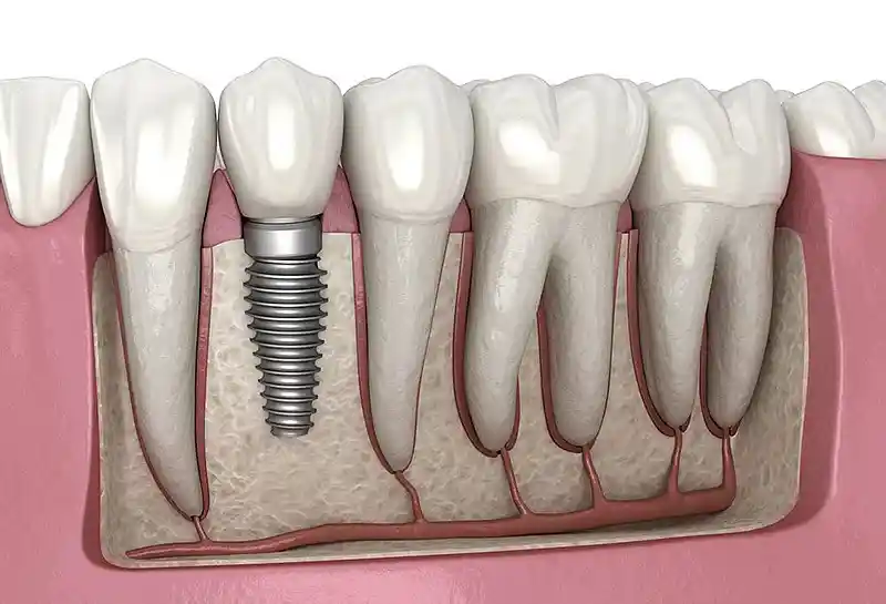
When planning implant surgery, the titanium artificial root to be placed into the edentulous site must be selected in a size appropriate for the vertical bone distance; panoramic radiography is the first and most critical step of this planning. Utilizing the millimetric measurement tools on the film, the clinician measures the height of the alveolar bone. To prevent the implant from penetrating into the maxillary sinus cavity in the upper jaw, and to avoid the risk of causing permanent lip numbness by striking the mandibular nerve canal in the lower jaw, anatomical hazard zones are scanned via the panoramic film to determine safe surgical corridors.
- Vertical Bone Measurement: Assessing the vertical millimetric boundaries of the bone space where the implant will be placed,
- Proximity Relationships: Planning the positioning of the implant at safe angles that will not damage adjacent tooth roots,
- Advanced Investigation Decision: Serving as a criterion to refer the patient for a 3D dental tomography scan if the transverse thickness (width) of the bone appears questionable on the panoramic film.
How Does a Panoramic X-Ray Support Orthodontic Treatment?
Before commencing orthodontic wire or clear aligner treatments, it is essential to know not only the crown portions of the teeth but also their root alignments within the bone and their developmental status; panoramic X-rays establish the primary foundation for these processes. In children during the mixed dentition period, the presence of permanent tooth germs beneath un-shed primary teeth, their eruption paths, and whether any congenital tooth agenesis exists are screened with this film. Whether roots are parallel to each other and whether any fibrotic or pathological barrier stands in the way of orthodontic forces inside the jawbone are verified with this wide angle before the sessions.
- Determination of Dental Age and Development: Analyzing the eruption chronology of permanent teeth and their root development levels.
- Root Parallelism Control: Mapping out root orientations so that teeth can be moved at correct angles during orthodontic brace treatment.
- Tracking of Impacted Teeth: Determining the locations of teeth delayed in eruption and formulating plans to guide them onto the orthodontic arch.
Why is a Panoramic X-Ray Important in Jaw Surgery?
In oral and maxillofacial surgery clinical practices, panoramic radiography is of vital importance for clearly monitoring the anatomical boundaries of pathological lesions and charting the operative strategy. Cysts or benign tumors can grow insidiously inside the jawbone, eroding the bone tissue from within; while this destruction presents no external symptoms, it is instantly caught on a panoramic film as dark-colored radiolucent areas. Furthermore, it is the first mapping tool a surgeon consults to determine where a fracture line is located along the mandible in jaw fractures.
- Pathology Screening: Early detection of cysts, tumors, and osteomyelitis (bone inflammation) foci within the jawbone.
- Surgical Margin Determination: Locating healthy teeth and nerve boundaries that must be preserved during the curettage (enucleation) of a lesion.
- Trauma Management: Topographic evaluation of cracks and fractures resulting from impacts sustained by the jawbone.
How is a Panoramic X-Ray Used in the Evaluation of Impacted Teeth?
Wisdom teeth (third molars) and canine teeth (eyeteeth), in particular, tend to remain impacted beneath the bone or gums when they fail to find space in the dental arch; panoramic radiography serves as the operational guide for these hidden teeth. Thanks to the film, the position angle of the impacted tooth inside the bone (horizontal, vertical, mesioangular, or distoangular) is clearly diagnosed. The most critical stage is its proximity to the mandibular nerve canal running underneath the roots of lower wisdom teeth; the surgeon examines this distance to measure the index of nerve injury and subsequent permanent lip numbness while removing the root, allowing them to select a minimally traumatic technique.
- Position Diagnosis: Determining whether the impacted tooth stands completely vertical or entirely horizontal parallel to the jawbone.
- Adjacent Tooth Risk: Assessing whether the impacted structure exerts pressure on the root of the healthy molar in front of it, causing resorption or decay there.
- Pericoronitis Analysis: Monitoring for cyst formation or chronic infection development within the follicle sac surrounding the impacted tooth.
Is Preparation Required Before a Panoramic X-Ray?
A panoramic X-ray scan is a non-invasive diagnostic step executed with zero prior preparation and high practicality, requiring no fasting, medication intake, or injection of fluids into the body beforehand. The sole and most uncompromising medical preparation prior to entering the scanning room is the removal of all metal objects located in the head, neck, and oral regions. When X-rays encounter metal, they cannot pass through, creating bright white lines and shadowing known as “artifacts” on the digital film; since this condition can camouflage real pathologies behind teeth and bones and sabotage the diagnosis, strict adherence to rules is a clinical necessity.
The primary objects that must be completely removed prior to the scan include:
- Removable partial dentures with metal clasps or false teeth present in the mouth,
- Earrings, nose studs, piercings in the ears, and all metal necklaces around the neck,
- Glasses, hearing aids, metal hairpins, and metal clip-in hair extensions.
How Long Does a Panoramic X-Ray Scan Take?
The panoramic X-ray application is the fastest radiological procedure that fits perfectly into the busy rhythm of modern urban life, consuming almost none of the patient’s time in the clinical workflow. The net duration during which the device head revolves around the patient and actively performs the scan by emitting X-rays consists of a very short window of only 10 to 15 seconds. Including escorting the patient into the room, removing metal objects, dressing them in a lead apron, and positioning them with millimetric accuracy in the device, the total clinical time is comfortably completed in 2–3 minutes at most.
- Active Irradiation Time: 10 – 15 Seconds (The circular rotation period of the device arm).
- Digital Screen Delivery Speed: It takes approximately 5–10 seconds for the image to clear on the screen as sensor data streams into the software.
- Time Convenience: The era of waiting for films to be developed in darkrooms is completely over; images are instantly before the clinician.
What Should Be Considered During a Panoramic X-Ray Scan?
The vital rules a patient must follow during that 10-15 second active process when the scanning arm begins to rotate directly determine the clarity, resolution, and quality of the film. The most critical medical rule is that while the device is in motion, the patient must stand completely still like a statue, avoid moving their head, refrain from swallowing, and keep their tongue pressed flat against the palate. A minor head movement or swallowing reflex at the micron level during the exposure creates a “motion artifact”—namely, waviness and blurring—in the digital sections, rendering the film unreadable and forcing a repeat scan, causing the patient to receive unnecessary radiation.
- Absolute Immobility: Standing motionless until the scan finishes by focusing the eyes on a fixed point,
- Tongue Position: Pressing the tongue completely flat against the upper palate; this action erases the air-space shadow above the upper tooth roots,
- Postural Balance: Holding firmly onto the device handles to balance body weight homogeneously and keeping the shoulders straight.
Is a Panoramic X-Ray a Safe Imaging Method?
Yes, although it involves ionizing radiation, digital panoramic radiography is a highly safe imaging method developed within the framework of modern technological standards and “ALARA” (As Low As Reasonably Achievable) principles. Because the sensitivity of digital receptor sensors is exceptionally high, a microscopically low X-ray dose is sufficient to obtain a clear image. When compared to the natural background radiation amounts we absorb from the sun, soil, or transcontinental flights in daily life, the radiation load emitted by a single digital panoramic X-ray is safe and negligible.
- Lead Apron Shielding: It is mandatory to wear a lead apron during the scan to protect the patient’s vital organs and thyroid gland.
- Advanced Dose Calibration: The device automatically adjusts the beam power to the absolute minimum according to the patient’s age, weight, and bone structure.
Can a Panoramic X-Ray Be Used on Children?
Yes, panoramic radiography can be used with high safety and frequency on pediatric patients in pediatric dentistry (pedodontics) and preventive orthodontics practices. The development of children’s jawbones and the eruption schedule of permanent teeth underneath primary teeth constitute a highly dynamic process; thanks to panoramic films, missing teeth or impaction problems that may arise at an early stage are detected. During scans of pediatric patients, the “child protocol” mode is selected from the device software; this mode narrows the scanning field and radically reduces the irradiation time and dosage power compared to adults, ensuring full protection.
- Pediatric Dose Management: Selecting ultra-low radiation levels to safeguard the sensitive tissues of children,
- Diagnosis of Eruption Disorders: Preventing permanent teeth from growing in incorrect directions and damaging adjacent tooth roots at an early stage,
- Gag Comfort: Offering the comfort of extraoral imaging in situations where children find it difficult to accommodate small films inside their mouth.
Which Dental Treatments Does a Panoramic X-Ray Facilitate Planning For?
Wide-angle digital panoramic X-ray data facilitates the formulation of initial diagnoses and the faultless planning of roadmaps across nearly all specialties of dentistry. Presenting the entire jaw in a single panorama provides the clinician with a holistic perspective, enabling the scheduling of sequential treatments. The table below summarizes the primary dental treatments that are directly supported and streamlined by panoramic radiography:
| Facilitated Dental Treatment | Diagnostic Convenience Provided by the X-Ray | Achieved Clinical Success Outcome |
|---|---|---|
| Multiple Bridge and Prosthetic Applications | Assessing the root lengths and bone support levels of the abutment teeth to be utilized as supports. | The load capacity that the prosthesis will carry is calculated accurately, ensuring longevity. |
| Widespread Decay / Root Canal Treatments | General screening of secondary decay hidden underneath existing fillings and root-tip abscesses (lesions). | Clarifying at the beginning of the session which tooth requires a filling and which requires root canal treatment. |
| Periodontal (Gum) Surgeries | Mapping the depth boundaries of horizontal and vertical bone resorption foci throughout the jaw. | Planning which regions bone operations or curettage treatments should be concentrated on. |
How is Digital Treatment Planning Performed with a Panoramic X-Ray?
The digital treatment planning workflow commences with the instantaneous transfer of radiological data captured by the panoramic device sensor into the clinic’s shared computer network in “JPEG” or “DICOM” format. Opening the film in an advanced dental imaging software interface, the clinician adjusts brightness, contrast, and sharpness settings to reveal micro-decay details invisible to the naked eye. Utilizing the software’s millimetric calibration measurement tools, the vertical distances of edentulous spaces are measured; the patient’s intraoral photographs and clinical examination findings are synthesized over this digital X-ray image to shape a personalized phased treatment algorithm and budget plan in a computer environment.
- Digital Measurement Filters: Millimetrically cataloging bone height and root lengths from the screen,
- Image Processing Capabilities: Isolating suspicious lesion areas more clearly with the help of color scales or negative filters,
- Holistic Archiving: Preserving the patient’s pre- and post-treatment films in their digital file to monitor developmental processes chronologically.
How Are Panoramic X-Ray Results Evaluated?
Evaluating the acquired panoramic X-ray film is a systematic reading process conducted by combining the dentist’s expertise in radiological anatomy and clinical diagnostic vision. The clinician follows a specific hierarchical sequence while examining the film on the screen; typically starting from the upper right jaw to screen all teeth sequentially, followed by inspecting bone densities, sinus floor outlines, and finally, the symmetry of the right-left temporomandibular joint condyles. Fillings and prostheses appearing radiopaque (white/bright) and decay and abscess foci appearing radiolucent (dark/black) are differentiated to report the patient’s intraoral pathology map.
- Anatomical Structure Inspection: Determining the distance boundaries of sinus cavities in relation to tooth roots and their degree of sagging.
- Radiological Density Differentiation: Detecting healthy bone tissue versus spongy bone zones resorbed due to infection based on darkness variations.
Can One Return to Daily Life Immediately After a Panoramic X-Ray?
Yes, a panoramic X-ray scan is a completely non-invasive diagnostic step that induces no fatigue, numbness, or restrictions on the patient’s motor skills, nervous system, focus capacity, or general functionality. No needles are pricked into the skin, no local anesthesia is administered, and no medicated contrast fluids are used during the procedure; therefore, the exact minute the session concludes, the patient can resume their normal daily, occupational, social, or academic life rhythm uninterrupted without the burden of an IV catheter or bandages. There are no barriers to driving or eating after the procedure.
- Zero Recovery Burden: The freedom to step out of the room as soon as the scan finishes and return to normal life flow,
- Practical Process Management: The convenience of stopping by the polyclinic even during break hours on working days to complete the scan within minutes.
What Are the Frequently Asked Questions About Panoramic X-Rays?
The most common practical anxieties arising in the minds of patients who are advised to undergo a panoramic film scan at the beginning of treatment processes, along with their clear, uncensored medical answers, are presented below:
Will I feel pain during the panoramic X-ray scan, or will the radiation hurt me?
No, you will absolutely feel no pain, ache, or soreness. The procedure is performed entirely from outside the mouth (non-invasive); the mechanical arm of the device does not even touch you physically as it revolves around your head. As for X-rays, they possess a physical nature imperceptible to human senses (sight, hearing, touch), so you will feel no heat, ache, or electricity during the scan.
Does a panoramic X-ray film show all cavities in my mouth with 100% certainty?
A panoramic X-ray perfectly displays large cavities throughout the mouth, damage beneath fillings, and intraosseous abscesses; however, because it features a wide two-dimensional angle, it can sometimes miss incipient microscopic “interproximal cavities” where two teeth touch due to overlapping tooth shadows. In such instances, to view suspicious teeth more clearly, your physician may support the diagnosis with small intraoral bite-wing X-rays.
I have metal fillings, porcelain bridges, or implants in my mouth; can I still have an X-ray?
Yes, fixed bridges, metal fillings, or titanium implants in your mouth pose absolutely no medical barrier to undergoing a panoramic X-ray; since these structures harbor no magnetic field, they bear no risk of flying out, displacing, or heating tissue like in MRI devices—they are completely safe. Only the metal portions form bright white patches on the film; the expert clinician possesses the clinical reading experience to filter behind these flares.
Can breastfeeding mothers undergo a panoramic X-ray, and is it necessary to pump and discard milk afterward?
Yes, breastfeeding mothers can safely undergo a panoramic X-ray if needed. The X-rays emitted by the device are energy waves moving at the speed of light; the moment they strike the body, they pass through and dissipate, harboring no radiation character that accumulates, gets stored in the body, or compromises the biological structure of breast milk. Therefore, there is absolutely no need to pump breast milk and throw it in the trash after the scan; the mother can continue breastfeeding her baby as soon as she leaves the room.
Do I have to wait for days to receive the results after having a panoramic X-ray?
You absolutely do not need to wait. In modern digital-sensor panoramic devices, the exact second the scan finishes, data streams into the computer software via cable, and the entire film becomes ready in high resolution on the computer screen within an average of 5–10 seconds. Since the images hit your physician’s screen directly over the shared network, you can transition directly to the examination and planning stage without losing any time.
Who Are We? & Panorama Ankara Oral and Dental Health
Panorama Ankara is an institutional oral and dental health center offering health services at international standards to its patients by combining an academic expert staff and the latest technological digital infrastructure under a single roof, particularly in oral, dental, and maxillofacial surgery, implantology, orthodontics, and aesthetic dentistry. Attaching vital importance to running diagnostic processes error-free prior to treatment, screening hidden pathologies early, and drawing up the most accurate personalized roadmaps, our center welcomes its patients with high-resolution and ultra-low radiation dose premium Digital Panoramic X-Ray systems.
Across all radiological screenings and clinical interventions performed under the roof of Panorama Ankara, absolute sterilization, high patient safety, transparency, and honesty principles are operated with uncompromising determination as per our charter. In our clinical practices, sensor technologies that calibrate the ionizing beam power to the safest levels based on the patient’s age and bone structure are applied under the supervision of our expert medical staff. Our goal is not to conduct risky processes with arbitrary patterns, but to analyze every patient’s jawbone quality, sinus boundaries, and the positions of their impacted teeth bare to the eye on the computer screen before the operation, achieving maximum session comfort and the highest treatment success graphs without damaging the skin and surrounding vascular pathways. If you are tired of your missing teeth, unremitting pain, or loss of your smile when looking in the mirror and wish to initiate your treatment process with a scientific guarantee in an expert center environment, you can safely protect your health and smile integrity by contacting the expert staff of Panorama Ankara for detailed digital panoramic analyses.