What Are Cyst Operations?
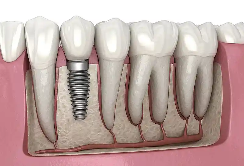
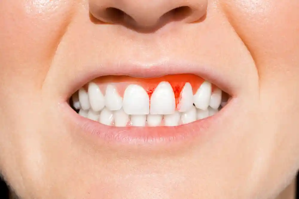
Cyst operations (cystectomy) are a surgical treatment protocol for a hidden pathological bag lined with epithelium and its semi-fluid debris contents, growing insidiously inside the oral, jaw, and facial structures. Although jaw cysts do not metastasize aggressively like tumors, they continuously expand along a linear axis due to osmotic pressure differences, melting the surrounding living jawbone cells (osteolysis). Surgical intervention is based on a hierarchy of milimetrically scraping and completely eliminating the cyst wall (lumen) from the surrounding healthy bone cortex with the help of micro-blades and scraping curettes, rather than just draining the cyst fluid, in order to zero out the risk of recurrence in the future.
- Enucleation (Complete Removal): The operation of removing the cyst pathology in one piece, along with its pathological capsule, without tearing its wall.
- Crest and Bone Preservation: Micro-surgical craftsmanship respecting the integrity of adjacent anatomical nerve and vascular channels while removing the leziyon.
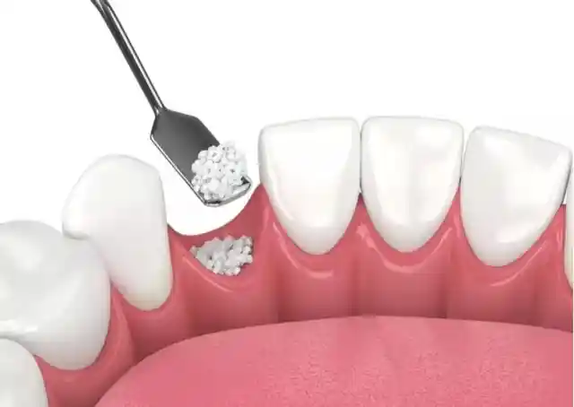
- Biological Defect Rehabilitation: Filling the hole left by the cyst in the bone with regenerative materials in order to maintain skeletal rigidity.
Why Do Cysts in the Oral and Maxillofacial Region Form?
The formation of pathological cavities in the jaw skeleton and surrounding mucosal bed occurs as a result of the activation of embryonic cell remnants hidden between tissues during facial bone development and tooth-related (odontogenic) inflammation agents with an insidious synergy. The most common cause is “radicular cysts” formed at the root tip by deep cavities that are neglected without performing root canal treatment. The chronic inflammation at the root tip triggers the body’s defense mechanism to surround the area with an epithelial membrane; over time, as fluid accumulates inside this bag, it swells and melts the jawbone from the inside like a balloon.
- Neglected Pulpal Necrosis: Continuous pumping of toxins to the root tip with the death of the dental nerve and the cystification of periapical tissue,
- Impacted Tooth Remnants: The pathological evolution of the developmental follicle surrounding the crown portion of unerupted wisdom or canine teeth into a “dentigerous cyst”,
- Embryonic Epithelial Remnants: The triggering of Malassez or Serres epithelial cells remaining from the mother’s womb along the fusion lines (suture lines) of the jawbones,
- Genetic Predisposition: The proliferation of multiple aggressive cyst foci (keratocysts) in the jaw in some syndromic cases (such as Gorlin-Goltz syndrome).
In Which Cases Are Cyst Operations Performed?
The elimination of jaw cysts through surgical operation is definitively indicated as a strict medical rule according to the volumetric size of the lesion, the degree to which it threatens skeletal integrity, and the histological damage it inflicts on adjacent living tooth roots. These insidious structures, which do not give any pain signals during the initial phase, chew through the jawbone to the outer cortical boundaries if not detected during routine controls, giving rise to risks of spontaneous jaw fracture; surgery is definitively resorted to in order to establish these skeletal collapse barriers.
The primary situations where jaw cyst surgeries are definitively selected in clinical operation practices are as follows:
- In cases where the cyst exerts pressure on the lower jaw main nerve line (inferior alveolar nerve), creating a risk of permanent numbness and loss of sensation in the lower lip,
- When the lesion in the upper jaw pushes the maxillary sinus and nasal floor layers, anatomically deforming the airways and the nasal bone,
- When it leads to the insidious melting of the roots of adjacent healthy teeth (root resorption) or causes teeth to shift axes and shake due to the growth pressure of the cyst,
- In cases where the expanding pathology thins the outer boundary (cortical base) of the jawbone, creating a risk of pathological fracture even under the slightest chewing pressure.
Who Are Cyst Operations Suitable For?
Cyst treatment operations planned in the jaw surgery polyclinic are medically suitable for children and adult individuals of all age groups whose cystic pathology focus in the intraoral bone or soft tissue is registered through radiological and clinical examinations. Since the system is carried out with modern minimally invasive surgical disciplines, it is completed with a high safety profile in every client whose general body immunity is stable. However, for patients with uncontrolled severe systemic disorders (those who have recently undergone bypass surgery, acute leukemia patients, or those using strong heavy IV medications of the bisphosphonate group that block the rate of bone destruction), it is mandatory to obtain written consultation approval from the relevant medical specialty prior to the procedure.
| Cyst Type / Skeletal Spread | Surgical Intervention Protocol | Expected Tissue and Cellular Output |
|---|---|---|
| Small and Medium-Sized Periapical Cysts | Local Anesthesia + Complete Enucleation | The cyst is completely eliminated; the cavity is filled with a fresh blood clot and left to ossify. |
| Giant Volumetric Skeletal Cysts (Thinned Bone) | Marsupialization (Decompression) | A hole is opened inside the cyst to reduce fluid pressure; as the bone grows from the inside, the cyst shrinks, preserving the bone. |
| Aggressive / Recurrence-Prone Cysts (Keratocyst etc.) | Enucleation + Carnoy’s Solution (Chemical) | After the cyst is removed, the bone walls are cauterized with a special solution; hidden micro-cell remnants are killed. |
| Active Draining and Acute Abscessed Cyst Cases | Medical Drainage + Medication Course (Must Be Postponed) | First, the abscessed fluid is drained; after the acute inflammation is suppressed with antibiotics, the definitive surgery schedule begins. |
How Are Intraoral Cysts Diagnosed?
Intraoral cyst diagnosis processes are a precise medical diagnostic algorithm carried out not by random guesses, since they do not mimic pain signals in the dental pulp, but through the sequential verification of radiological, clinical, and histopathological analyses. Lesions are usually detected coincidentally on classical X-rays taken with complaints of toothache. However, drawing fluid from the lesion with a syringe during or before the surgery (aspiration biopsy) or taking a micro-piece and forwarding it to the pathology laboratory is the only definitive way of diagnosis in order to detect the exact histopathological type (whether it is malignant or benign) and to map out the intra-tissue character map.
- Radiolucent Boundary Scanning: Monitoring bone-melting cyst areas on X-ray films as dark, black, and round shadows,
- Cortical Expansion Examination: The cyst expanding to swell the outer wall of the jawbone, giving a ping-pong ball elasticity (parchment crackling) during manual palpation,
- Tooth Vitality Mapping: Scanning the teeth associated with the cyst using vitality devices to mathematically register the origin root of the infection.
What Evaluations Are Performed Before Cyst Operation?
The clinical evaluation carried out before the surgery schedule is finalized is the most vital pre-operative analysis phase that traces the milimetric extension lines of the cyst wall inside the bone. The physician examines the patient’s gingival flap flexibility and mucosal quality during the examination; because sealing the bone surface tightly after the operation (primary closure) is the only way to halt wound site infections. At this stage, the patient’s general blood clotting times and laboratory biochemistry values are checked, completing the systemic risk profile management without error.
- Adjacent Root Relationship Cataloging: Scanning the root integrity and cementum attachment qualities of the sound teeth around the cyst.
- Vascular Pathway Planning: Analyzing the distance of the greater palatine or mental neurovascular bundles within the jaw skeleton to the lesion.
- Volumetric Cc Calculation: Determining the dose of bone powder to be used during the surgery based on the size of the hole that will remain in the bone after the cyst is cleared.
How Is Cyst Operation Planned?
The planning of the cyst surgical workflow is carried out through a phased surgical hierarchy according to the skeletal spread diameter of the lesion, the patient’s biological age lines, and the post-operative implant infrastructure schedule. The bone skeleton is not battered with hasty moves while planning the process; because preserving the bone volume is the key to future chewing function success. In planning, the operation technique and control schedules are meticulously logged into the surgical card according to the type of cyst (whether aggressive or shallow).
- Phase 1 (Prophylaxis and Hygiene): Zeroing out the risks of microb leakage into the socket through ultrasonic scaling of all calculus in the surgical site.
- Phase 2 (Surgical Intervention): The process of opening the mucosa, tracing the intra-bone window, scraping the cyst capsule, and transferring it to pathology laboratory tubes.
- Phase 3 (Regenerative Reconstruction): The phase of reinforcing the cleared cavity without gaps using growth-factored PRF membranes and xenograft mineral powders.
- Phase 4 (Follow-up and Ossification): The cycle of subjecting the patient to radiological follow-ups at the 3rd and 6th months to register new bone formation.
Which Surgical Methods Are Applied in Cyst Operations?
In maxillofacial surgery practices, two main evidence-based surgical treatment methods designed according to the pathological character of the cyst and the degree of destruction it creates in the jawbone are deployed. The surgeon does not impose a single method on the patient; they field the protective technique that will batter the tissue the least according to the tangent line of the lesion to the nerve channels. The choice of techniques is the main factor that zeroes out the risks of permanent facial numbness remaining in the patient after the surgery.
- Enucleation Protocol: Scraping the cyst bag from the surrounding bone as a whole and closing the hole; provides definitive and permanent clearance,
- Marsupialization (Decompression): In giant cysts, a medical tube (drain) is placed inside the cyst to continuously leak fluid outward; as the intra-cyst pressure is zeroed out, the jawbone grows from the inside, shrinking the cyst, and the remaining small piece is enucleated months later,
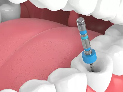
- Apicoectomy Combination: The method of cutting the root tips of the teeth causing the cyst with 3-millimeter sections (apicoectomy) to dry out the inflammation factory from the root.
How Is Jaw Cyst Operation Performed?
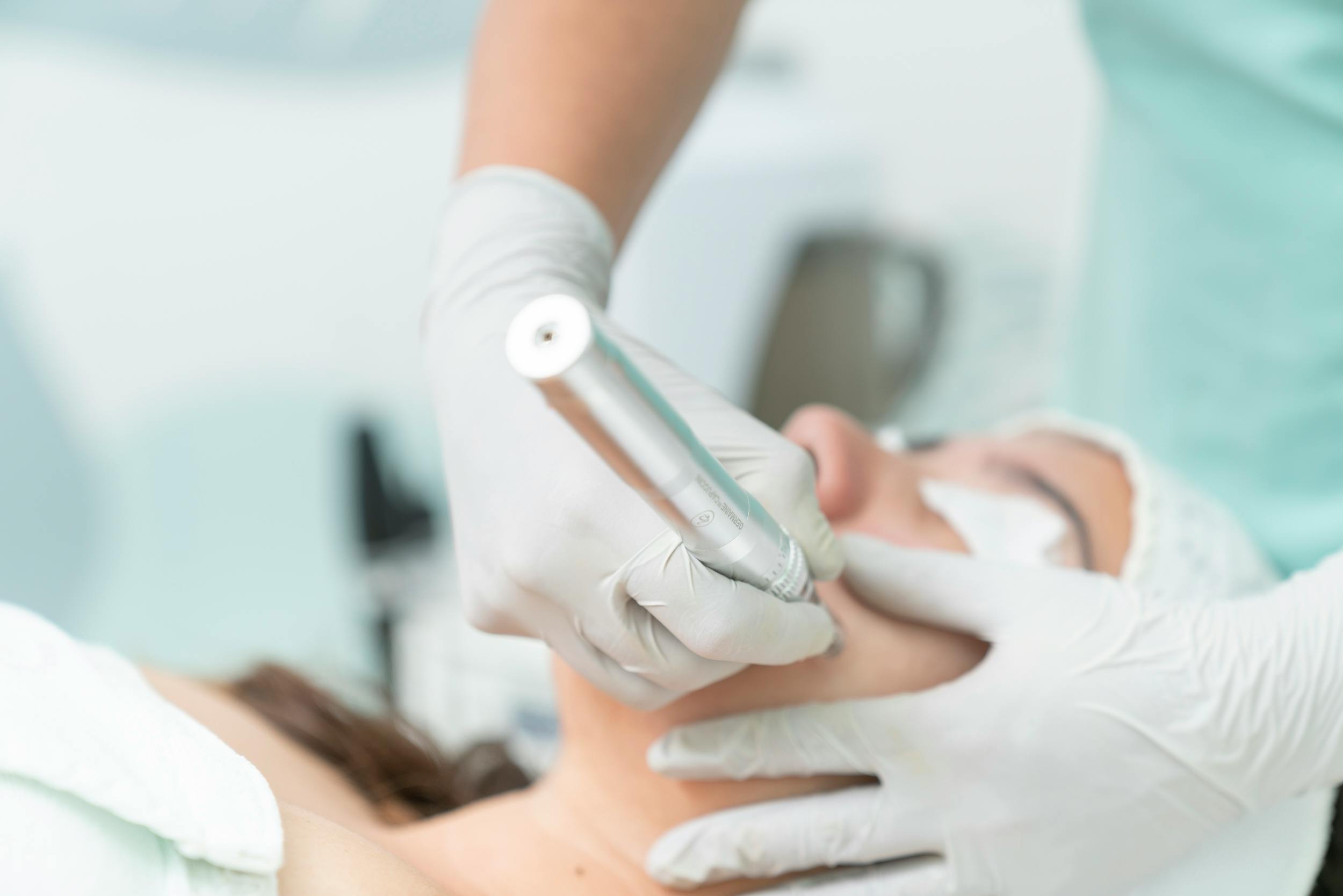
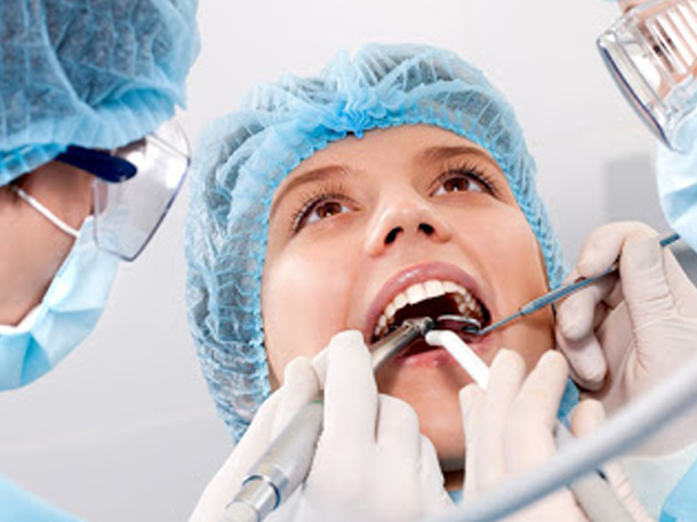
Jaw bone cyst surgeries are highly technical focus-driven advanced surgical procedures carried out painlessly under digital local anesthesia blockade in polyclinic operating rooms subject to full sterilization protocols. First, the gum tissue in the projection of the lesion is loosened along a smooth line with the help of a scalpel and lifted over the periosteum bone membrane (a flap is opened). A micro bone window (ostectomy) is opened on the exposed deflated bone base with the help of smart surgical burs; micro-surgical curette tips infiltrating through this window scrape the cyst capsule from the bone smoothly and remove it in one piece. After the cleaned intra-bone cavity is washed with antiseptics and grafted, the mucosa is closed without tension using primary sutures.
- Opening the Bone Window: Micro-drilling calibration that will apply minimum trauma to the intact bone roof behind the cyst.
- Scraping the Capsule (Decortication): The phase of microscopically shaving the bone wall without leaving a single cell remnant on the lumen wall.
- Primary Leakproof Closure: Combining the tissue margins seamlessly with microscopic suture threads to prevent oral fluids from leaking into the bone.
Why Are Digital Imaging Methods Important in Cyst Operations?
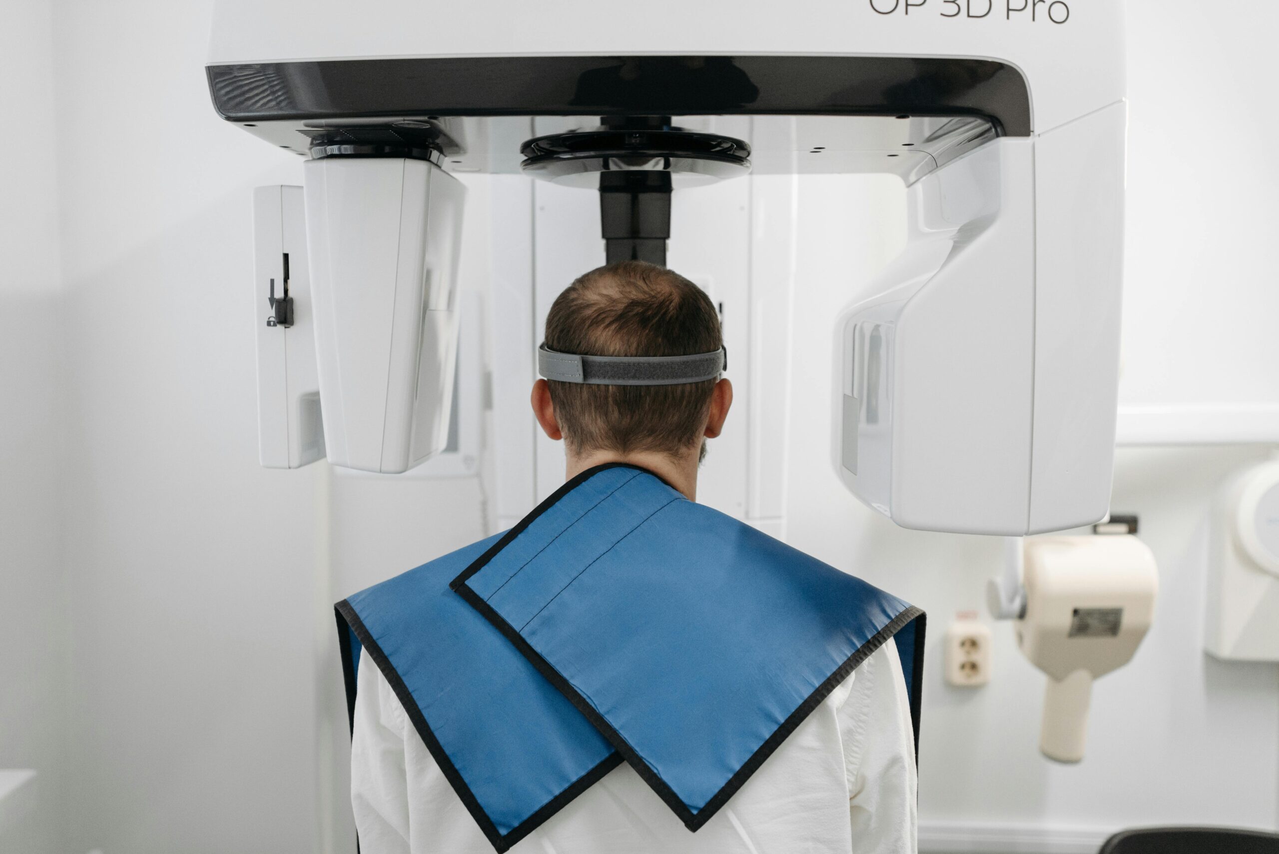
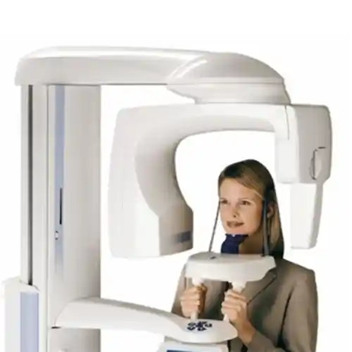
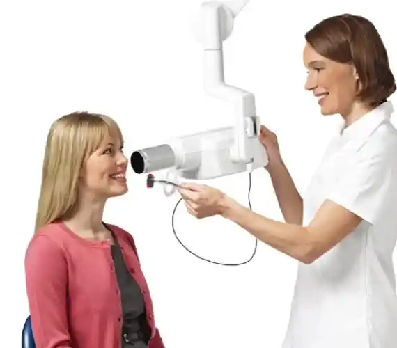
Digital 3D imaging systems in jaw cyst surgeries register surgical safety at the highest note by zeroing out the depth blindness and superimposed error margins brought by two-dimensional classical X-rays. Thanks to CBCT 3D dental tomography scanning, the surgeon examines the volume of the cyst inside the bone, whether it perforates the cortical walls, and its exact milimetric distance to the lower jaw main nerve channel in three axes (axial, coronal, sagittal) on a 1:1 scale; this digital accuracy prevents blind shots from being fired during surgery, definitively breaking the complication chain of permanent facial paralysis and nerve rupture.
- Volumetric Simulation: Tracing the surgical route by 3D modeling the melting boundaries of the cyst in the jawbone on the computer.
- Milling / Surgical Guide Integration: The luxury of producing robotic navigation plates that show exactly where the bone will be drilled in lesions at very critical boundaries.
How Long Does a Cyst Operation Take?
The active chairside session duration of cystectomy operations varies depending on the skeletal diameter of the lesion, its positional depth inside the bone, and the complexity of the grafting (bone powder transplantation) steps. The complete enucleation phases of a shallow radicular cyst involving a single tooth root are usually completed in a comfortable session of 20 to 30 minutes; however, in wide developmental cyst surgeries wrapping around multiple teeth and thinning the bone walls, the operation time is meticulously shaped between 45 to 60 minutes per session on average, as pin fixations and milimetric suturing steps will be required.
- Small Defect Surgeries: 20 – 30 Minutes (Fast scraping and antiseptic irrigation period; a schedule that does not tire the patient).
- Large Reconstructive Surgeries: 45 – 60 Minutes (Advanced craftsmanship duration containing bone block fixations and PRF locks).
What Anesthesia Methods Are Used During Cyst Operation?
Anesthesia technologies with high vascular safety are deployed in modern jaw surgery to eliminate the patient’s psychological stress level at the moment of surgery and to render pain transmission lines completely insensitive. In standard cases, painless local anesthetic solutions infiltrated at a rate suitable for tissue resistance with the help of computer-controlled digital anesthesia devices are the primary rule. However, for cases with the highest dental phobia level, surgery room anxiety, or aggressive cases where huge lesions will be cleared in a single session, sedation (semi-sleep) or general anesthesia (narcosis) infrastructures are successfully integrated to manage the process in a full relaxation mode.
- Deep Mandibular Blockade: The security of locking the main nerve trunk providing intra-bone transmission directly from the root in lower jaw lesions.
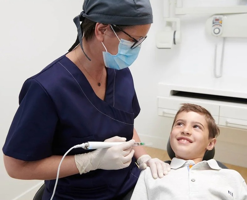
- Computerized Injection (The Wand): The comfort of infiltrating the solution at micro-vessel speed without creating pressure pain in the tissue.
How Does the Healing Process Proceed After Cyst Operation?
Although it contains intra-bone surgical intervention, the tissue recovery phase after the operation proceeds with extremely predictable and linear steps thanks to minimally invasive micro-surgical approaches. Forming a temporary micro-edema (swelling) in the cheek area during the first 48 hours following the surgery due to intra-bone stimulation and vascular sealing steps is a completely natural physiological wound repair reflex given by the extracellular matrix. The healing process flows smoothly; at the 10th day, the mucosal sutures close tightly, and the complete weaving of the intra-bone cavity with new cortical bone trabeculae requires an average ossification schedule of 6 months.
- First 48 Hours (Edema Peak): The phase where the swelling is most visible; the process is comfortably absorbed with intermittent cold packs applied from outside the cheek.
- 1st Week Turning Point: Epithelial coverage is completed, sutures are removed without leaking during the clinical control, and facial swelling completely subsides.
- 6th Month Final Restoration: The phase of transitioning into the phase fully ready for implantation, with graft particles leaving their place to living, vascularized real human bone.
What Should Be Considered After Cyst Operation?
The care discipline that the patient will follow in the home environment after the procedure is of vital importance to make the success of regenerative jaw surgery permanent, prevent the displacement of the bone powders placed in the bone hole, and protect the suture lines. Since the surgery bed temporarily transitions into an extremely delicate phase against external intraoral pressure fluctuations during the first days, it is a medical necessity for the patient to preserve the homeostasis balance.
The aspects that must be meticulously followed at home during the critical period after the cyst surgery are as follows:
- For the first 24 hours following the surgery, the mouth should not be vacuumed and spit out in order to protect the clot integrity and not to revive leaking bleeding; saliva should be swallowed instead.
- For the first 1 week, hard, crusty, grainy foods must be cut off to protect the micro-integrity of the sutures from mechanical tears; nutrition should be soft with a puree texture at room temperature.
- For at least 3 days, smoking, tobacco products, and alcohol consumption must be completely cut off so as not to paralyze the capillary vessel repair speed and growth factors at the wound site.
- For the first 48 hours, hot baths, saunas, Turkish baths, and heavy sports activities that will raise facial capillary pressure and increase cheek swelling should be postponed.
How Should Oral Care Be Performed After Cyst Operation?
Post-operative home hygiene processes must contain a delicate balanced discipline that will limit the intraoral pathogenic bacterial population to prevent graft infections but will also not mechanically batter the fresh suture lines. That exact local mucosal line where the operation was performed should strictly not be brushed with normal hard toothbrushes for the first 10 days; while other teeth throughout the mouth are cleaned as standard, the hygiene of the procedure area is entrusted to chemical protection.
- Antiseptic Mouthwash Shield: Performing gentle rinsing morning and evening with chlorhexidine-based medical mouthwashes without touching the toothbrush, starting 24 hours after the surgery.
- Transition to Ultra Soft Brush: Utilizing filamented micro-surgical brushes manufactured specifically for sensitive post-operative periods to clean adjacent teeth in that area starting from the 10th day.
What Are the Contributions of Cyst Operations to Oral and Maxillofacial Health?
The removal of pathological cystic cavities by scraping them from the jawbone is the clearest preventive medicine investment that saves the oral health ecosystem from destruction on a linear line. It does not just end a local infection focus; it definitively breaks the risk chain of the lesion growing insidiously in the lower layers of the jawbone from completely melting the jaw skeleton and creating a spontaneous bone fracture. When the chronic toxin load in the tissues is relieved, root resorption damage of adjacent sound teeth is halted, and bone quality is taken into permanent safety.
- Skeletal Rigidity Reinforcement: Regaining the mechanical chewing power of the jaw by grafting the bone hole emptied by the cyst.
- Nerve Line Isolation: Eliminating the risks of facial numbness by removing the chronic pressure on the main mandibular nerve channel.
Why Are Regular Controls Important After Cyst Operation?
Periodic control appointments following the surgical intervention involve the most vital examination steps that monitor intra-bone ossification quality and stop the possibilities of the lesion recurring in the long term early in the journey. Some cyst types (especially odontogenic keratocysts) may exhibit a tendency to regenerate (recur) from micro-wall remnants due to their histological character; the physician monitors the biological state of the jaw with radiological data during these sessions, safely finalizing the schedule of permanent implant prosthetic phases.
Which Treatments Can Be Planned Together with Cyst Operations?
Cystectomy surgeries can be planned in a combined synergy with other modern instruments of dentistry in order to preserve intraoral hard and soft tissue integrity and to regain facial aesthetics. While infrastructure bone destruction is reinforced with surgery, form losses in the upper layers can be treated simultaneously with different technologies; the combination schedule is designed according to the result of the pre-operative facial analysis to be performed by the specialist physician.
- Dental Implantology: Completing the missing teeth in that area with titanium screws after the cyst is cleared and bone powders mature (following a 6-month waiting period),
- Root Canal Treatment (Endodontics): Keeping the infected teeth that are the source of the cyst in the mouth by finishing their root canal treatments under a microscope before or during the surgery,
- Regenerative Bone Grafting: The processes of filling the skeletal cavity formed by the removal of the cyst with PRF (living fibrin) and bone powders within the same session.
How Is Long-Term Follow-up of Cyst Operations Conducted?
The post-operative long-term follow-up of jaw cysts is conducted within the framework of a periodic “radiological and clinical monitoring” protocol registered in accordance with medical standards. After the permanent delivery is finished, the patient is called for periodic control appointments at the 3rd month, 6th month, 1st year, and once a year for the first following 3 years. The process is kept safe by monitoring down to micron resolution how much of the grafted bone hole has turned into living human bone and whether any new dark shadow (sign of recurrence) has awakened on the lesion boundary lines via low-radiation digital panoramic X-rays or CBCT tomography slices taken during these sessions.
What Are the Frequently Asked Questions About Cyst Operations?
The most common practical concerns arising in the minds of clients planning surgical processes to get rid of cysts in the jawbone and their polyclinic medical counters are presented below:
Does jaw cyst surgery hurt a lot in the operation chair during the session?
No, absolutely not; you will not feel the slightest pain or ache. Prior to the procedure, strong clinical anesthetic gels are applied to the gingival mucosa, followed by local solutions delivered at a rate matching tissue resistance with computer-controlled digital anesthesia units to provide full insensitivity; since nerve transmission lines are completely shut down throughout the session, you only perceive the light touch of the instruments and the water coolness, and the procedure is completed with high comfort.
Will you completely extract my teeth where the cyst is located after the cyst is cleared?
No, the primary goal in modern conservative jaw surgery is not to extract teeth, but to keep them in the mouth. Root canal treatments are applied to the teeth associated with the cyst under a microscope before or during the surgery, and only the inflamed cyst bag at the root tip is scraped and removed during the operation; the tooth itself continues to maintain its viability and chewing function inside the jawbone. Only fractured teeth that have lost 100% of their bone support are sacrificed.
Is there a risk of jaw cysts turning into cancer in the future if they are not cleared or if the surgery is postponed?
Radicular (tooth-derived) or dentigerous cysts, which are most commonly seen in the oral and maxillofacial region, are “benign” structures due to their histological character and do not show a tendency to evolve directly into cancer. However, if the lesion is neglected and not cleared for years, it completely melts the jawbone, leading to a fracture of the jaw, or very rarely, epithelial cells in the cyst wall may undergo metamorphosis, triggering more aggressive tumor structures called “ameloblastoma”; postponing should not be done.
Why do you send the piece removed after the cyst surgery to the pathology laboratory for examination?
This situation is an uncompromised necessity registered in accordance with medical ethics and international surgical standards. Some insidious structures that look like a simple tooth inflammation cyst on an X-ray film may turn out to be a more aggressive charactered keratocyst or the beginning of a tumor when examined under a microscope; pathology registration is mandatory to give the lesion a definitive name, to map out the post-operative recurrence risk schedule without error, and to offer the patient 100% correct diagnosis assurance.
Can I return to my normal professional and daily life immediately on the day the cyst operation is completed?
Yes, cystectomy surgeries are completely local micro-surgical steps that do not disrupt consciousness, do not affect motor manual skills, and do not involve the weight of general anesthesia; therefore, you can get up from the chair the exact second the session ends and continue your normal daily, professional, social, or academic life rhythm uninterruptedly from where it left off; there is no clinical barrier to driving or participating in business meetings immediately after the procedure.
Who Are We? & Panorama Ankara Oral and Dental Health Center
Panorama Ankara is a licensed oral and dental health center offering institutional health assurance to its patients by combining an academic specialist staff and the latest global bio-technological digital infrastructure under a single roof in all specialty branches of dentistry, primarily oral, dental, and maxillofacial surgery, advanced implantology, microscopic endodontics, periodontology, and aesthetic dentistry. Instead of abandoning pathological cyst foci growing insidiously in the jawbone to the simplicity of rough analog force that breaks the bone, our center attaches vital importance to reaching permanent oral integrity by centering “microscopic tissue-preserving surgery” principles with evidence-based medical disciplines, and operates seamless 3D Digital Diagnostic Modeling and Jaw Cyst Operations ecosystems with calendar discipline from the very beginning to the end of the clinical operation workflow.
Under the roof of Panorama Ankara, absolute sterilization, high patient safety, transparency, and honesty principles are operated with full compliance with medical codes in all diagnostic, radiological scanning, design, and macroscopic surgical interventions required by regulations. In our clinical practices, while analyzing jawbone limits and the depth curves of the stain with 3D tomographies in milimetric sections, smart surgical cauteries that coagulate vascular integrity and ultrasonic piezo surgery units that do not damage surrounding hard tissue are applied with milimetric precision under the supervision of our expert jaw surgeon staff. Our aim is not to impose old-fashioned, slow, bone-battering, and distressing analog operation patterns on our patients that take weeks to heal, but to analyze each individual’s jaw skeletal quality, occlusion mechanics, and nerve channels on the virtual screen in 3D before the operation, achieving maximum session comfort, seamless comfort standards without sutures, and healthy, smooth oral functions that you will use with an unshakeable confidence for a lifetime. If you are tired of your missing teeth, persistent root leaks, or jaw swellings when looking in the mirror, yet feel anxious about surgical risks or fear of pain and wish to start your smart digital treatment process from scratch in an expert corporate center environment with scientific guarantee; you can safely protect your health and smile integrity by contacting the expert staff of Panorama Ankara for detailed 3D scanning and digital planning analysis.