What Are Bone Grafts and Membranes?
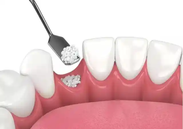
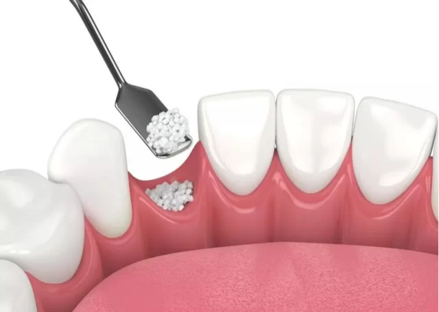
Bone grafts and membranes are the most fundamental biomaterial combination that realizes the principles of “Guided Tissue Regeneration” (GTR) and “Guided Bone Regeneration” (GBR) in oral and maxillofacial surgery. Tooth extractions, traumas, cysts, or chronic periodontitis infections cause irreversible volumetric collapses in the jawbone. Bone graft (bone powder) has a micro-porous structure and acts as a three-dimensional scaffold that guides the body’s own bone-building cells (osteoblasts) in the area where it is placed. The membrane, on the other hand, is a medical film that prevents connective tissue cells from infiltrating the bone mineralization area, thus creating a sealed barrier for the graft particles to safely transform into genuine bone tissue.
- Osteoconductive Matrix: Establishing a biological scaffold network where new bone cells can attach and proliferate.
- Selective Cell Filtration: Mechanically preventing rapidly dividing epithelial cells from entering the bone cavity and undermining osteogenesis.
- Bio-Integration Profile: Integration of certified sterile bio-materials of medical standards that do not disrupt the body’s homeostasis balance.
In Which Cases Are Bone Grafts Applied?
Jawbone grafting operations are applied as a clinical and anatomical necessity in cases where the existing hard tissue volume is insufficient to surround the dental implant screws with a cortical shield of at least 1.5 – 2 millimeters thickness from each wall. The alveolar bone, which loses its function after teeth are extracted, enters a rapid resorption (melting) process due to gravity and facial pressures. In cases where the vertical height or horizontal width of the bone regresses to a level that jeopardizes the stabilization of the implant, grafting protocols are strictly utilized to carry the surgical corridors to safe limits.
The primary clinical situations where bone graft surgical processes are definitely deployed are as follows:
- In cases where the maxillary sinus cavities sag downward (requiring sinus lifting) in the upper jaw molar region due to long-term toothlessness,
- For the purpose of socket preservation to prevent the empty space (alveolar socket) from collapsing and losing volume after tooth extraction,
- In filling large anatomical defects remaining in the hard tissue after operations for persistent cysts, tumors, or trauma developing within the jawbone,
- In horizontal bone widening (ridge split) surgeries where the jawbone becomes excessively narrow transversely and takes on a knife-edge morphology.
What Is a Membrane Application?
Membrane application is the process of adapting protective medical films to the surgical site, which cover and protect the placed bone powders like a hat, provide mechanical stabilization, and filter soft tissue cell transitions in regenerative oral surgery. In human physiology, gingival epithelial cells have the capacity to divide and move approximately 10 times faster compared to bone-forming osteoblast cells. If the area where the graft is applied is not isolated with a membrane, gingival cells rapidly infiltrate that area, fill the spaces between the bone powders, completely sabotage the bone mineralization process, and lead to soft connective tissue formation. The membrane definitively cuts off this leakage.
- Resorbable Membranes: Membranes usually obtained from bovine or porcine collagen, which are absorbed by the body after healing and do not cause the burden of a second surgery.
- Non-Resorbable Membranes: These are titanium-reinforced, stitched synthetic membranes; they protect the bone like a mold in large vertical volume constructions and are surgically removed from the line months later.
- Micro Pin Fixation: The phase where the complete immobility of the membrane is verified by nailing it to the jawbone with the help of titanium micro tacks (pins).
Who Are Bone Grafts and Membranes Suitable For?
Hard tissue regeneration treatments are suitable for any adult individual over the age of 18 who is hindered by bone insufficiency, wishes to achieve the comfort of fixed implant teeth, does not have uncontrolled systemic pathologies, and has completed intraoral hard and soft tissue development. The system exhibits maximum bio-compatibility and a high safety profile in every patient whose bone metabolism functions healthily. However, patients with uncontrolled severe diabetes, individuals who have recently received radiotherapy to the head and neck region, or patients using strong intravenous bisphosphonate group drugs that suppress bone metabolism are not suitable candidates for these surgical bone construction processes.
| Patient Group / Skeletal Condition | Degree of Surgical Suitability | Expected Bone and Cellular Response |
|---|---|---|
| Advanced Bone Resorption Due to Chronic Toothlessness | Completely Suitable / Primary Indication | Provides full osseointegration to implant screws by increasing the width and height of the bone. |
| Sensitive Patients Demanding Rapid Healing and Minimal Swelling | Completely Suitable / PRF Supported | Accelerates the postoperative wound closure rate and reduces edema thanks to PRF membranes. |
| Those Who Continue Heavy Smoking Tobecco | Risky / Controlled Suitable | Cigarette smoke narrows the capillaries; it radically reduces the attachment success and mineralization lifespan of the graft. |
| Presence of Active Jawbone Infection (Osteomyelitis) | Not Suitable (Infection Phase) | Grafting steps are postponed until the active microbial infection inside the bone is completely cleared. |
How Is the Evaluation Performed Before a Bone Graft?
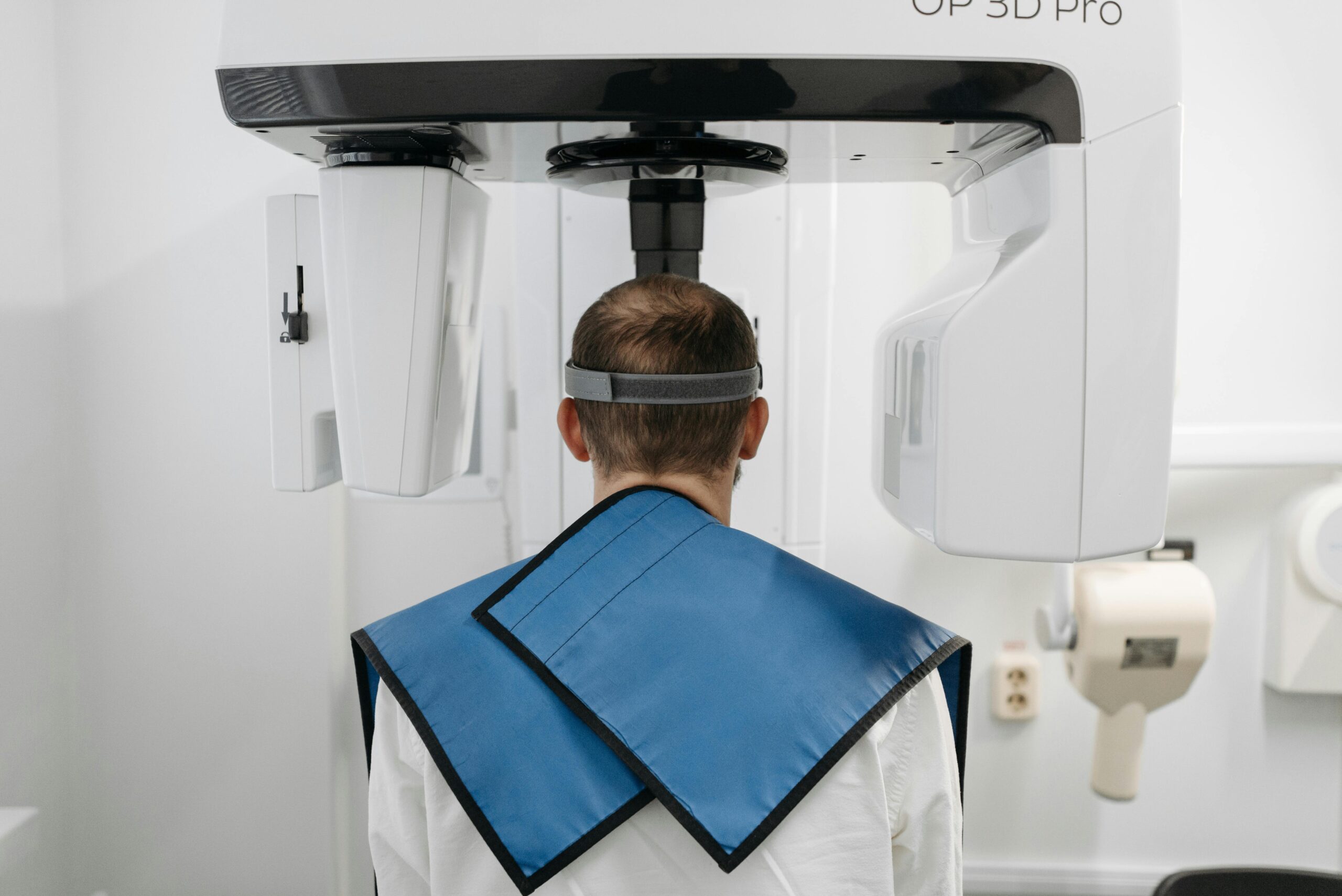
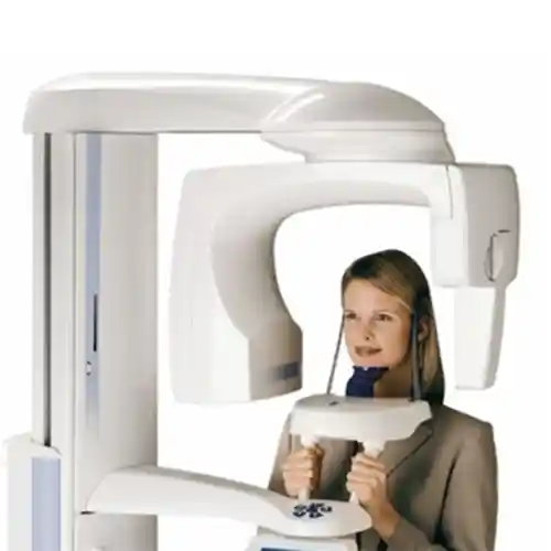
The clinical evaluation carried out before moving on to the operation steps is the most vital analysis phase that draws the millimetric boundaries of the bone injection and guarantees the long-term unshakable permanence of the prosthesis. During the examination, the surgeon examines the thickness (biotype) and attachment quality of the gingival tissue with the help of periodontal probes. At this stage, a CBCT 3D dental tomography scan is requested from the patient in order to overcome the limitations of two-dimensional classical X-rays; bone width, vertical height, and density are measured on a 1:1 scale through tomography slices, and the required graft volume (syringe dose in cc) is rationally determined before the surgery.
- Bone Defect Morphology: Selection of the most accurate material combination according to the shape of the resorption (one-walled, multi-walled defect classification).
- Anatomik Border Isolation: 3D mapping of the lower jaw main nerve canal (inferior alveolar nerve) and upper jaw sinus floor height limits.
- Systemic Analysis Audit: Screening the patient’s HbA1c (blood sugar) values and bone metabolism rate parameters with laboratory tests.
How Are Bone Graft and Membrane Applications Planned?
The planning of the bone graft and membrane workflow is carried out through a phased algorithm based on the vertical or horizontal chronicity of bone loss, the patient’s general immune potential, and suitability for simultaneous implant placement (immediate implantation). Hasty steps are avoided during the planning process; because the transformation of graft powders into the patient’s own living, vascularized genuine bone requires an ossification schedule that biologically takes months. In the planning, the maturation phases of bone gain are meticulously scheduled into the patient’s social calendar.
- Phase 1 (Purification and Preparation): Scaling teeth, filling cavities to reduce the intraoral bacterial load, and certifying a sterile operation ground.
- Phase 2 (Surgical Phase): Opening the bone with flap surgery, performing cortical perforations, graft-membrane integration, and tight suturing.
- Phase 3 (Ossification Waiting Period): The phase where the tissue is left undisturbed for 4 to 6 months for the powder particles to evolve into living human bone.
- Phase 4 (Restoration): Placing titanium screws into the newly formed hard bone bed and mounting the superstructure crown veneer.
What Are the Types of Grafts Used in Bone Grafting?
The materials used in bone dusting procedures are classified into four main groups according to the biological raw material sources from which they are obtained and their histological mechanisms of action within the jawbone. Which material will be selected is calibrated by the specialist physician according to the size of the defect and the targeted mineralization rate. All of the materials are certified products that have passed high bio-compatibility tests in medical standards.
- Autogenous Grafts (Gold Standard): Living bone blocks scraped and taken from the patient’s own body (from the chin or posterior jawbone); since it contains cells, it is the fastest mineralizing type.
- Allografts (Human-Sourced): Freeze-dried sterile human bone powders obtained from medical bone banks.
- Xenografts (Animal-Sourced): The most frequently used safe bone powders obtained from bovine or porcine bones, purified of organic proteins, leaving only the calcium mineral skeleton.
- Alloplastic Grafts (Synthetic): Calcium phosphate or hydroxyapatite-based bio-ceramic crystals produced artificially in a laboratory environment.
How Do Membrane Applications Support Bone Formation?
The supporting power of barrier membranes on bone cell architecture is based on the principles of “selective cell filtration” and “surgical area stabilization”. When the membrane is laid over the bone powders, it isolates that area from the pressure of intraoral mobile mimetic muscles, preventing the bone particles from displacing (migration). Thanks to its microscopic porous structure, while it cuts off harmful cell leaks, it allows the smooth infiltration of blood flow, nourishing fluids, and oxygen molecules into the area, which are vital for mineralization; this situation certifies smooth ossification by protecting the sub-tissue homeostasis balance.
“Membrane application is a protective medical umbrella drawn over your bone powders; it provides a safe working room for bone-building cells by preventing epithelial cells from leaking inside.”
How Do Bone Grafts Contribute to Implant Treatment?
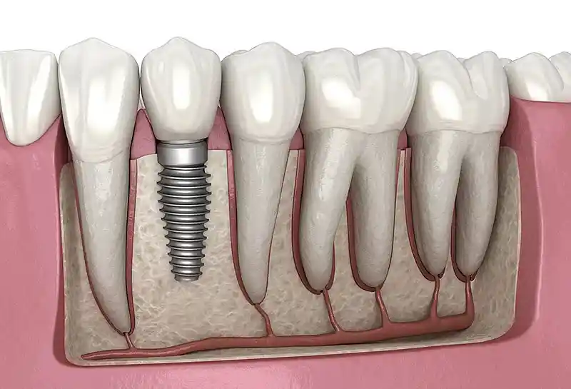
Long-term clinical success in dental implant surgeries depends directly on maintaining the bone tissue surrounding the titanium screw as a cortical shield with a thickness of at least 1.5 – 2 mm; bone grafts are the most uncompromising guarantee of this reinforcement. If an implant is placed into a thin or melted jawbone with forced attempts, the threads of the screw will soon be exposed, the gums will turn purple, and an insidious implant inflammation called “peri-implantitis” will be triggered, causing the screw to loosen and fall out. Grafting thickens thin bone walls, ensuring 100% interlocking (osseointegration) of titanium surfaces with bone cells and laying the groundwork for the implant to remain in the mouth for a lifetime.
- Horizontal and Vertical Bone Gain: Ensuring that the implant remains completely embedded and safe within the bone membrane.
- Aesthetic Pink Line Support: Establishing a perfect pink aesthetic harmony at the collar level of the crowns by preventing the recession of the gums fed by the bone.
How Is Graft Application Performed in Cases of Jawbone Insufficiency?
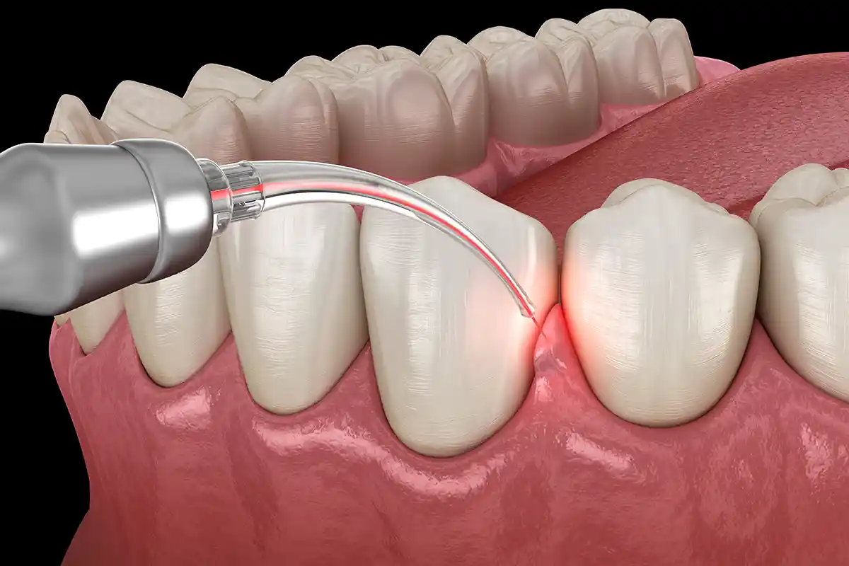
Graft injection in cases of jawbone insufficiency is a sensitive jaw surgery procedure performed under local anesthesia block in fully sterilized polyclinic operating rooms. First, the gingival tissue in the area where resorption occurs is loosened along a smooth line with the help of a scalpel or cautery and lifted over the bone membrane (periosteum) (a flap is opened). After opening micro-holes (cortical perforations) to stimulate blood supply on the exposed flat bone surface, putty or powder-form graft materials are millimetrically placed into the defect area; the membrane is laid over the powders and closed with sutures, ending the operation.
- Cortical Perforation: Micro-drilling of the bone to ensure stem cell leakage from the underlying living spongy bone to the graft area.
- Block / Powder Selection: Calibration of preferring screwed bone blocks (onlay graft) in severe resorptions, and granule powders in superficial resorptions.
Why Is Digital Planning Important in Bone Graft and Membrane Applications?
Digital planning in regenerative jaw surgery zeroes out the margin of error that human eyes and analog measurements may bring, certifying the execution of evidence-based and fully predictable operational algorithms. Thanks to the processing of 3D dental tomography data (DICOM) in special surgical software, the three-dimensional volume of the bone defect is calculated millimetrically. The surgeon simulates on the screen from which angle, at what depth they will work, and how many cc of graft they will use before starting the surgery; this digital foresight definitively prevents encountering unexpected surprises during the operation.
- 3D Navigation Safety: Zeroing trauma risks by tomographically marking intra-bone anatomical nerve pathways and vascular networks.
- Precise Material Selection: Virtual mapping of the most suitable membrane shape and pin fixation coordinates for the defect morphology.
How Long Do Bone Graft and Membrane Applications Take?
Bone graft and membrane combination operations require more meticulous time management in terms of active chairside session duration compared to a standard tooth extraction, as they include millimetric suturing and fixation steps that require a high depth of focus from the surgeon. The active surgical phase of the process is smoothly shaped, usually between 30 to 60 minutes per session, depending on the volume of the bone defect to be intervened and the width of the area; since the digital anesthesia block phase carried out during the session completely blocks the feeling of pain, time flows with comfort for the patient.
- Single Tooth Socket Grafting: A comfortable session of approximately 30 minutes, including cleaning and powder placement.
- Extensive Advanced Sinus Lifting Surgeries: An operation time of approximately 45 – 60 minutes as it involves skeletal shaving and membrane fixations.
How Does the Recovery Process Proceed After Bone Graft and Membrane Applications?
The tissue recovery phase after the operation proceeds with highly predictable and comfortable steps, despite containing surgical flap sutures, thanks to minimal invasive micro-surgical techniques. During the first 48 hours following the surgery, the formation of a temporary micro-edema (swelling) in the cheek area due to intra-bone stimulation and vascular stabilization steps is a completely natural physiological reaction given by the extracellular matrix. The healing process flows on a linear line; at the end of the first week, the mucosal sutures reach the smoothness to be removed, and the underlying mineralization cycle enters the maturation phase in an insidious silence.
- First 48 Hours (Peak Edema Phase): The phase where swelling is most visible; this process is easily absorbed with intermittent cold packs applied from outside the cheek.
- 7th to 10th Days: Epithelial coverage is completed, sutures are removed without tingling in the clinical control, and cheek swelling completely subsides.
- 4th Month and After (Ossification): Passing to the phase fully ready for implantation, with powder particles leaving their place to living, vascularized real human bone.
What Should Be Considered After a Bone Graft?
In order to make the success of the regenerative bone operation permanent, prevent the displacement of the bone powder particles placed under the tissue, and protect the suture lines, the care discipline that the patient will follow at home after the procedure is of vital importance. Since the newly transplanted bone powders are not yet fully mineralized in the first days and are open to leaks, it is a clinical rule for the patient to show full loyalty to pressure and hygiene rules.
The medical rules that must be meticulously followed at home during the critical period after the procedure are as follows:
- For the first 24 hours after the procedure, saliva should be swallowed instead of vacuuming and spitting out in order to protect the clot balance (spitting triggers bleeding).
- For the first week, hard, crusty, grainy foods should be cut off to protect the suture lines from mechanical tears; soft food in puree consistency at room temperature should be consumed.
- In cases where sinus lifting is performed, in order to prevent upper jaw bone pressure fluctuations, nose cleaning should not be done by blowing hard for at least 2 weeks, and the mouth should be kept open when sneezing.
- For at least 1 month, sauna, Turkish bath, solarium, and heavy sports activities that will increase leakage and bruising risks by increasing the capillary pressure of the tissue should be postponed.
How Should Oral Care Be Performed After a Membrane Application?
Postoperative home hygiene of the area where the membrane is laid must include a precisely balanced protocol that will not disrupt the micro-integrity of the sutures, will not dislodge the barrier membrane, but will also eliminate bacterial plaque accumulations that can produce infection on the wound edges. That exact local mucosal line where the operation was performed should definitely not be brushed with normal hard toothbrushes for the first 10 days; while other teeth throughout the mouth are cleaned regularly, the hygiene of the procedure area is entrusted to chemical antiseptics.
- Antiseptic Mouthwash Shield: Gentle rinsing morning and evening with chlorhexidine-based medical mouthwashes for the first week to prevent plaque accumulation mechanically without contact.
- Switching to Ultra Soft Brush: After the sutures are removed, the use of filamented micro-surgical brushes produced only for sensitive postoperative periods to clean the tooth surfaces in that area.
How Can PRF Support Be Used in Bone Graft Applications?
In modern regenerative surgery, bone dusting procedures are brought to revolutionary success charts by combining them with 100% autologous PRF (Platelet-Rich Fibrin) technology obtained by centrifuging blood taken from the patient’s own arm vein. The separated living yellow fibrin clot gel is blended with bone powder particles to bring it into the “Sticky Bone” form. This smart combination prevents the chalky powders from scattering, making it easier to place them in the defect area like a mold; at the same time, thanks to the intense growth factors it contains, it accelerates the postoperative wound site cell division rate and carries healing comfort to the peak.
- Biological Adhesive Power: Eliminating the risk of recurrence of graft particles coagulating and escaping into facial spaces.
- Triggering Rapid Angiogenesis: Fibrin networks accelerating the weaving of micro-capillary bed structures that will feed the new bone mass.
What Are the Advantages of Bone Grafts and Membrane Applications?
Among holistic maxillofacial restorations, the bone graft and membrane workflow is the most visionary surgical solution that brings patients who have given up hope on dental treatment due to bone resorption to permanent fixed tooth comfort. Thickening knife-edge thinned jawbones with a power beyond non-surgical solutions makes this method one of the most popular and evidence-based processes of modern implantology.
- Makes the Impossible Possible: It safely carries borderline cases, where implants were said to be impossible due to lack of bone, to the treatable phase.
- High Bio-Safety: It eliminates tissue rejection or allergic reaction risks thanks to the use of certified materials in medical standards.
- Rapid and Comfortable Wound Closure: It minimizes postoperative infection, pain, and abscess complication coefficients thanks to the membrane shield.
- Permanent Bone Infrastructure: The newly formed bone structure functions as a permanent protective shield skeleton around titanium screws for life.
Which Treatments Can Bone Graft and Membrane Applications Be Planned With?
Hard tissue regeneration surgeries can be planned in an integrated combination with other medical surgical and restorative dental protocols in order to holistically elevate facial aesthetic architecture, bone rigidity, and masticatory function quality. While lower structural bone foundations are reinforced by surgery, form losses in upper layers can be treated simultaneously with different technologies.
- Dental Implant Treatment: A chain of placing titanium screws in the same session with bone dusting or after the bone matures.
- Sinus Lifting (Sinus Elevation): Operations where the sagging sinus membrane is lifted up and the cavity underneath is filled with graft and PRF.
- Surgical Extractions of Impacted Teeth: The process of filling huge cavities left in the jawbone after large wisdom teeth extractions with PRF and graft to ensure rapid mineralization.
How Do Bone Grafts and Membranes Support Implant Success?
The molecular-level interlocking of titanium implant screws with the jawbone (osseointegration biology) depends on all surfaces of the screw remaining embedded in a stable, living, and vascularized bone bed. The combination of graft and membrane reinforces the bone walls thinned by insidious resorption, blocking the stress tensions that may occur in the implant neck region; this mechanical support prevents the implant from loosening under hydraulic chewing forces, prevents gingival recession, and certifies that the restoration remains problem-free in the mouth for decades.
- Axial Load Distribution: The capacity of the thickened bone skeleton to homogeneously distribute hydraulic chewing forces to the implant body.
- Marginal Bone Preservation: Establishing the peri-implantitis infection barrier by preserving the cortical bone crest height around the implant.
What Are the Frequently Asked Questions About Bone Grafts and Membranes?
The most common practical anxieties arising in the minds of patients planning to apply for these regenerative surgical components due to jawbone insufficiency and their uncensored medical answers are presented below:
Do these bone powders (grafts) used cause tissue rejection or an allergic reaction in my body?
No, absolutely not. Xenograft (animal-sourced) or allograft (human-sourced) bone powders used in modern clinical practices are subjected to very high-temperature thermal and chemical processes in international medical laboratories; all organic proteins, cells, and DNA remnants within them are completely purified, leaving only a sterile calcium mineral skeleton. Therefore, the risk of the body perceiving this powder as a foreign enemy and initiating tissue rejection or an allergy reaction is medically zero; the bone integrates with full compatibility.
Will the membrane film placed with the graft be surgically removed from my mouth after healing?
This situation varies according to the type of membrane chosen by our surgeon at the time of the surgery. In the vast majority of cases, “bio-absorbable” (resorbable) membranes produced from bovine or porcine collagen are preferred; these smart films are completely dissolved and absorbed by the body within a few months after completing their duty, and do not create the burden of a second removal surgery. Only non-resorbable titanium-reinforced membranes used in very large vertical bone block constructions are practically removed from the line months later while placing the implant.
After the operation, I feel that the bone powders placed in my mouth are spilling out or coming to my tongue grain by grain, is this normal?
Yes, it is completely normal and expected to have small, chalk-like gritty particles come into your mouth when you spit or touch your tongue to the suture lines for the first few days following the operation. A few excess particles of thousands of micro bone powders placed in the surgical area may leak out between the sutures into the mouth; this situation definitely does not affect the success or volume of the main bone mass inside, there is nothing to worry about.
How long does it take to fit permanent porcelain teeth to the area where the graft and membrane were placed?
The transformation of the placed bone powders into living, vascularized real bone tissue capable of carrying chewing loads (ossification biology) requires a fixed waiting schedule of an average of 4 to 6 months depending on the age of the patient and the size of the defect. During this waiting period, the grafts mature; when the period is completed, the bone quality is confirmed with a new 3D tomography scan, and the mounting phase of titanium screws or porcelain crowns is safely initiated.
Can I return to my normal social and professional life, my job, immediately on the day the operation is completed?
Yes, graft and membrane applications are completely local micro-surgical procedures that do not involve general anesthesia and do not disrupt consciousness and motor skills; therefore, you can get up from the chair the exact second the session ends and continue your normal daily, academic, or professional life rhythm uninterruptedly from where it left off. Applying cold packs to manage the slight swelling that may occur on the cheek on the first day and avoiding hot foods are just comfortable details that will not affect your practical life.
Who Are We? & Panorama Ankara Oral and Dental Health Center
Panorama Ankara is a licensed oral and dental health center that offers institutional health assurance to its patients by combining an academic specialist staff and the latest global bio-technological digital equipment under a single roof in all specialty branches of dentistry, especially oral, dental and maxillofacial surgery, advanced implantology, orthodontics, periodontology, and aesthetic dentistry. Instead of condemning advanced resorptions occurring in the jawbone, cyst defects, and sinus sagging obstacles to the simplicity of surgical extraction or removable palate prostheses, our center attaches vital importance to keeping the bone in the mouth by revitalizing it at the cellular level with evidence-based medical disciplines, and uncompromisingly operates original Graft, Membrane, and PRF Supported Jawbone Restoration ecosystems that offer high success charts in the clinical operation workflow.
Under the roof of Panorama Ankara, absolute sterilization, high patient safety, transparency, and honesty principles are operated with an uncompromising determination in accordance with the regulations in all radiological, scanning, design, and micro-surgical interventions. In our clinical practices, while analyzing the jawbone quality of the patient with 3D tomographies in millimetric sections, smart surgical cauteries that coagulate vascular integrity and certified centrifuge units that separate living fibrin membranes from your own blood are meticulously applied under the supervision of our specialist jaw surgeon staff. Our aim is not to force implants on insufficient bone tissues with rote-learned patterns and cause failure risks, but to analyze the facial golden ratio boundaries and skeletal bone density of each individual on the digital screen before the operation, and to achieve maximum session comfort, seamless comfort standards without damaging surrounding soft tissues and nerve pathways, and smooth fixed tooth restorations that you will use with an unshakable confidence as if they were your own bone for a lifetime. If you are tired of your missing teeth when you look in the mirror, but postpone your treatments due to bone resorption or fear of pain, and want to start your smart regenerative treatment process from scratch in an expert corporate center environment with scientific guarantee; you can safely protect your health and smile integrity by contacting the expert staff of Panorama Ankara for detailed analysis in line with your 3D tomographic evaluations.