What is a mini dental implant?
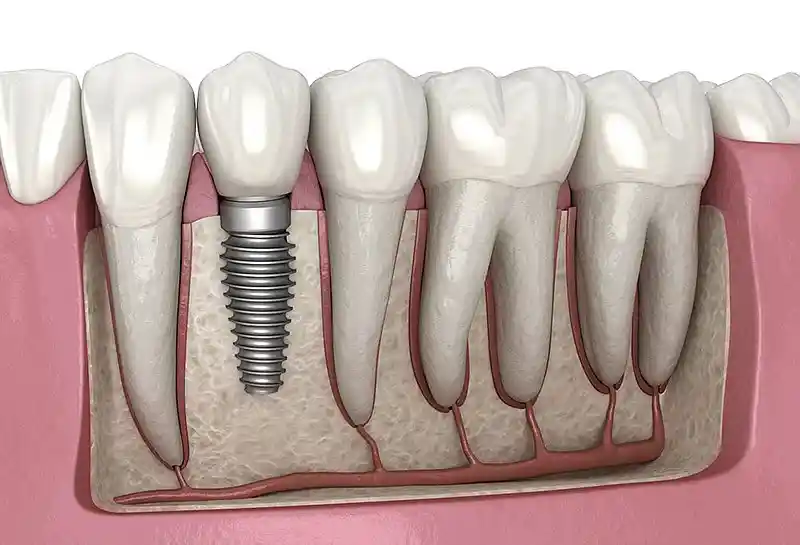
Unlike traditional two-piece implant systems, mini dental implants (MDI) are monoblock structures where the screw body and the prosthetic abutment are produced as a single piece. These screws, with diameters generally ranging from 1.8 mm to 2.9 mm, are manufactured from pure titanium or titanium alloys. They typically feature a spherical (ball-top) head at the apex, allowing the prosthesis to lock onto the implant via a snap-on mechanism. Because they are placed directly into the bone by opening a microscopic pilot hole without cutting the gum tissue, they are classified under the minimal invasive (tissue-preserving) implant category.
- Narrow Diameter Structure: Thanks to their millimetric thicknesses between 1.8 – 2.9 mm, they adapt to thin bone ridges.
- Monoblock Design: Since the screw and abutment parts are single-piece, they harbor no mechanical risks such as intermediate screw loosening or breakage.
- Ball-Head: They hold onto the metal sockets inside the removable prostheses tightly, reducing the vertical movement of the prosthesis to zero.
| Component | Structural Feature | Clinical Function |
|---|---|---|
| Thread Design | Self-tapping aggressive threads | Provides high initial hold (primary stability) even in insufficient bone. |
| Surface Feature | SLA or roughened titanium surface | Supports rapid fusion (osseointegration) of bone cells to the screw. |
In which situations are mini dental implants preferred?
Mini dental implants are preferred in situations where the jawbone has resorbed excessively (due to long-term edentulism) in the lower and upper jaws, and insufficient bone thickness remains to place standard implants. They are life-saving in systemic situations where elderly patients cannot tolerate heavy surgical procedures such as large bone grafting (bone powder) or sinus lifting. They are evaluated as a primary option in clinical presentations where the patient complains about their existing removable complete denture (false teeth) launching out of the mouth while speaking or eating, but also fears major surgeries. Additionally, they are used to create temporary anchorage (support) points in orthodontic treatments or to place teeth in very narrow gaps, such as in the lower anterior incisor region.
- Atrophic Jawbones: Advanced-level resorptions where bone thickness in the horizontal direction has fallen below 4-5 mm.
- Medical Risk Factors: Controlled systemic diseases where major surgical operations are contraindicated.
- Economic and Time Constraints: Patients not wishing for bone-growing procedures requiring long healing periods.
| Clinical Indication | Bone Condition | Reason for Preference |
|---|---|---|
| Prosthesis Stabilization | Ridges with height but insufficient width | Immediacy of stabilizing existing prosthesis (immediate loading) |
| Narrow Gap Restoration | Constriction in the lower anterior tooth region | Adaptation to narrow anatomical fields where standard screws will not fit |
Who is suitable for mini dental implant treatment?
This treatment method is suitable for every adult individual who has completed bone growth and whose general health status can tolerate a simple tooth extraction procedure under local anesthesia. Elderly patients who are completely edentulous, use removable prostheses, and suffer from sore spots and speech disorders created by the prosthesis are the most ideal candidates. It can be safely applied in individuals who do not have heavy smoking habits, possess the consciousness to maintain oral hygiene, and have chronic conditions like diabetes or hypertension that are kept under control. Patients with osteoporosis, provided they have sufficient local cortical density in the jawbone, can also be included in the scope of this treatment.
- Totally Edentulous Adults: All age groups wishing to stabilize their complete dentures.
- Those Avoiding Bone Grafts: Patients not wishing to have bone taken from another part of their body.
- Those with Anatomical Obstacles: Individuals whose distance to the nerve canal or sinus cavity does not allow for standard screws.
| Patient Group | Suitability Level | Clinical Rationale |
|---|---|---|
| Uncontrolled Diabetes and Heavy Smokers | Low Suitability | Risk of delayed wound healing and implant-site infection |
| Elderly Individuals with Mobile Prostheses | High Suitability | Minimal surgical trauma and rapid restoration of function |
What are the differences between mini dental implants and standard implants?
The most fundamental difference between mini dental implants and standard implants is their structural dimensions and consequently the changes in surgical application methodologies. Standard implants usually consist of two or more pieces, have diameters ranging from 3.3 mm to 6 mm, and require cutting the gums to fully expose the bone for placement. Mini implants are single-piece (monoblock), have a diameter of less than 3 mm, and are screwed directly into the bone through the tissue without cutting the gums (flapless). While a waiting period of 2 to 6 months without loading is observed for standard implants to fuse to the bone, immediate prosthesis integration (immediate loading) can usually be performed in mini implants since primary retention is very high.
- Diameter Difference: Standard implants (3.3-6.0 mm) vs. Mini implants (1.8-2.9 mm).
- Surgical Technique: Standard implants involve flaps (gum opening) and sutures, whereas mini implants are applied flapless.
- Superstructure Connection: While screwed abutments are used in standards, mini implants possess inherent spherical heads.
| Criterion | Standard Implant | Mini Dental Implant |
|---|---|---|
| Required Minimum Bone Width | At least 5.5 – 6 mm | 3.5 – 4 mm is sufficient |
| Suture and Surgical Incision | Mandatory | Generally not required (Sutureless) |
How is the evaluation conducted before mini dental implant treatment?
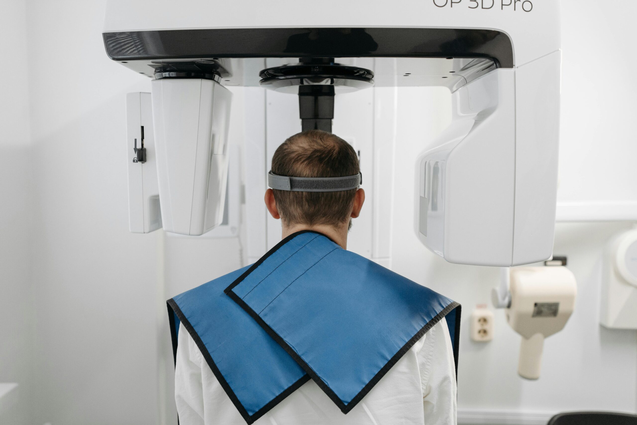
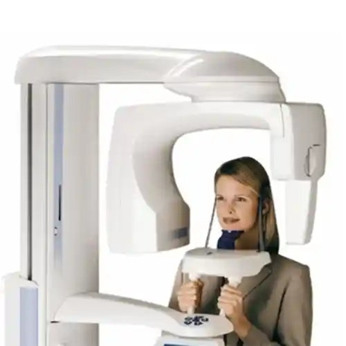
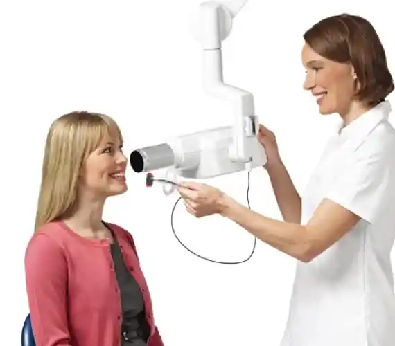
The pre-treatment evaluation process encompasses both the review of the patient’s general systemic status and the microscopic examination of jawbones. In the first stage, the patient’s medical history, medications used (especially bone resorption medications or blood thinners), and the health of intraoral soft tissues are examined in detail. Subsequently, radiological evaluation is moved to, analyzing bone density, vertical height, and critical anatomical proximities such as the lower jaw nerve (nervus alveolaris inferior) via two-dimensional panoramic X-rays or three-dimensional cone-beam computed tomography (CBCT). The structure of the patient’s existing prosthesis is also examined to check whether it possesses sufficient acrylic thickness to accommodate the placement of mini implant-supported sockets.
- Anatomical Boundary Analysis: Measurement of sinus floor and mental foramen (nerve exit hole) distances.
- Soft Tissue Thickness: Determination of the gum thickness where the implant neck part will seat.
- Prosthesis Compatibility: Determining whether the old prosthesis is suitable for modification to add snap-ons.
| Diagnostic Tool | Inspection Criterion | Clinical Benefit Provided |
|---|---|---|
| 3D Tomography (CBCT) | Bone volume and bone quality (Hounsfield unit) | Error-free determination of the exact angle where the implant will be placed |
| Intraoral Physical Examination | Mucosal health and inter-jaw distance | Planning prosthesis thickness and vertical dimension |
How is mini dental implant treatment planned?
The primary goal in planning mini dental implant treatment is to capture the most accurate biomechanical distribution that will ensure the prosthesis is carried in a balanced manner. To stabilize the prosthesis in a completely edentulous lower jaw, 4 mini implants are usually planned to be placed at equal intervals between the two mental foramina (nerve holes); because the bone structure is more porous and softer in the upper jaw, this number is increased to between 4 and 6. The lengths of the implants (generally 10, 13, or 15 mm) and the parallel angles at which they will be placed are calculated one by one on the patient’s radiological data in a computer environment. At this stage, the bite line (occlusal plane) of the teeth is also taken into account, aiming for chewing forces to arrive at the screws at a perpendicular angle.
- Number Determination: Planning a minimum of 4 for the lower jaw and 4 to 6 for the upper jaw.
- Size Selection: Determining the appropriate implant length according to the vertical height of the bone.
- Force Distribution: Ensuring implants are not too close to or too far from each other.
| Application Region | Ideal Implant Number | Planning Strategy |
|---|---|---|
| Lower Jaw (Mandibula) | 4 Mini Implants | Symmetric alignment between the two front nerve exits |
| Upper Jaw (Maxilla) | 4 – 6 Mini Implants | Positioning in front of the sinuses by spreading over a wide area |
How is the mini dental implant application performed?
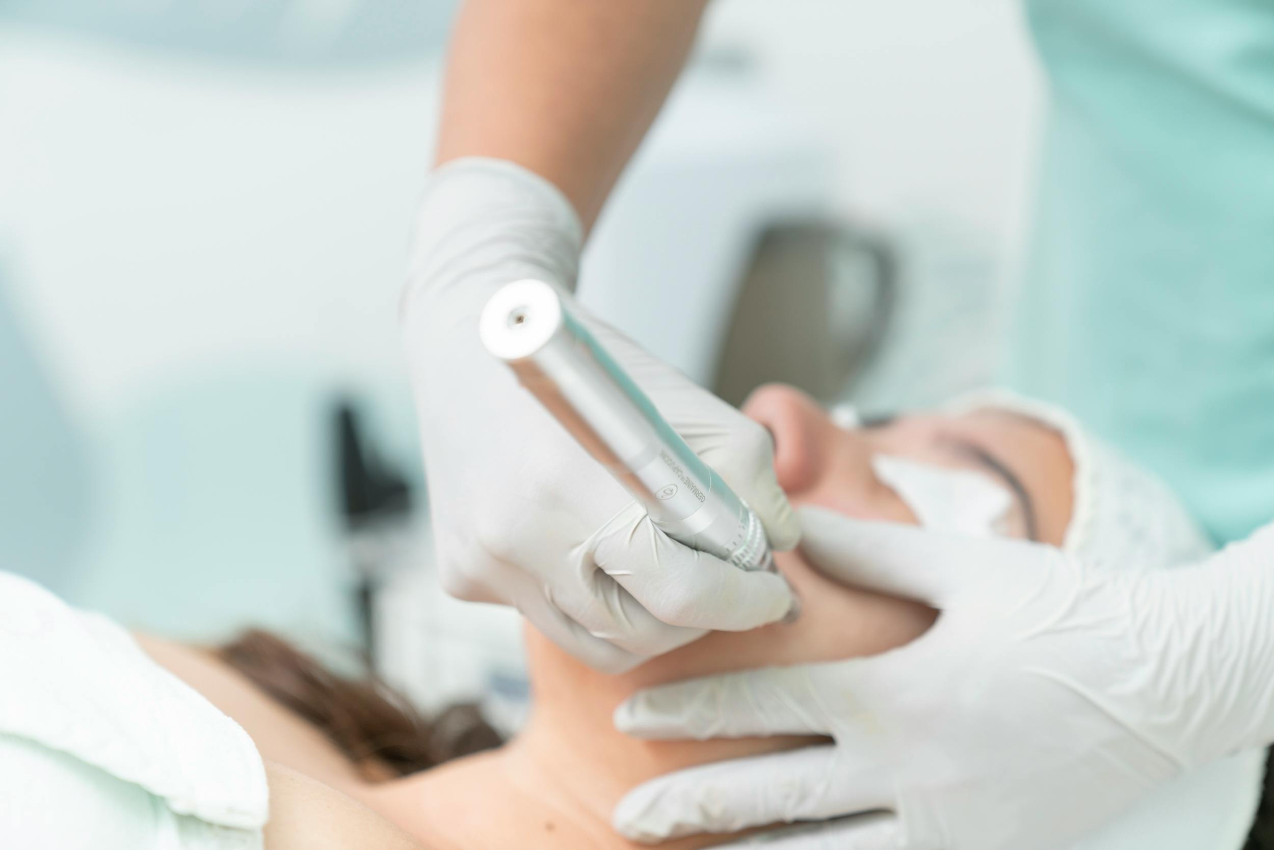
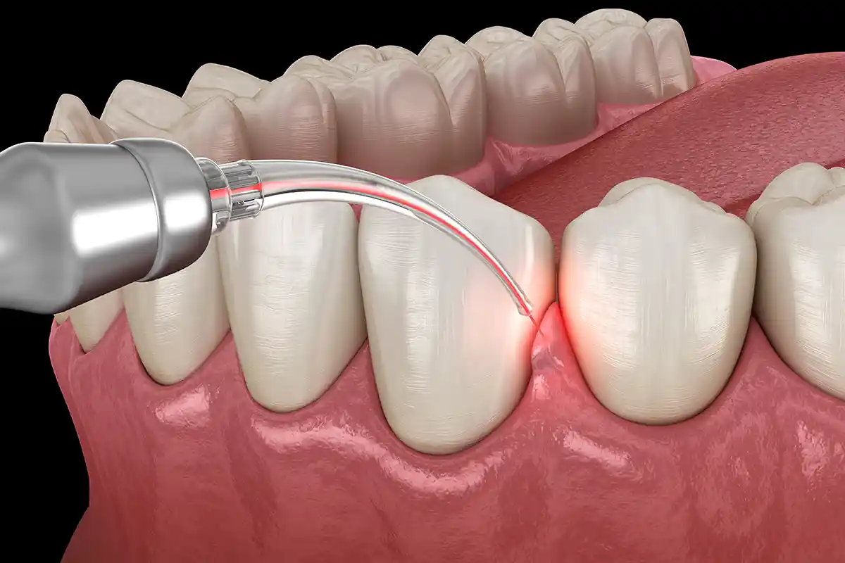
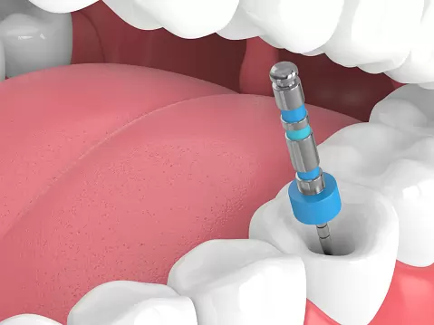
The mini dental implant operation is a short, entirely intraoral procedure where surgical trauma is at a minimum level. Before the operation, the region where the implants will be placed is completely numbed with local anesthesia, so the patient feels no pain or stinging. Without making any incision in the gums (flapless technique), a small pilot hole is opened at a microscopic scale in the bone using specialized implant drills (drill) directly over the tissue. After the mini implant is placed into this hole, it is first screwed into the bone using motorized surgical rounds, then tightened into the bone with manual torque wrenches (ratchets) until it is compressed; the operation is completed such that the implant head remains 2-3 mm above the gingiva.
- Local Anesthesia: Blocking of nerves only in the operation region.
- Transmucosal Intervention: Reaching the bone directly through the mucosa without lifting the gum.
- Torque Control: Reaching primary stability by compressing the implant into the bone with a force of at least 30-35 Ncm.
| Surgical Stage | Equipment Used | Application Process |
|---|---|---|
| Opening Pilot Hole | Micro-surgical drill tip | Initial socket of 1-2 mm depth is created in the bone. |
| Compressing the Implant | Manual Torque Wrench | The screw is locked into the bone with maximum tightness by opening its own thread. |
In which regions can mini dental implants be applied?
The primary and safest application field of mini dental implants is the anterior region of the lower jaw (between canine-to-canine). Since the bone structure in this region is quite hard (cortical), it ensures a very strong hold of narrow-diameter screws to the bone and completely prevents lower jaw removable dentures from shifting due to tongue movements. In the upper jaw, they can be applied at the root levels of the anterior and lateral incisors remaining in front of the sinus cavities. Since chewing loads are very high in posterior regions where large molars are located anatomically, mini implants are rarely preferred as solo crown carriers in these fields; however, they can be positioned as auxiliary elements in narrow posterior ridges where vertical bone remains to support the removable prosthesis.
- Mandibular Anterior Region: The lower jaw front region, the area with the highest success rate.
- Maxillary Anterior and Premolar Region: Narrow bones along the front and small molar tooth levels of the upper jaw.
- Narrow Inter-Root Spaces: Spaces where natural tooth roots are too close to each other and standard screws will not fit.
| Anatomical Region | Bone Quality | Application Suitability |
|---|---|---|
| Lower Jaw Front Region | Type 1 – Type 2 (Hard and Dense) | Excellent (Primary Preference) |
| Upper Jaw Posterior Region | Type 4 (Soft and Spongy) | Limited / Must be Planned Carefully |
How do mini dental implants increase the stability of removable prostheses?
Mini dental implants increase the stability of removable prostheses by means of a mechanical locking system (overdenture retention) also known as a “snap-on.” Metal sockets are placed on the base of the patient’s existing or newly made prosthesis to fit the spherical head of the implant remaining open in the mouth. Inside these metal sockets are “O-rings,” which are high-durability rubber or elastic gaskets; when the patient places the prosthesis in their mouth and presses, these elastic gaskets flex around the ball head of the implant and grip it tightly. This mechanism stabilizes the prosthesis to the bone ridge by resisting vertical dislodgement forces (speaking, yawning, chewing sticky foods).
- O-Ring Technology: The retentive force provided by the elastic gasket surrounding the spherical head.
- Lateral Load Resistance: The implant bodies physically blocking the prosthesis from shifting left and right.
- Balancing Tissue Support: Distributing chewing force both to the gums and to the bone via implants.
| Retentive Part | Material Structure | Role in Stabilization |
|---|---|---|
| Spherical Abutment Head | Monoblock Titanium | Acts as a fixed male part to which the prosthesis will lock. |
| O-Ring Matrix | Medical Rubber / Silicone | Grips the ball by flexing, prevents the prosthesis from launching out of the mouth. |
Can mini dental implants be used in patients with limited bone structure?
Yes, the greatest reason for existence and design purpose of mini dental implants in the medical world is to serve patients with advanced limited bone structure. While a horizontal width of at least 5.5 – 6 mm of jawbone is required to place a traditional implant, mini implants can be placed even in razor-thin bone ridges (knife-edge ridge) of 3.5 mm width without fracturing the bone. This saves the patient from advanced surgical operations such as osteoplasty, bone augmentation (grafting), or block bone transplantation, which are months-long, costly, and painful. In situations where bone height is also at the limit, short mini implant models can be preferred to avert anatomical risks.
- Eliminating Bone Powder Necessity: Direct application without needing artificial bone materials.
- Cortical Bone Utilization: Receiving maximum support from the hard layer (cortex) outside the thin bone.
- Adapting to Atrophy: Flexibility to modify the implant angle according to the natural resorbed form of the jawbone.
| Bone Thickness | Surgery Required (Standard) | Surgery Required (Mini) |
|---|---|---|
| 3.5 – 4.5 mm (Very Narrow) | Bone Expansion / Split + Graft | Direct Flapless Application |
| < 3.0 mm (Extremely Critical) | Block Bone Transplant | Contraindicated (Not applicable) |
How is digital planning used in mini dental implant treatment?
Digital dentistry technologies are utilized to render the flapless surgical procedure performed in a sutureless and blind manner entirely safe and predictable. The process begins with scanning the inside of the patient’s mouth with 3D digital scanners (intraoral scanner) and taking a jaw tomography. These two data are superimposed in computer software to create the digital twin of the patient’s jawbone and gums; the physician places implants virtually in the software at the most ideal angle relative to nerves and bone boundaries; in accordance with this digital planning, custom “surgical guide plates” are produced in 3D printers and the implants are screwed to the planned point without even a millimetric deviation by seating this guide in the patient’s mouth on the day of surgery.
- 3D Data Integration: Fusion of soft tissue (STL) and bone (DICOM) data on the computer.
- Virtual Surgery: Simulating the surgery on a computer screen before physically performing it.
- Guided Surgery: Zeroing the surgeon’s margin of error thanks to templates emerging from the 3D printer.
| Digital Phase | Technology Used | Clinical Advantage |
|---|---|---|
| Intraoral Scanning | Optical Intraoral Scanner | Comfortable impression taking without need for plaster impression trays |
| Guide Plate Production | SLA / DLP 3D Printers | Prevents the implant from losing its direction within the bone. |
How long does mini dental implant application take?
One of the most striking features of mini dental implant application is that operation durations contain extremely short processes compared to traditional surgeries. Because the gums are not cut, wide cavities are not opened in the bone, and sutures are not placed at the end of the procedure, the time to place a single mini implant into the bone is between 5 to 10 minutes on average. A standard application of 4 mini implants for lower jaw prosthesis stabilization is terminated entirely within 30 to 45 minutes, including the numbness time of the local anesthesia. Opening the snap-on sockets inside the patient’s existing prosthesis and placing the elastics immediately after the surgery requires a laboratory/clinical workmanship of approximately 20-30 minutes.
- Single Screw Placement Duration: Approximately 5 – 10 minutes.
- Full Jaw (4 Implants): Approximately 30 – 45 minutes of surgical time.
- Prosthesis Modification Duration: 30 minutes of mechanical adjustment within the same session.
| Procedure Step | Average Time Spent | Patient Experience |
|---|---|---|
| Anesthesia and Preparation | 10 Minutes | Regional numbness is awaited, pain sensation is blocked. |
| 4 MDI Placements | 20 – 30 Minutes | Only a slight vibration and screwing pressure are felt. |
How does the healing process progress after mini dental implant treatment?
Since it is performed with the flapless (sutureless) surgical technique, the healing period of mini dental implants passes in a highly comfortable and rapid manner for patients. Since there is no suture pain or wide surgical wounds after the operation, patients can usually return to their normal daily social lives the next day. In the first 2-3 days, observing slight stinging, sensitivity, or capillary edema (swelling) along the gingival margins where the implants are placed is a completely natural and expected repair reflex, and this situation is easily suppressed with mild painkillers. While the microscopic complete fusion of the implant with the bone (biological osseointegration) takes about 1.5 to 3 months, the patient can begin using their prosthesis from the very first day since the implants are mechanically compressed very tightly into the bone.
- First 24 Hours: Risk of slight stinging and very little swelling; kept under control with ice compresses.
- First 1 Week: Complete closure and healing of micro-entry holes on the gingival surface.
- 6th to 12th Week: Bone cells fully weaving between titanium threads to ensure permanent retention.
| Time Phase | Clinical Symptoms | Healing Support to be Applied |
|---|---|---|
| Post-Op Days 1 – 3 | Mild stinging, feeling of pressure | Consuming soft foods, analgesic usage |
| Post-Op Week 2 | Symptom-free, normal gum color | Cleaning implant necks via soft brushing |
What should be considered after mini dental implant treatment?
In the early period following the operation, specific rules must be adhered to with sensitivity to prevent micro-movements of the implants within the bone and not reduce the success rate. Strictly away from excessively hot, spicy, acidic, or hard-shelled foods should be kept for the first few days following the operation. If the physician delivered the prosthesis to the patient on the day the implant was placed, the patient should not perform excessively hard tugging movements while removing or inserting the prosthesis for the first 1-2 weeks, seating the snap-on mechanism gently parallel to its axis. Habits that could trigger bleeding by creating negative pressure inside the mouth, such as spitting, drinking beverages with a straw, or smoking, should be paused for the first 48 hours.
- Gentle Prosthesis Use: Not applying lateral leverage force to the implant bodies while inserting/removing the prosthesis.
- Compliance with Prescribed Medication: Usage of antibiotics or antiseptic gargles recommended by the physician in accordance with their schedule.
- Protection from Trauma: Protecting the jaw region from external impacts and hard chewing attempts.
| Potential Risk Factor | Clinical Damage It Causes | Precaution to be Taken |
|---|---|---|
| Hard Chewing in Early Days | Risk of failure to fuse (fibrosis) due to early-period micro-movement | Liquid and puree-based nutrition regimen for the first 2-3 weeks |
| Neglecting Oral Care | Bacteria leaking from the gum line dropping the implant | Washing sutureless areas with specialized gargles after every meal |
How should the maintenance of mini dental implants be performed?
To ensure the longevity of mini dental implants, both the titanium heads remaining exposed inside the mouth and the retentive sockets inside the prosthesis must be cleaned meticulously. The patient must remove the removable prosthesis from their mouth after every meal, purify the metal sockets and O-ring elastics inside the prosthesis of food remnants using specialized prosthesis brushes. The implant heads, which are exposed above the gum, must be brushed at least twice a day with circular motions using a soft-bristled brush and toothpastes containing no abrasive substances. Bacterial plaque accumulation in these regions can lead to peri-implantitis (gum inflammation around the implant), causing bone resorption and loss of the implant.
- Brushing Around the Implant: Cleaning around the heads emerging from the gum using a sweeping motion.
- Interdental Brush Usage: Cleaning areas where screws are close to each other with micro-brushes.
- Chemical Cleaning: Soaking prostheses in specialized implant-supported prosthesis cleaning tablets once a week.
| Maintenance Area | Application Method | Recommended Frequency |
|---|---|---|
| Intraoral Titanium Heads | Soft brush + Antiseptic solution | Twice a day (Morning – Evening) |
| Prosthetic O-Ring Elastics | Wiping with cotton swab or soft brush | After every main meal |
What are the factors affecting the lifespan of mini dental implants?
The lifespan of mini dental implants inside the mouth can continue for many years when combined with correct planning and ideal patient care; however, since they are structurally narrow-diameter, they are more sensitive to certain external factors compared to standard implants. The most critical factor is the patient’s oral hygiene; bone losses formed around screws that are not cleaned well rapidly destabilize the narrow-bodied implant. The presence of uncontrolled bruxism (night teeth clenching and grinding) in the patient can impose excessive lateral load on the implants, leading to metal fatigue in the titanium body or intra-bone fractures. Additionally, failure to replace the O-ring elastics inside the prosthesis as they loosen over time causes the load to be distributed unevenly between implants, shortening their lifespan.
- Changes in Systemic Health: Situations of heavy osteoporosis or diabetes that escape control with advancing age.
- Occlusal Overloading: Uncontrolled chewing force applied by natural teeth in the opposing jaw onto the implant-supported prosthesis.
- Regular Physician Control: Correcting early functional load deviations during controls performed every six months.
| Contributing Factor | Negative Effect | Solution to Extend Lifespan |
|---|---|---|
| Night Teeth Clenching (Bruxism) | Micro-cracks and bone destruction | Use of night protective guard (splint) |
| Excessive Smoking | Disrupts tissue nourishment by reducing blood flow | Complete cessation or minimization of tobacco products |
What are the advantages of mini dental implant treatment?
Clinical advantages offered by mini dental implant treatment include solutions that maximize patient comfort in both the surgical phase and the prosthetic phase. Its greatest advantage is eliminating the graft (bone powder) surgery necessity in patients with bone insufficiency, along with the associated additional costs and 6-month waiting periods. Because it is applied with a sutureless and incisionless technique, surgical complaints such as bleeding, bruising, and pain are almost never experienced. Economically, it is much more budget-friendly compared to standard implant treatments; furthermore, if the patient’s existing old prosthesis is compatible, it can be adapted over the implant without entering the cost of fabricating a new prosthesis.
- Minimally Invasive Surgery: A scalpel-free, sutureless, painless, and rapidly healing operation model.
- Immediate Function (Same-Day Loading): The ability to eat with a stabilized prosthesis within just a few hours after surgery.
- Cost Advantage: Economical treatment budget compared to standard surgical protocols and advanced bone surgeries.
| Feature | Condition in Traditional Treatment | Advantage in Mini Implant Treatment |
|---|---|---|
| Waiting Period | 3 – 6 Months (Bone fusion duration) | Immediate use on the same day or within a few days |
| Surgical Trauma | High (Gums are opened, sutures placed) | Very Low (Entry solely the size of a needle prick) |
With which prostheses can mini dental implants be used?
Mini dental implants are used in clinical practices primarily to enhance the retention of implant-supported removable total (complete) dentures termed “overdentures.” Furthermore, in partial tooth deficiencies in the lower jaw anterior region that are excessively narrow (e.g., in lower single incisor deficiency), since standard screws will not fit, mini implants are successfully tasked with carrying single porcelain or zirconium crowns (fixed crowns). In certain temporary situations, they can also be planned as temporary loading elements to carry “temporary transfer prostheses” so that patients undergoing major implant surgeries do not remain completely edentulous during the 6-month healing period.
- Snap-On Total Prostheses: Denture prostheses seating onto implant tops in completely edentulous mouths.
- Narrow Field Fixed Crowns: Single fixed crowns of lower lateral or central teeth with insufficient bone width.
- Temporary Hybrid Bridges: Temporary fixed bridge systems keeping the patient with teeth while essential implants fuse.
| Prosthesis Type | Mode of Connection to Implant | Clinical Purpose of Use |
|---|---|---|
| Removable Total Denture | O-Ring / Rubber Snap Socket | Definitively preventing the prosthesis from shifting while chewing |
| Fixed Single Crown | Cementation (Bonding) | Completing the missing tooth as fixed in narrow-boned anterior regions |
How do mini dental implants support oral functions?
Mini dental implants directly and positively restore the most important oral functions, which are chewing, speaking, and swallowing. While chewing efficiency of patients using standard removable prostheses is at the level of 20-30 percent relative to natural teeth, this rate jumps to 70-80 percent levels when the prosthesis is locked to the bone with mini implant support; thus, the patient can comfortably grind hard foods such as meat, nuts, or apples. Because air and fluid leakage from under the prosthesis is prevented, sounds produced by the tongue striking the palate and teeth, such as “S, T, Z,” become clear, lisping comes to an end. Additionally, since the prosthesis is prevented from moving continuously and wounding the gums, chronic intraoral pains are completely eliminated.
- Chewing Force Restoration: The ability to transfer force directly to the bone via implants without putting pressure on the tissue under the prosthesis.
- Phonetic Correction: Prevention of the palate shifting inside the mouth and clicking sounds during speech.
- Nutritional Diversity: The freedom to have a rich diet without being dependent solely on liquid or soft foods.
| Oral Function | Status of Non-Implant Prosthesis | Status After Mini Implant |
|---|---|---|
| Chewing Performance | Limited (Swallowing without fully grinding food) | High (Easily crushing meat and hard vegetables) |
| Diction / Speech | Timid and lisped due to fear of prosthesis shifting | Clear, fluent, and confident word pronunciation |
What are the frequently asked questions about mini dental implants?
Will I feel a lot of pain during mini implant application?
No, the operation is conducted entirely under modern local anesthesia techniques and the relevant jaw region is thoroughly numbed. Since the gum tissue is not cut and deep drilling processes are not performed in the bone, the patient experiences only a slight sensation of touching and screwing during the procedure, strictly not feeling acute surgical pain.
How often should the O-ring elastics inside the prosthesis be changed?
The elastic gaskets (O-rings) inside the prosthesis may lose their flexibility and loosen over time depending on the repeated insertion/removal of the prosthesis every day and the effect of oral fluids. To ensure the prosthesis preserves the tightness of the first day, it is recommended that these elastics be renewed via a simple procedure in a clinical environment once every 1 to 1.5 years.
Can we fit my existing old false teeth over the mini implants?
If the acrylic structure of the patient’s existing removable prosthesis is sound, and its vertical dimension and closure are within clinically correct boundaries, there is no need to fabricate a new prosthesis. Sockets can be opened in the areas where the implant tops meet on the inner surface of the existing old prosthesis, and snap-ons can be adapted on the same day.
Is there a risk of mini implants breaking or coming out of the bone?
As in every implant system, the risk of breakage or loss of osseointegration theoretically exists in mini implants depending on excessive uncontrolled loads. However, these risks are at a minimum level in applications performed at the correct anatomical region, tightened with correct torque, and conducted in the number determined by the physician (e.g., 4 pieces for the lower jaw).
| Core Question Area | Clinical Answer Summary |
|---|---|
| Operation Pain | Zero pain thanks to local anesthesia, slight stinging after the procedure |
| Elastic Change Frequency | Once every 12 – 18 months on average, depending on usage frequency |
| Old Prosthesis Compatibility | If the prosthesis is sound, it can be used by modifying its inner surface. |
Who We Are? Panorama Ankara Oral and Dental Health Polyclinic
As Panorama Ankara, we provide high-standard services in the field of advanced implantology by combining the latest technological developments in dentistry and the clinical experience of our expert academic physician staff. In our modern polyclinic in Ankara, we conduct mini dental implant treatments for our patients who have jawbone insufficiency or who avoid major surgeries due to advanced age, with a comfortable protocol. Via our artificial intelligence-supported 3D tomography systems and digital intraoral scanners, we transfer our patients’ jaw structures to the computer environment millimetrically, and perform sutureless mini implant applications flawlessly by preparing custom surgical guide templates; our mission is to restore our patients’ quality of life to the highest level by bringing them to their stabilized, functional, and aesthetic prostheses within the same day, without exposing them to long surgical waiting periods.
- Advanced Diagnostic Infrastructure: 3D dental tomography devices analyzing bone density at the micro level.
- Rapid and Comfortable Protocol: Implant placement and adaptation of existing prosthesis to a snap-on system within the same session.
- Expert Surgical Staff: Physician team with extensive case experience in the field of minimally invasive and flapless implant surgeries.
| Health Institution | Service Region | Featured Digital Clinical Solution |
|---|---|---|
| Panorama Ankara Oral and Dental Health | Ankara | Custom-Made 3D Surgical Template-Supported Flapless Mini Implantology |