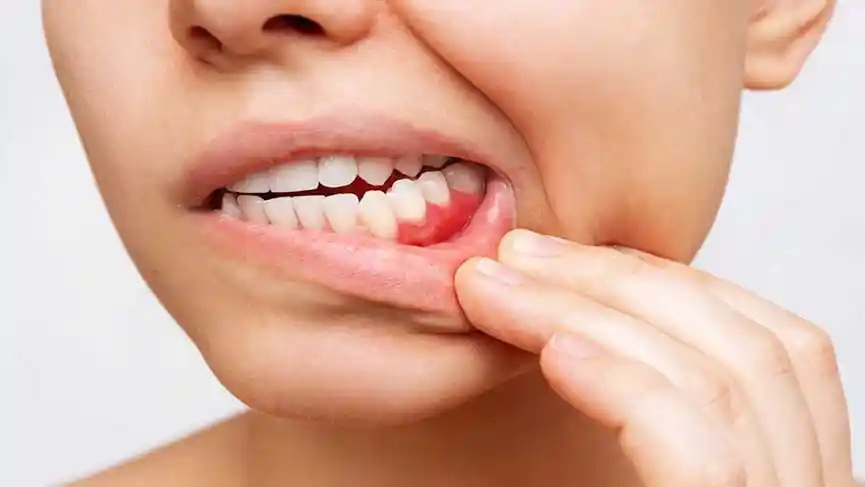
What Is Gum Recession Treatment?
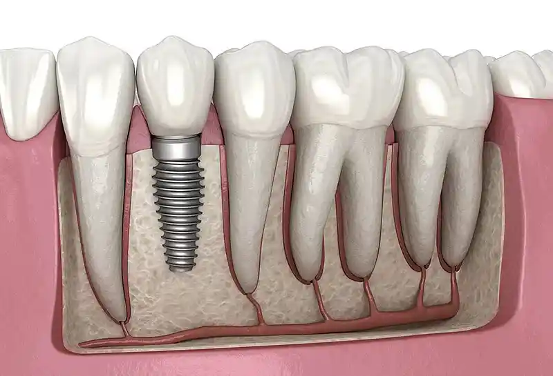
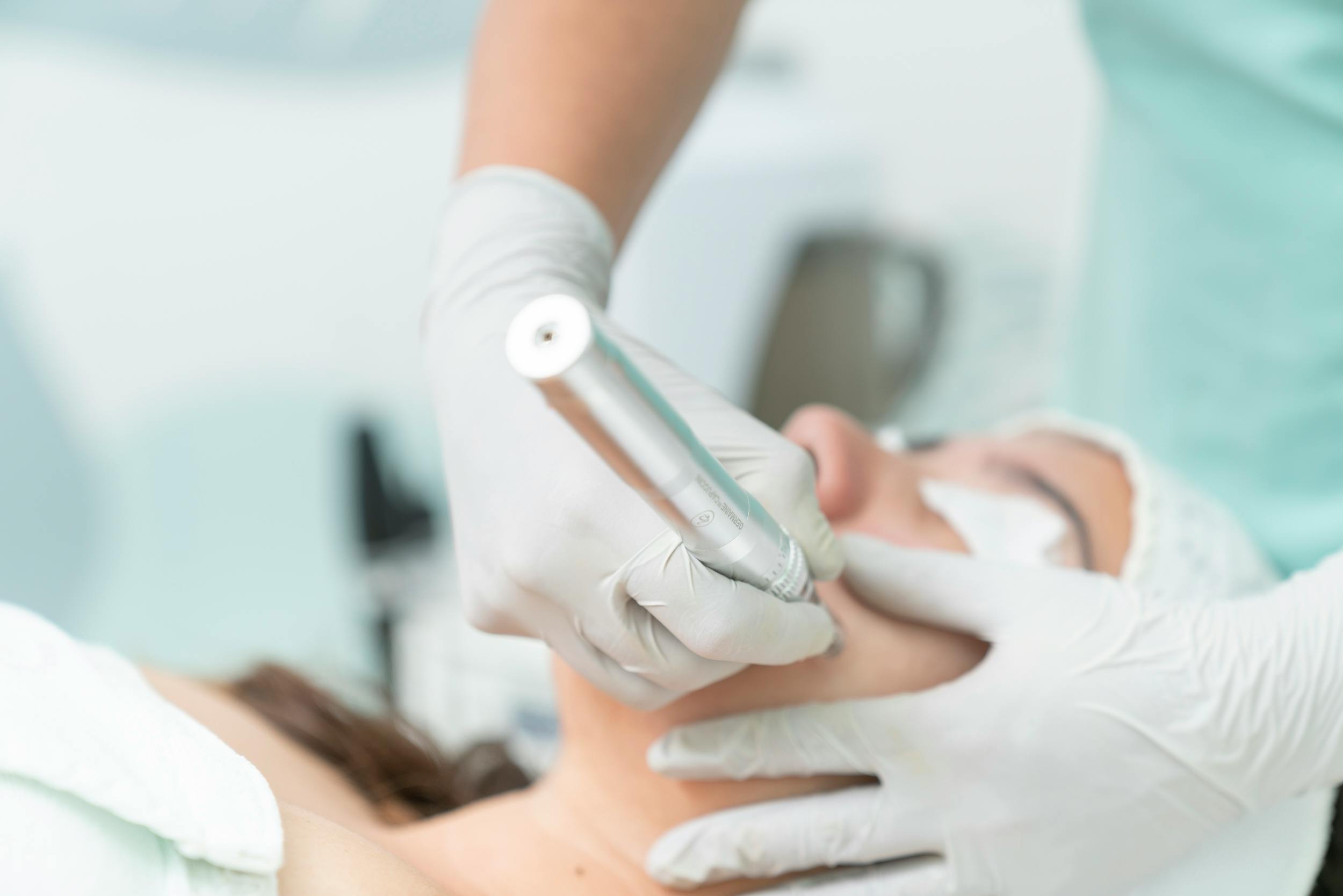
Gum recession treatment (periodontal plastic surgery and prophylaxis) is an integrated treatment discipline aimed at halting and restoring the process by which the attached gum tissue that surrounds the tooth root like a shield shifts downward or upward due to alveolar bone loss or mechanical trauma. When the gum recedes, the tooth’s protective enamel layer comes to an end and is replaced by sensitive dentin (root) surface, which contains microscopic pores; this leaves the tooth exposed to decay and loosening. Treatment is based on eliminating the pathological and biomechanical root causes underlying the recession to keep the tissue stable, and in advanced stages, surgically covering the root surface with new pink mucosal bands.
- Causal Treatment: Eliminating the inflammation-producing biofilm layer and faulty force loads.
- Root Surface Protection: Isolating the exposed cementum layer from outside factors, reducing decay risks to zero.
- Mucogingival Surgery: Increasing tissue thickness and volume to build biological resistance against future waves of recession.
Why Does Gum Recession Occur?
Recession of the gum margin is a multifactorial (multi-cause) chronic periodontal pathology triggered synergistically by the destructive activities of intraoral pathogens and the patient’s daily mechanical habits. The most common cause is calcified tartar buildup between and around the necks of the teeth pushing down on the connective tissue fibers like a mechanical wedge and eroding the alveolar bone. Conversely, even in individuals with very good oral hygiene, brushing the teeth horizontally and with excessive pressure using hard-bristled brushes can trigger recession by mechanically tearing and wearing away the gum, especially in facial structures with a thin mucosal biotype.
- Chronic Periodontitis: Bacteria in tartar triggering bone loss and collapsing the gum’s support base,
- Faulty Brushing Trauma: Physically destroying the upper epidermal barrier by sawing the teeth with hard scraping motions,
- Bruxism (Teeth Clenching): The chewing muscles applying bending (abfraction) force on the teeth and cracking the neck bone,
- Anatomical and Genetic Variations: Muscle attachments (frenula) holding the gum from above and continuously pulling it downward.
What Symptoms Indicate Gum Recession?
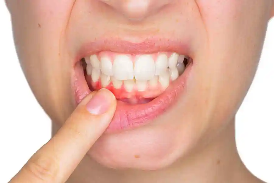
The process of mucosal tissue recession progresses silently in its early stages, without sending any pain or ache signal to the patient, due to the slow erosion of bone cells; symptoms only become noticeable once the sensitive layers of the root surface begin coming into contact with oral fluids. The most obvious visual symptom is that the tooth crowns appear elongated, and the yellowish, matte-colored root body can be seen with the naked eye along the line where the tooth meets the gum. At this stage, when floss or the tongue catches on these areas, millimetric ledges (notches) can be felt.
- Thermal Sensitivity Shocks: A sudden aching sensation, especially when drinking cold water or breathing in windy weather,
- Aesthetic Balance Disruption: The front tooth line losing its smooth arc form when smiling and appearing asymmetric,
- Chronic Food Impaction: Food getting stuck in the “black triangle” gaps that open up as the triangular tissue (papilla) between two teeth recedes,
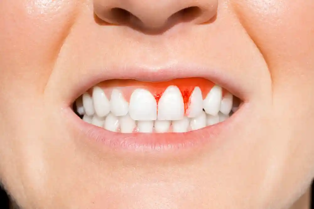
- Localized Gum Bleeding: Redness and bleeding while brushing, resulting from the inability to clean the mucosal pocket along the recession line.
Who Is Gum Recession Treatment Suitable For?
Gum recession treatment protocols are medically suitable for any adult patient over the age of 18 who has an aching complaint at the tooth roots, in whom the process of bone loss has begun, and who aims to prevent teeth from loosening and being lost prematurely. Since it does not involve a systemic anesthesia process, it can be safely carried out in all cases where the patient’s general health condition allows local anesthesia. If surgical grafting stages are planned, the patient must fully commit to oral hygiene discipline and must completely stop smoking — since it radically lowers wound-repair success by disrupting the oxygenation quality of capillary vessels beneath the tissue.
- Those Experiencing Dentin Sensitivity: Patients whose quality of life is diminished by chronic aching in response to hot-cold foods.
- Orthodontic Candidates/Patients: Those looking to prevent tooth movement from further receding the gum during wire treatment.
- Those with Tooth-Length Asymmetry: Individuals aiming to equalize gum boundaries by establishing pink aesthetic harmony.
How Is Gum Recession Diagnosed?
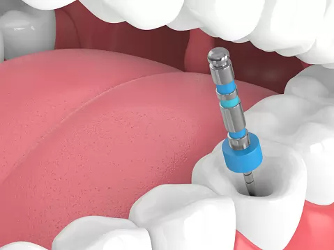
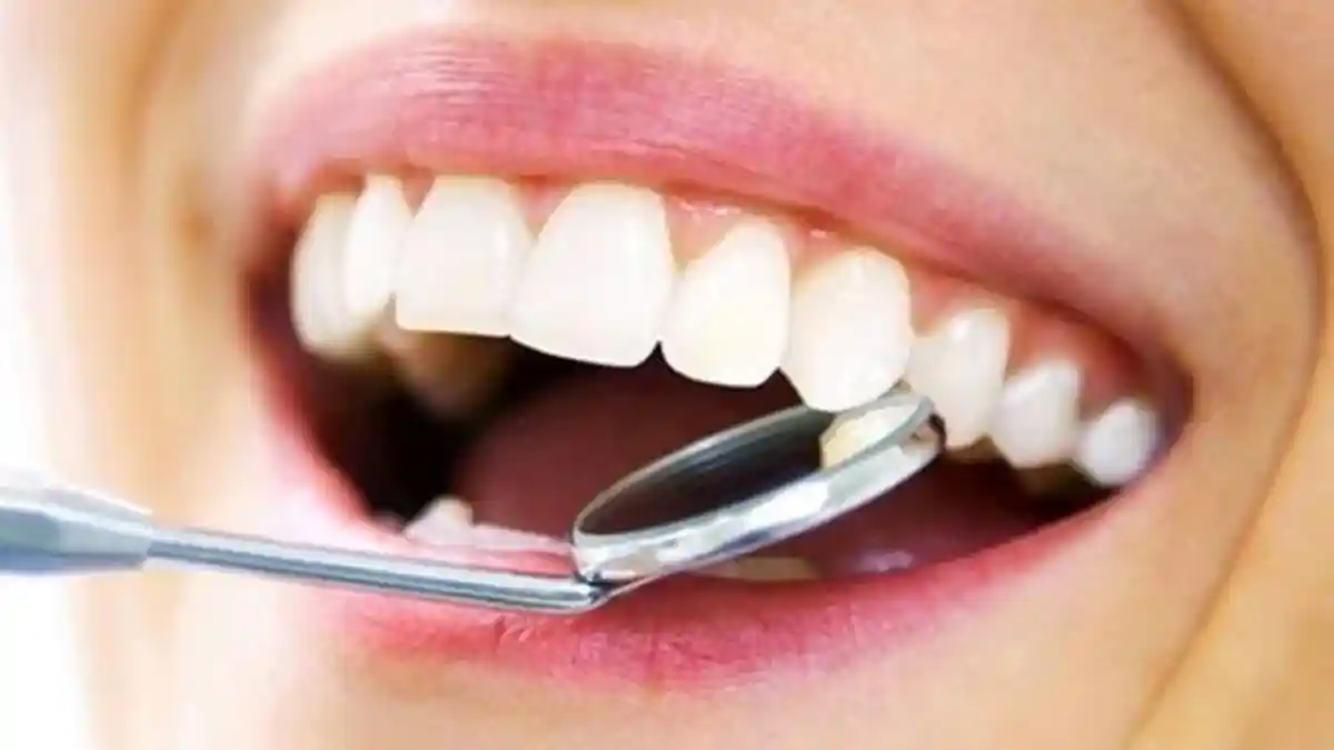
The diagnosis of gum recession carried out in clinical settings is not just a visual assessment; it includes scientific documentation steps that measure the millimetric depth of tissue loss and bone damage. During the examination, the dentist inserts a special medical instrument called a “periodontal probe,” which has a millimetric scale, into the gum sulcus, measuring the amount of recession (in millimeters) and the strength of the gum’s attachment to the tooth. According to the international periodontal scale known as the “Miller Classification,” the degree of recession (Class I, II, III, IV) is determined, mathematically clarifying before the operation whether the tissue can be surgically covered 100%.
- Clinical Attachment Level (CAL) Measurement: Measuring the net vertical loss distance from the tooth’s enamel-cementum boundary to the gum margin.
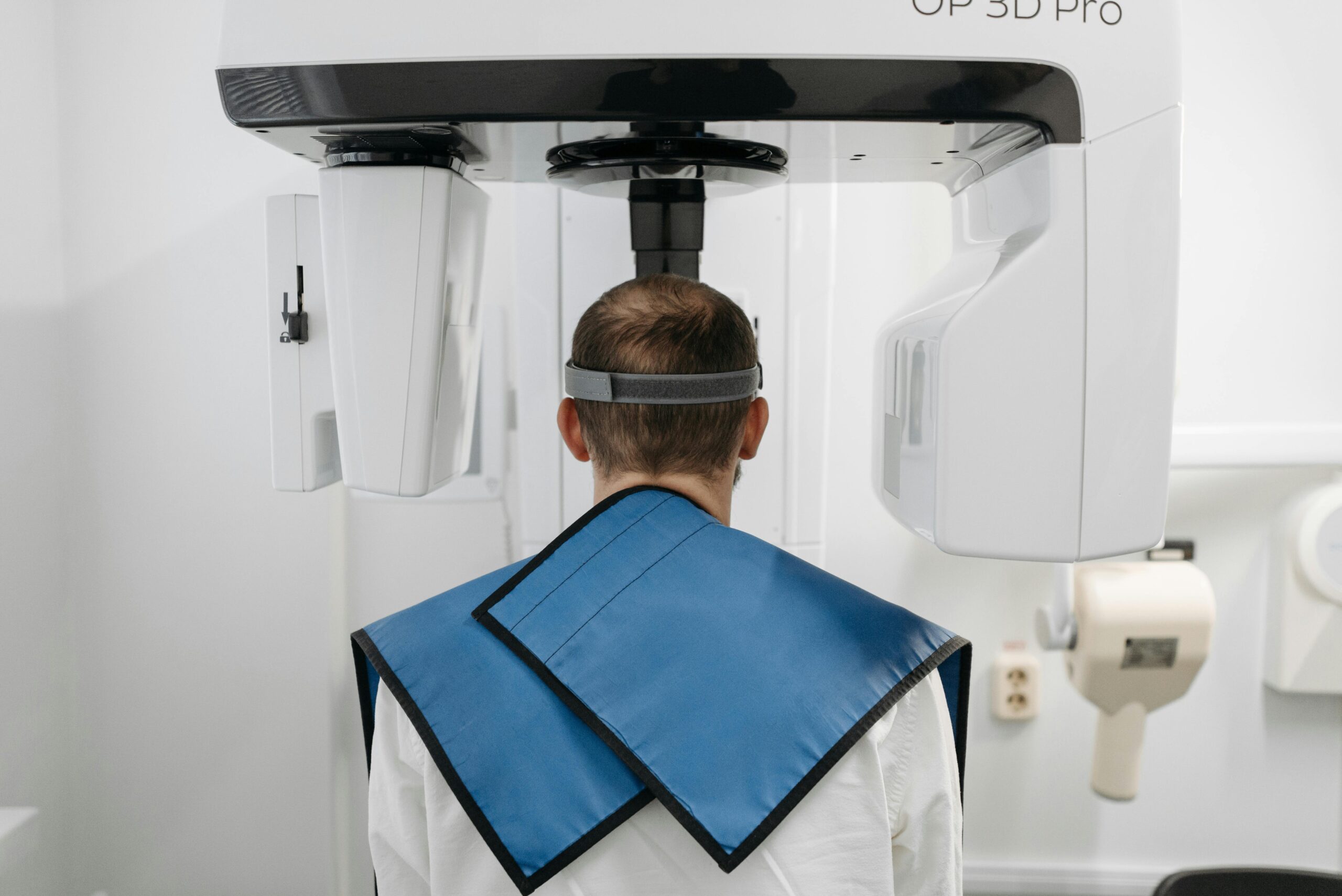
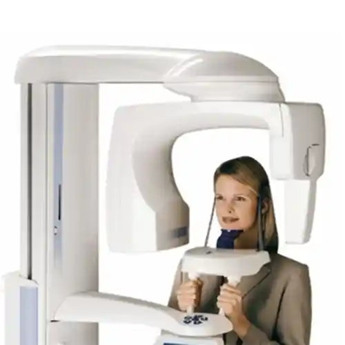
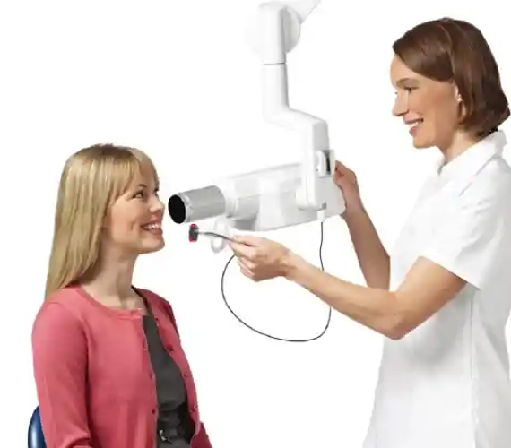
- Digital Panoramic and Periapical X-Ray: A 2D scan of the vertical and horizontal erosion map in the alveolar bone holding the tooth root.
- CBCT 3D Dental Tomography: In borderline cases, layer-by-layer determination of the integrity of the thin bone wall, especially on the cheek (buccal) side of the tooth.
How Is Gum Recession Treatment Planned?
Planning the gum recession treatment protocol is carried out through a phased schedule based on the patient’s biological age, the Miller class degree of the recession, and the intraoral mucosal biotype (thin or thick tissue structure). Direct surgical steps are not taken right away in the planning process, since the likelihood of surgical grafts taking hold in an inflamed, tartar-filled mouth is close to zero. In the planning stage, an “initial periodontal treatment” phase to calm the tissue is scheduled first, after which surgical or non-surgical restoration steps are shaped according to the extent of the damage.
| Treatment Phase / Step | Clinical Procedure Protocol Applied | Tissue and Biological Purpose |
|---|---|---|
| Phase 1: Hygiene and Hygiene Boosting | Ultrasonic scaling, Airflow stain removal, and root surface smoothing (curettage). | Removing bacterial plaque, reducing gum swelling, and firming the tissue. |
| Phase 2: Biomechanical Elimination | Providing correct brushing training, laser-cutting high frenula (frenectomy), making a night guard. | Eliminating mechanical stresses pulling the gum downward and bruxism pressures. |
| Phase 3: Plastic Surgery / Restoration | Free gingival graft (FGG), connective tissue graft (CTG) injection, or sensitivity fillings. | Permanently covering exposed root surfaces with new pink mucosal barriers. |
What Methods Are Used in Gum Recession Treatment?
In periodontology practices, evidence-based surgical and non-surgical treatment methods are employed based on the stage of recession and the chronicity of bone loss. In early-stage (Class I) cases, simply protecting the tissue is sufficient, whereas in advanced root exposure, tissue-transfer surgeries are the primary rule. Which method is chosen is decided based on the dentist’s millimetric pocket-depth and attachment analysis.
The main medical methods actively used to halt and repair gum recession are as follows:
- Root Planing: Scraping away microscopic tartar deposits stuck to the root surface with special Gracey curettes to smooth the cementum,
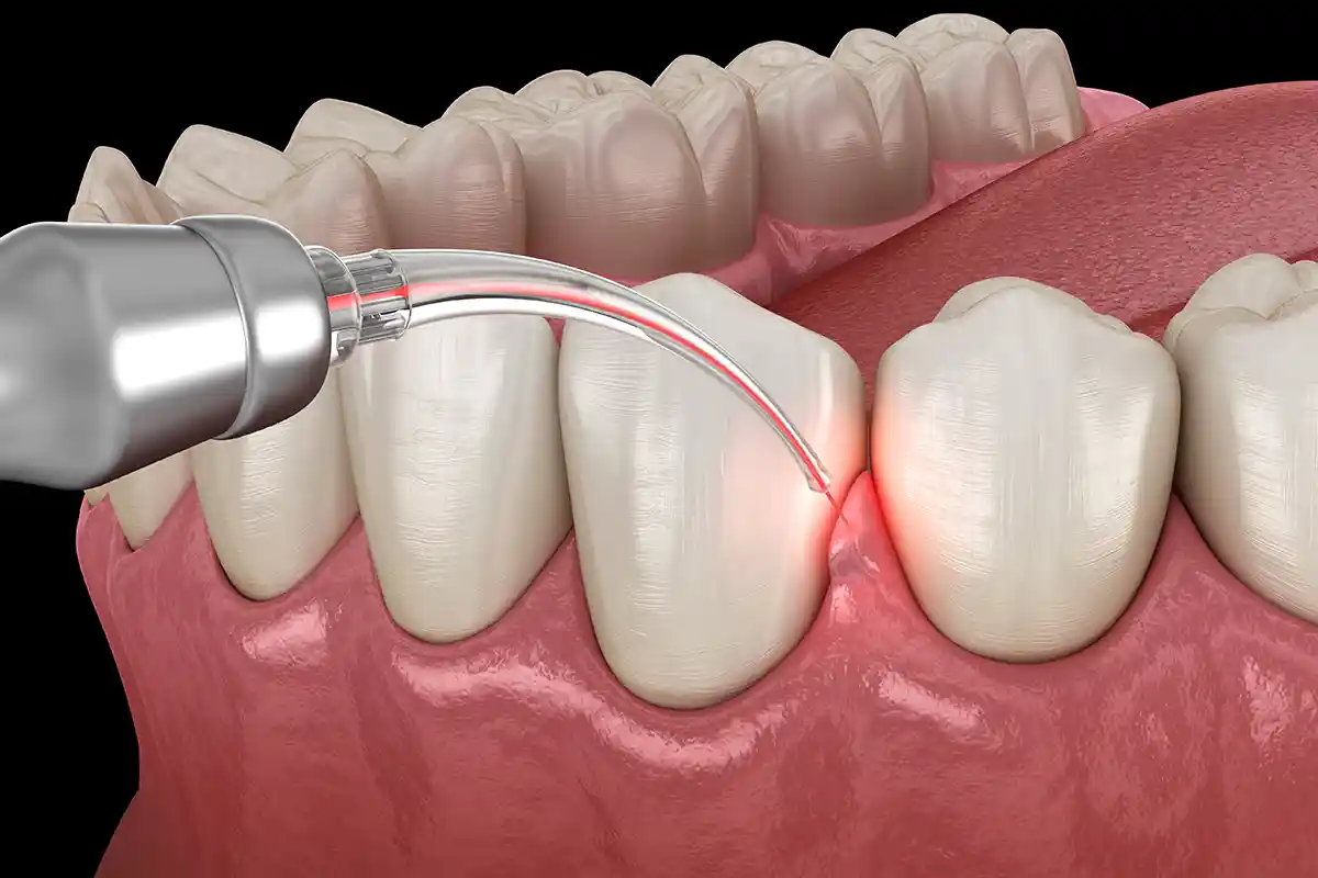
- Laser Periodontal Decontamination: Killing pathogenic bacteria inside deep pockets with radiation using Nd:YAG or Er:YAG laser beams,
- Glass Ionomer / Composite Neck Fillings: Camouflaging exposed root notches with biocompatible fillings in cases not suitable for surgery,
- Tunnel Technique and Sliding Flap Surgeries: Loosening healthy gum tissue next to the recessed area and surgically sliding it over the receded root.
How Is Gum Grafting Used in Gum Recession Treatment?
Gum grafting (mucosal transfer) is a micro-surgical tissue transplant procedure applied in order to cover exposed root surfaces and increase the amount of tight, keratinized tissue in that area. During the procedure, a thin, micron-thick piece of pink tissue (graft) is typically surgically removed from the patient’s own upper palate (hard palate mucosa). This freshly harvested tissue is transferred to the recessed root area using fine sutures. The graft taken from the palate begins exchanging blood with the capillary vessels of the root bed it is placed on within seconds (through the process of angiogenesis) and fuses there, creating a thick, unshakable new pink tissue barricade that will protect the root surface for a lifetime.
- Connective Tissue Graft (CTG): Transferring fibrous tissue taken from beneath the top layer of skin of the palate; provides maximum aesthetic color match, chosen for front teeth.
- Free Gingival Graft (FGG): A full transfer of the palate’s top skin layer; applied especially in the molar area to reinforce tissue thickness.
- Extracellular Matrices (Alloderm): The use of ready-made, sterile, lab-produced cellular membranes for patients who do not want tissue taken from their own palate.
What Are the Regenerative Applications Used in Gum Recession Treatment?
Regenerative applications are high-tech biotechnological treatment methods aimed at biologically regenerating, at the cellular level, the alveolar jawbone loss and severed periodontal fibers that underlie gum recession. In these procedures, after the surgeon gently loosens the gum, they apply “Enamel Matrix Derivatives” (protein gels similar to Emdogain) to the root surface. These smart proteins mimic the body’s cellular codes from the tooth-development years (the embryological stage), triggering the organic rebuilding of a new cementum layer, new periodontal fibers, and, most importantly, eroded jawbone on the root surface.
- Platelet-Rich Fibrin (PRF): Laying down living yellow clot membranes, rich in growth factors, separated from the patient’s own blood using a centrifuge, onto the wound bed.
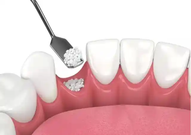
- Guided Tissue Regeneration (GTR): The phase of placing bone powder (graft) into eroded bone cavities, then covering it with sterile, biodegradable membrane barriers to prevent the gum from growing in.
Does Gum Recession Treatment Help Reduce Sensitivity?
Yes, the clinical outcome of gum recession treatment that patients notice most immediately, and that most improves their quality of life, is the radical reduction or complete elimination of those sharp “dentin sensitivity shocks” experienced when consuming hot-cold foods. The dentin layer on the root surface exposed by recession is filled with millions of microscopic capillary channels (dentin tubules) that open directly into the tooth’s living nerve chamber (the pulp). When, as part of treatment, root surfaces are covered with new gum grafts, or these micro channels are sealed at the cellular level using laser beams or biocompatible smart fillings, the reaching of external temperature fluctuations to the nerve endings is definitively blocked.
- Sealing of Dentin Tubules: Melting and watertight-welding the exposed micro nerve channels with laser energy.
- Biological Blanket Effect: Graft tissues acting as a thick heat-insulating layer over the root fibers, ending the aching sensation.
How Does Gum Recession Treatment Contribute to the Preservation of Teeth?
The biggest contribution this professional periodontal treatment model makes to tooth-preservation biology is halting the erosion rate of the alveolar jawbone that holds the teeth, breaking the chain of teeth loosening and being lost prematurely (early loss). Once the gum recedes, the root surface is exposed to the outside environment, and since the root’s cementum layer is much less resistant to acid attacks than enamel, it erodes rapidly, inviting “root decay.” Thanks to the treatment process, bacterial colonies around the root are destroyed and the tissue is thickened, preserving the skeletal stability of the teeth within the jawbone for a lifetime.
- Root Decay Barrier: Preventing the risk of the tooth root rotting from the inside and fracturing, by preventing cementum wear.
- Attachment Stabilization: Increasing vertical resistance by expanding the attachment area of the fiber networks connecting the tooth to the bone.
How Long Does Gum Recession Treatment Take?
The total duration of gum recession treatment varies depending on the nature of the method applied (non-surgical curettage versus surgical graft transfer) and the number of teeth being treated. Standard root surface smoothing and disinfection phases performed with devices are generally completed in a single clinical session lasting 20 to 30 minutes. If a microscopic gum graft operation is planned that involves taking tissue from the palate’s hard mucosal bed, the session duration averages a comfortable 45 to 60 minutes due to the meticulous surgical craftsmanship required.
- Non-Surgical Curettage Session: A practical half-hour period of ultrasonic and laser cleansing (does not disrupt social life).
- Micro-Surgical Grafting Session: An operation block lasting approximately 1 hour due to the tissue transplant and millimetric suturing steps.
How Does the Healing Process Progress After Gum Recession Treatment?
The tissue recovery phase after the procedure proceeds with extremely predictable steps, depending on the surgical extent of the method applied; after non-surgical curettage sessions, there is zero recovery burden since there are no stitches or incision marks on your skin, and the tissue is completely normalized by the next day. In graft surgeries in which tissue is taken from the palate, however, healing follows a linear path; both the donor site on the palate and the recipient site at the root area are rapidly covered with clot membranes within the first 24–48 hours, thanks to the skin’s homeostasis mechanism. On average, within 7 to 10 days, the stitches reach the point where they can be removed, and after 1 month, the newly transferred pink tissue band becomes fully biologically fused with the tooth.
- First 72 Hours (Graft Fixation): The phase in which the tissue must not be disturbed, and mild aching on the palate and micro swelling on the face may occur.
- Days 7 to 10: Epithelialization (top skin covering) is completed, and the stitches are comfortably removed from the line during a clinical checkup.
- The 3-Week Mark: The pink tissue’s color fully blends with the surrounding gums, and the capillary vessel networks complete a smooth integration.
What Should Be Watched For After Gum Recession Treatment?
The success of periodontal interventions, and especially of tissue-transplant surgeries, depends directly on the patient fully adhering to the medical protection protocols they will follow at home after the operation — since it takes the first 7–10 days for the transferred grafts to biologically fuse with blood vessels in the tissue bed. The smallest mechanical disturbance, hard brush stroke, or lip-pulling motion performed during this critical process can pop the stitches, cutting off the graft’s blood supply and causing tissue death (necrosis); the patient must be patient and gentle.
The medical points that must be carefully followed at home during the critical period after the operation are as follows:
- During the first 24 hours after the procedure, extremely hot, ice-cold, or acidic foods should absolutely not be consumed, in order not to disrupt the integrity of the clot.
- For the first 1 week, grainy, crumbly foods (crackers, chips) and overly hard, shelled foods should be avoided in order not to disturb the suture line, and a puree-like diet should be followed.
- The lips should not be pulled up by hand to inspect the operated area, and the jaw should not be stretched; this motion can mechanically tear the stitches.
- For at least 1 month, sauna, steam bath, tanning bed use, and heavy cardio sports that would raise subsurface vascular capillary pressure and trigger bleeding risk should be avoided.
How Should Oral Care Be Done After Gum Recession Treatment?
The home hygiene processes after the session must follow a medical discipline that supports the regeneration speed of skin and mucosal epithelial cells, protects the wound area from outside microbial attacks, but also does not disturb the stitches. The exact local area where the operation was performed must absolutely not be brushed with a normal toothbrush for the first 10 days. While the other healthy teeth in the mouth are brushed as usual, cleansing of the wound bed is entrusted to chemical solutions.
- Antiseptic Mouthwash Protocol: Rinsing the mouth morning and evening with chlorhexidine-containing solutions for the first 10 days, to prevent plaque buildup without mechanical brushing.
- Transition to a Surgical Brush: Once the stitches are removed after day 10, using special ultra-soft (surgical) bristled brushes made specifically for post-operative use to clean that area.
- Interdental Cleaning Discipline: Once the healing period is over (approximately after 1 month), beginning to clean the opened black triangles with soft interdental micro-brushes to prevent buildup.
Can Gum Recession Occur Again?
Once the treatment protocols are successfully completed, the tissue thickening and root-coverage effect achieved becomes histologically permanent; however, the dynamics of tissue recession in the human body and the oral ecosystem — driven by aging, gravity, and mechanical stressors — remain active for a lifetime. If the patient returns to their old faulty lifestyle habits after treatment, neglects oral hygiene and builds up new towers of tartar, or continues to scrape their teeth harshly like a saw, gum recession can definitely recur over the years; permanence directly correlates with the patient’s commitment.
- Mechanical Protection: Keeping the brushing technique fixed for life as a vertical sweeping massage “from the gum toward the tooth.”
- Bruxism Blockage: The requirement for patients with a teeth-clenching habit to regularly wear the protective night guards (splints) made for them while sleeping, to eliminate pressure on the neck bone.
What Should Be Considered to Prevent Gum Recession?
Preventing tissue recession before it even starts, in the comfort of one’s home, is the most rational and safest preventive approach for preserving the integrity of hard and soft tissue in the mouth. The only way to achieve this is to remove the microscopic bacterial plaque that irritates the gums before that critical 48-hour window passes and it mineralizes into hard tartar. When the right tools are integrated into one’s lifestyle, the need to see a periodontist for surgical purposes is eliminated entirely.
- Choosing soft or medium-firm nylon-filament brushes that won’t sand down the gums,
- Uncompromisingly flossing every night the narrow interfacial areas where teeth touch each other and where bristles can never reach,
- Completely giving up tobacco and cigarette products; since nicotine narrows the gum’s capillary vessels (causing vasoconstriction), feeding hidden recession,
- In the presence of a high frenulum (lip-cheek tie) muscle attachment, having it released early with a laser (frenectomy) to prevent it from pulling the gum downward.
What Are the Contributions of Gum Recession Treatment to Oral and Dental Health?
Periodically planned periodontal treatments are the most cost-effective anti-aging investment that extends the biological lifespan of intraoral tissues and halts the silent erosion of the jawbone. They do not only create a superficial aesthetic pink line; they protect the periodontal ligament fiber networks that connect the teeth to the jawbone. When the infection and mechanical stress burden in the deeper layers of the tissue is relieved, the appearance instantly gains a fresh, youthful look and lasting smoothness.
- Prevention of Root Decay: Bringing the vulnerable cementum layer under a protective shield against acid erosion.
- Elimination of Loosening Risks: Preserving the skeletal rigidity of the teeth within the jawbone by reinforcing alveolar bone support.
- Systemic Health Barrier: Definitively cutting off the chain of pathogenic microbe spread that seeps from inflamed gum vessels into the bloodstream and settles in the heart or joints.
Frequently Asked Questions About Gum Recession Treatment
Below are the most common practical concerns on the minds of individuals planning to seek this treatment to get rid of the aching in their tooth roots or to preserve their pink aesthetic boundaries, along with their clear, straightforward medical answers:
Does gum recession treatment hurt a lot in the operating room?
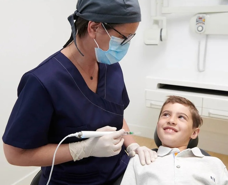
No, this is definitely not a procedure that causes pain or ache. At the start of the session, the most advanced local anesthetic solutions are infused with computer-controlled digital anesthesia devices at a rate suited to the tissue’s resistance, achieving full regional numbness (a block). Since the nerve pathways are completely closed off during the session, all you will perceive is the gentle touch of the instruments and the coolness of the water; the procedure is completed in complete comfort.
Can receded gums grow back up with special toothpastes or mouthwashes sold at pharmacies?
No, this is medically impossible and a form of commercial misinformation. Once gum tissue has receded and the alveolar bone beneath it has eroded, no toothpaste, herbal remedy, oil, or mouthwash can chemically stimulate that tissue to grow back to its original position; these products can only temporarily soothe inflammation and sensitivity in the gum. Recovering the receded area is only possible through micro-surgical tissue graft transplantation performed by a periodontist.
Will a permanent hole or gap remain in my palate after graft surgery where tissue was removed?
No, absolutely no permanent gap, cavity, or damage will remain on your palate. The hard palate mucosa is among the epidermal layers in human anatomy that can regenerate itself the fastest and most aggressively (with the highest epithelialization speed). The donor site from which tissue was taken is quickly closed by the skin’s repair cells within the first few days, and within about 2 weeks, that area fully returns to its original, smooth form with no surgical trace left behind.
Black triangle gaps have opened up between my teeth after the gum recession was cleaned — is this normal?
Yes, this is a natural clinical outcome resulting from the removal of thick tartar masses that had settled between the teeth, filling those gaps like a wedge. Those black triangles were not created by the instruments; on the contrary, it is that old tartar buildup that eroded the bone in those areas and created the gaps in the first place, and the real situation became visible once the tartar was removed. As your gums firm up with regular interdental cleaning, those areas will enter a normalization phase.
Can I go back to my normal social and professional life right away on the day the treatment is finished?
Yes, you can return to social life immediately after non-surgical curettage and laser sessions, as soon as the numbness wears off. In surgical cases where tissue is taken from the palate, since no general anesthesia is given and your consciousness is not affected, you can get up from the chair and go back home or to work right away; it is simply enough to follow practical medical precautions in the first few days, such as not stretching the jaw excessively while talking and avoiding hot foods.
Who We Are? & Panorama Ankara Oral and Dental Health
Panorama Ankara is a licensed oral and dental health center that brings together an academic team of specialists and the latest global digital technological infrastructure under one roof — in oral, dental, and maxillofacial surgery, implantology, orthodontics, cosmetic dentistry, and specialized periodontology (gum disease and surgery) — providing patients with institutional health assurance. Our center places vital importance on halting recession at the gum margin, silently progressing jawbone erosion, and dentin sensitivity shocks that sabotage quality of life — while they are still in their early stages — through evidence-based medical disciplines, and uncompromisingly operates original Laser-Assisted Gum Recession Treatment and Microscopic Graft Transplantation ecosystems, the safest link in modern micro-gingival plastic surgery, in its clinical prophylaxis workflow.
Across all radiological, screening, design, and micro-surgical interventions carried out at Panorama Ankara, the principles of absolute sterilization, high patient safety, transparency, and honesty are enforced without compromise, in accordance with our founding charter. In our clinical practice, intelligent piezoelectric units that remain passive toward hard enamel and cementum tissue while only breaking down pathogenic biofilm and tartar, along with CBCT 3D tomography planning systems that capture micron-level cross-sections, are applied with millimetric precision under the supervision of our team of expert periodontists. Our aim is not to offer our patients cookie-cutter package programs promising instant, temporary fixes; it is to analyze each client’s own authentic periodontal anatomy and attachment quality with a discerning eye, providing maximum session comfort without harming tissues or sensitive blood vessels, a suture-free comfort standard, and healthy pink gum integrity that will remain watertight for a lifetime. If you’re tired of looking in the mirror and seeing your elongated tooth lines and exposed, aching root surfaces, or worried about the risk of your teeth loosening and being lost, yet keep postponing your checkups due to a fear of needles or surgery, and you want to begin your professional treatment process from start to finish with scientific assurance at an expert center, you can get in touch with the expert team at Panorama Ankara and protect your health and the integrity of your smile through a detailed oral examination and periodontal analysis.