What Is Root Canal Treatment Under a Microscope?
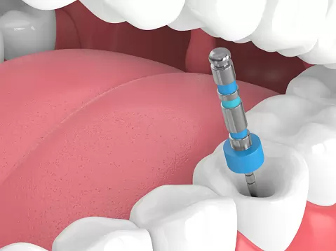
Root canal treatment under a microscope (micro-endodontics) is an advanced endodontic discipline aimed at treating the complex, branching, microscopic-scale anatomy of the root canal system by making it visible. A tooth’s root canals are dark, winding tunnels as thin as a strand of hair; in conventional methods, the dentist has to try to scan these tunnels in the dark relying only on manual skill and tactile perception. Dental microscope technology illuminates these dark chambers with high-intensity coaxial light waves and magnifies the field of view many times over, turning the operative area into a surgical map. This allows remnants of living nerve tissue, anatomical variations, and hidden infection foci to be cleaned away with cellular-level precision, giving the tooth a second biological lifespan.
- High Optical Magnification: Progressively magnifying the treatment area between 4x and 25x for greater detail.
- Shadow-Free Illumination: Integrated LED/halogen light sources that send parallel beams directly into the tooth’s pulp chamber.
- Evidence-Based Intervention: Treating based on directly observed reality rather than guesswork-driven blind injections or cleaning steps.
How Is Root Canal Treatment Performed Under a Microscope?
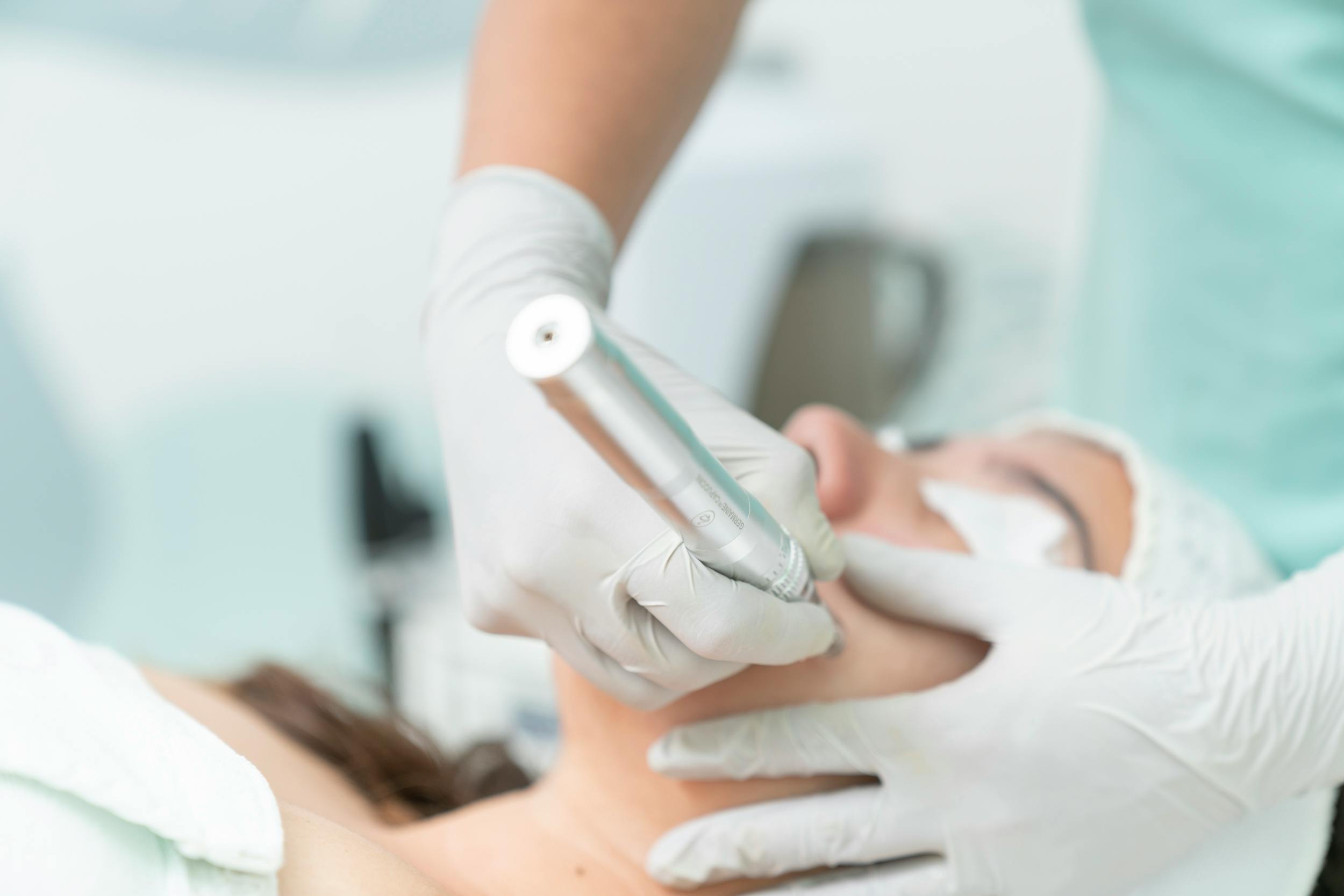
Root canal treatment carried out under a microscope is a micro-surgical procedure performed in modern clinical settings subject to full sterilization protocols, using patient-chair ergonomics designed to match the microscope’s optical axis. First, to ensure the patient’s comfort during the procedure, the tooth and surrounding bone tissue are fully numbed using digital or conventional local anesthesia methods. The tooth is then isolated with a “rubber dam” (a waterproof medical rubber sheet) to keep it separate from saliva bacteria and moisture. The dentist positions the dental microscope head at the entrance of the patient’s mouth and begins looking through the eyepiece; from this point on, the pulp chamber is opened and the anatomical entrances of the canals are scanned under the microscope, with rotary instruments and ultrasonic tips used to clean infected tissue at the micron level before the canals are sealed with biocompatible gels.
- Rubber Dam Isolation: Setting up an absolute barrier that prevents oral fluids and bacteria from entering the treatment corridor.
- Microscopic Access Cavity: The phase of opening the most minimal possible opening needed to see the canal entrances, without unnecessarily cutting into the tooth’s healthy crown structure.
- Chemical-Mechanical Disinfection: Ultrasonically flushing the widened canals with special antiseptic solutions (sodium hypochlorite) while monitoring under the microscope.
- 3D Thermoplastic Filling: Three-dimensionally sealing the cleaned spaces with heated gutta-percha material so no leakage gap remains at the root tip.
In Which Situations Is Root Canal Treatment Under a Microscope Preferred?
Micro-endodontic protocols are the preferred first choice whenever conventional root canal treatments fall short due to the limitations of two-dimensional X-rays or the naked eye, and in the presence of anatomical blind spots or complications. Tooth roots don’t always follow neat, tapered lines; C-shaped canals, severely calcified (blocked) tunnels, and root curvatures reaching up to 90 degrees can only be safely treated with the aid of microscopic vision. This technology is also the only real solution in borderline cases such as correcting past faulty procedures or dealing with broken metal fragments lodged inside a tooth.
The main situations in clinical practice where microscopic root canal treatment is definitively the treatment of choice are:
- Scanning for extra hidden canals — such as the MB2 (second mesiobuccal) canal in upper molars — which are frequently missed and lead to sneaky, lingering pain,
- Millimetrically sealing perforations — cases where the root wall of a tooth was accidentally punctured during a past root canal treatment — using bio-ceramic materials,
- Safely opening tunnels in canals that have become blocked with calcification (calcified canals) due to chronic inflammation, using ultrasonic tips,
- Definitively diagnosing the presence of vertical or horizontal microscopic cracks in a tooth’s root during root canal treatment sessions.
Who Is Root Canal Treatment Under a Microscope Suitable For?
Microscope-assisted root canal treatment is medically suitable for adults and children of any age who have lost tooth vitality due to deep decay or trauma, with the aim of saving the tooth from extraction and keeping it in the mouth. Because the procedure is performed under anesthesia and offers tissue-friendly optical magnification, it can be safely planned regardless of the patient’s systemic condition — including patients with heart or blood pressure issues. It is the most appropriate endodontic approach especially for patients with molars that have complex anatomy, or for conscientious patients who view tooth extraction as an absolute last resort. However, planning may need to be adjusted for patients with advanced neurological conditions who have difficulty keeping their head steady during the procedure.
| Clinical Indication Scheme | Treatment Approach Under the Microscope | Biological Success Rate Achieved |
|---|---|---|
| Anatomically Curved / Complex Roots | Scanning the curved canal with flexible nickel-titanium files while monitoring under the microscope. | 98%; risks of root perforation and ledge formation are eliminated entirely. |
| Failed Previous Root Canal Treatments | Melting away and cleaning out old root fillings under the microscope to dry out the infection. | 95%; the tooth is kept in the mouth without the need for surgical extraction. |
| Large Cyst / Abscess at the Root Tip | Microscopic disinfection and medication of the lesion at the root tip, working through the canal. | High; bone loss is halted and tissue regeneration (renewal) is triggered. |
| Acute Jaw Lock / Patients Unable to Open Their Mouth | Micro-intervention with a limited mouth opening, since the field of view can be narrowed with the microscope. | Medium-High; carried out with the help of special mouth props to reduce joint fatigue. |
What Are the Advantages of Root Canal Treatment Under a Microscope?
The advantages the dental microscope brings to modern endodontic clinics both maximize diagnostic accuracy and protect the tooth’s structural lifespan. The pulp chamber, which looks like a dark well to the naked eye, is fully illuminated under the microscope in complete detail; this prevents the dentist from taking steps based on guesswork or damaging the tooth through trial and error. The smooth progress of the treatment process directly improves both the patient’s time in the chair and the quality of comfort afterward.
- Tissue-Friendly Conservation: Maximum conservation of the tooth’s healthy crown and root dentin tissue while decay is cleaned away, so the tooth doesn’t become thin.
- Complete Diagnostic Power: Microscopic cracks that would be hidden on conventional X-ray films are caught instantly, preventing unnecessary treatments.
- Long Treatment Lifespan: Since no bacterial residue is left behind in the canals, the likelihood of recurrence (reinfection) after the procedure is minimal.
- Visual Proof Comfort: Integrated cameras allow the procedure to be video recorded, providing transparent data to the patient or the lab.
How Does Root Canal Treatment Under a Microscope Differ From Conventional Treatment?
In conventional root canal treatment, the dentist proceeds in the dark by looking at a two-dimensional X-ray film and interpreting the resistance felt from the instrument against the tooth’s root (tactile perception); this analog approach carries a risk of error — particularly in complex canals — such as missing a canal or perforating the root. Root canal treatment under a microscope, however, moves this entire process into a fully visual, predictable, and evidence-based dimension. The microscope shows the dentist the pulp stones inside the canal, the location of broken fragments, and the entrance openings of lateral canals with map-like clarity; in other words, the blind spots of the conventional method are completely illuminated under the digital microscope.
- Field-of-View Resolution: The difference between the 1x view offered by the naked eye in the conventional method versus the 25x magnification power offered by the microscope,
- Illumination Quality: Eliminating the loss of intraoral light caused by the shadow of the cheek and shoulders, replaced by the microscope’s coaxial in-lens LED light,
- Flexibility and Mechanical Safety: A radical reduction in the rate of medical files getting stuck and breaking inside the root, since the anatomical boundaries of the canals can be observed.
What Assessments Are Done Before Root Canal Treatment Under a Microscope?
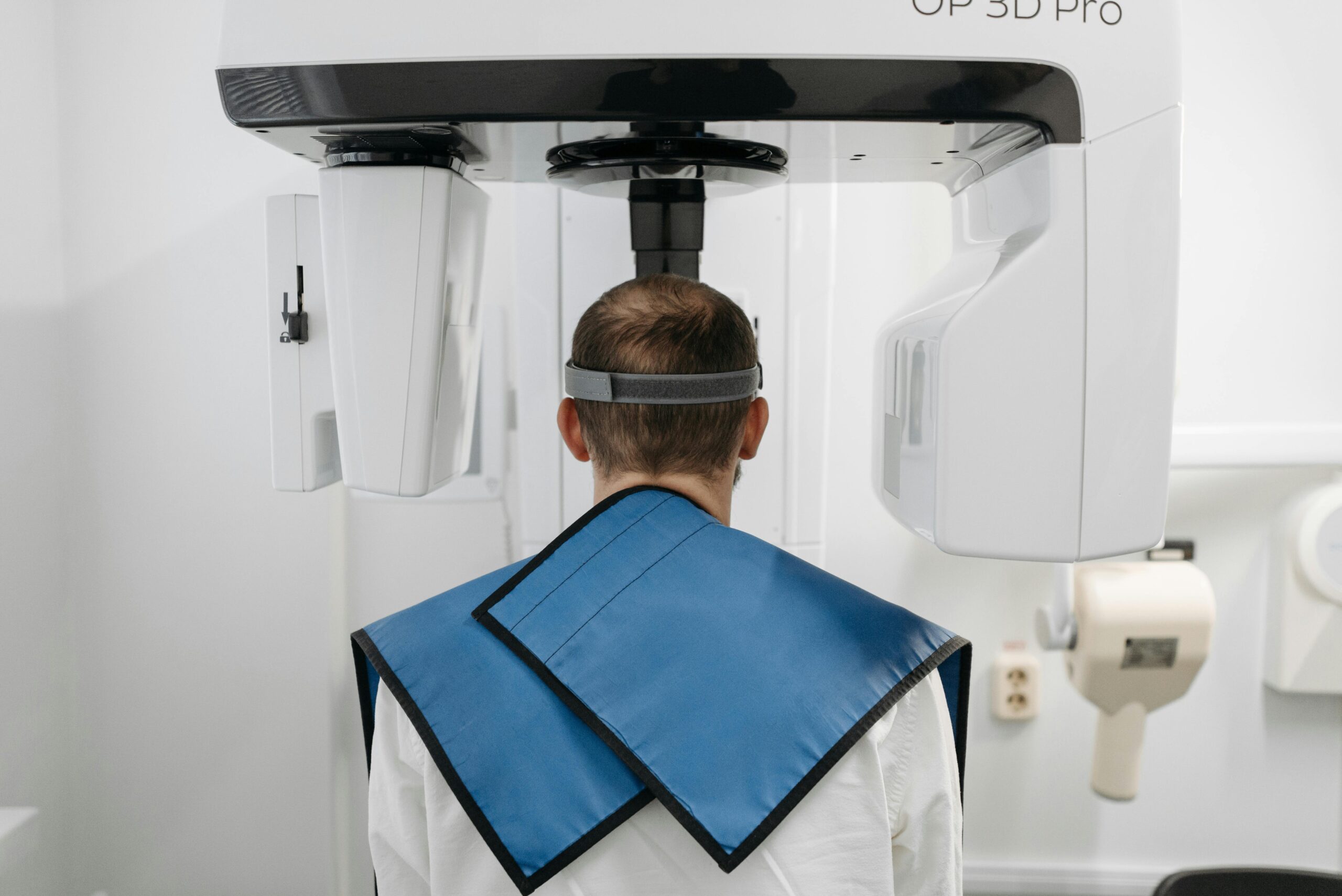
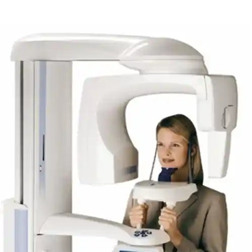
The clinical assessment performed before moving to the treatment session is a vital analysis phase that measures the tooth’s potential for being saved (its prognosis) and lays out the operative strategy. During the examination, vitality tests are performed on the tooth and the resistance of the periodontal tissues is screened. The most vital digital support at this stage is CBCT 3D dental tomography mapping, which goes beyond two-dimensional panoramic films to scan the tooth roots in millimetric cross-sections in three dimensions; the data obtained from the tomography scan reports the working route under the microscope to the dentist in advance.
- 3D Volumetric Scanning (CBCT): Determining the number of root canals and the volume, in cm³, of any cysts within the bone.
- Dentin Thickness Check: Measuring the amount of healthy tissue remaining in the crown of the tooth to determine the type of restoration to be done afterward (a filling or a porcelain crown).
- Facial Boundary Screening: The process of clinically documenting the degree of acute inflammation in the gum and surrounding mucosal barrier.
How Is the Dental Microscope Used During Root Canal Treatment?
Clinical use of the dental operating microscope is a highly focused piece of micro-surgical craftsmanship in which the dentist fixes their eyes on the lens eyepieces and coordinates all hand movements based on the greatly magnified image on the screen. The dentist positions the device’s ergonomic head so it sits about 20–30 cm above the entrance of the patient’s mouth and calibrates the focal distance. The microscope’s magnification level is changed according to the stage of treatment; for example, low magnification (4x) is chosen while opening the pulp chamber, while the maximum magnification phase (25x) is used when searching for a microscopic crack inside the canal or removing a broken instrument.
- Coaxial Light Transmission: Beams emerging from the exact center of the lenses illuminate the base of even the narrowest canal tunnel without creating shadows.
- Binocular Eyepiece Intelligence: Giving the dentist depth perception (a sense of 3D dimension) for comfortable, millimetric hand-eye coordination.
- Ultrasonic Integration: Melting away calcifications in narrow areas monitored through the microscope using specially angled ultrasonic device tips designed to match the microscope’s viewing angle.
How Are Root Canals Visualized During Treatment Under a Microscope?
The internal architecture of the root canals is visualized live and dynamically as the microscope’s high-intensity light waves reflect off the enamel and dentin walls and return to the eyepiece lenses. When the dentist looks down at the tooth from above, the power of the microscope lets them see not just the entrance opening of the canal, but the full integrity of the inner wall of that dark tunnel extending toward the root tip, in clear, defined lines. This optical imaging perfectly distinguishes old filling residue left inside the canal, anatomical ledges, and strips of infected living nerve tissue on the screen by creating light-dark contrast.
- Reflective Micro Mirrors: The use of special rhodium-coated intraoral micro mirrors that don’t distort the microscope’s light, allowing a direct view into the canals of the back molars.
- High-Resolution Monitor Transfer: The phase of transferring the image from the eyepiece simultaneously onto large clinical screens, ensuring synchronization between the assistant and technician.
Can Root Canal Treatment Under a Microscope Be Used to Remove Broken Canal Instruments?
Yes, microscope technology is the strongest and most established method available in the field of endodontics for safely removing metal instruments (fragments of nickel-titanium files) that broke and got stuck during past treatments due to the narrow or curved structure of the root canals, blocking the canal corridor. In conventional methods, since the location of the broken instrument can’t be seen, there is often no option left but to extract the tooth; under the microscope, however, with 25x magnification, the upper end of the broken metal fragment can be identified with total visual clarity. Watching under the microscope, the dentist melts away the dentin surrounding the fragment using micro ultrasonic tips and removes the broken instrument from the root with special forceps, saving the tooth from being condemned to extraction.
- Visual Localization: Precisely determining at which millimeter of the root (the coronal, middle, or apical third) the broken fragment is located.
- Ultrasonic Loosening: Bypassing the calcified tissues surrounding the metal fragment without damaging the tooth, while monitoring under the microscope.
- Reopening the Canal Corridor: Once the instrument is removed, reinforcing the sealing line that reaches the root tip to finish off the infection.
Why Is Root Canal Treatment Under a Microscope Preferred for Retreatment Procedures?
Root canal retreatment procedures involve removing and starting over the treatment of teeth with old fillings that have failed, where the abscess at the root tip hasn’t resolved; the anatomical risk factor here is very high, and the microscope is preferred because it manages these risky restorations smoothly. In the old treatment, the canals may not have been fully filled, may be blocked with hardened artificial materials, or the canal’s original path may have been diverted, perforating the side wall. The microscope shows the dentist the exact boundary line of those old solid filling materials (gutta-percha or hard cements), allowing instruments to slip in safely, and starts the healing process by thoroughly clearing away the tooth’s past anatomical errors.
- Cleaning Old Filling Material: Microscopically scraping away stubborn old paste residue harboring chronic bacteria that has stuck to the canals.
- Confirming the Anatomical Path: Returning to the original canal line by monitoring artificial ledges created in the past under the microscope.
- Perforation Repair: Sealing artificial holes opened in the root wall with bio-ceramic (MTA) materials, watertight, under the microscope’s view.
How Long Does Root Canal Treatment Under a Microscope Take?
Root canal treatment under a microscope requires a somewhat longer active chairside session time compared to conventional manual treatments, along with patient, careful time management, due to the millimetric positioning of the microscope head, high-resolution optical focusing, and the meticulous use of micro-surgical instruments. The active cleaning and filling phases generally take between 45 and 90 minutes per session, depending on the complexity of the case in the tooth (presence of a broken instrument, canal calcification, etc.) and the number of root canals in the tooth; however, this time is a proven safety period that prevents the tooth from being extracted.
- Single-Canal Front Teeth: A single session averaging 45–60 minutes, including disinfection and filling processes.
- Multi-Canal Molars: A clinical visit of roughly 60–90 minutes due to the need to scan for anatomical variations.
- Complicated Cases (Broken Instrument/Retreatment): A meticulous micro-surgical phase where the duration may vary depending on how quickly the fragment can be removed.
Can Root Canal Treatment Under a Microscope Be Completed in a Single Session?
Yes, in modern endodontic practice, root canal treatment under a microscope can be smoothly completed in a single session in all suitable cases where the tooth’s pulpal and periapical (root tip) infection status allows it. Thanks to the microscope’s high disinfection power, canals can be fully cleared of bacteria within seconds, so in teeth without acute pain or with only a living (vital) pulpitis complaint, scanning, cleaning, and permanent root filling can all be finished in the same chair session. However, in cases with active inflammatory discharge (abscessing) at the root tip or a stubborn chronic lesion, the process is scheduled over 2 sessions, placing medical calcium hydroxide paste in the canal to calm the tissue in between.
- Criteria for a Single Session: Living nerve inflammation, stable dental conditions without an actively discharging abscess at the root tip.
- When Multiple Sessions Are Required: Stubborn infections where pus/fluid is continuously draining from the canal and tissue homeostasis cannot be established in one go.
What Should Be Watched For After Root Canal Treatment Under a Microscope?
Making the success of the micro-surgical session under the microscope permanent, protecting the root-tip tissues that are entering the healing phase, and preventing the fracture of the tooth’s crown structure holding a temporary filling — these all depend on medical rules the patient will follow at home after the procedure. Since the tooth’s nerve network has been removed, no pain is felt, but because the surrounding bone attachments will be under a healing stimulus, avoiding mechanical load on the area in the first few days directly improves the treatment’s success.
The points that must be carefully observed during the critical period after the procedure are as follows:
- No food whatsoever should be consumed during the first 2 hours, until the effect of the anesthesia (numbness) has fully worn off, to prevent the risk of biting the tongue or cheek.
- Until a permanent porcelain crown or upper restoration is placed on the treated tooth, hard nuts or shelled foods should not be chewed on that side in the meantime (the tooth is prone to fracture).
- Anti-inflammatory pain relievers prescribed by the dentist should be taken regularly in the first few days to relieve subsurface tissue aching.
- Oral hygiene routines (brushing and flossing) should continue uninterrupted, with gentle movements that don’t disturb the treated area, starting from the evening of the procedure.
How Does the Healing Process Progress After Root Canal Treatment Under a Microscope?
The tissue recovery phase after the procedure progresses extremely quickly, predictably, and comfortably, since no surgical incisions are made in the scalp or gums; however, because the inflamed tissues at the root tip are scraped away with ultrasonic waves, the bone cells in that area mounting a repair response is a natural part of healing. During the first 3–5 days, it is completely normal to experience mild aching, sensitivity, or a “sense of height” (periodontal sensitivity), especially when pressure is applied to the tooth or when the teeth touch each other. The healing process moves along a linear path; as the extracellular matrix is cleared away, the abscesses inside the bone shrink and are replaced by fresh bone tissue.
- First 72 Hours: The peak point of micro-aching sensation while chewing (this phase is comfortably managed with pain relievers).
- The 1-Week Mark: Acute stimulation in the tissues fully ends, and the tooth regains its normal functional chewing power in the mouth.
- The 6-Month Check-Up: The phase in which an X-ray confirms that the area of bone loss (the dark shadow) at the root tip has been refilled with new white bone tissue.
What Factors Affect the Success Rate of Root Canal Treatment Under a Microscope?
The success of microscope-assisted endodontic treatments depends not just on the use of high-tech lenses, but on the flawless simultaneous operation of a range of multidisciplinary medical criteria. The single most uncompromising success criterion is the near-100% elimination of the bacterial population inside the canals and the creation of a watertight seal at the root tip. As long as there isn’t a systemic immune deficiency or neglect of home care on the patient’s part, the visual quality offered by the microscope raises the treatment’s clinical accuracy rate to its highest level.
- Quality of Microbial Disinfection: The ability of antiseptic solutions, activated ultrasonically, to reach even the most microscopic lateral canals.
- Watertightness of the Root Filling: The gutta-percha material locking into place with millimetric precision, without overflowing past the root tip or falling short.
- Upper Coronal Restoration: Definitively stopping oral fluids from seeping downward by capping the tooth with zirconium or porcelain fillings (onlays) once treatment is complete.
Which Teeth Can Root Canal Treatment Under a Microscope Be Applied To?
Microscopic endodontic protocols can be safely and successfully applied to all groups of teeth with an anatomical root structure in both the lower and upper jaw — front teeth, canines, premolars, and molars alike. Since front teeth typically have a single, wide canal, the process is usually completed more quickly; the large molars at the very back of the mouth, however, are the group of teeth that most need the microscope’s magnification and illumination power, due to their complex, branching micro-anatomy with 4 or 5 canals. No matter how far back the tooth is positioned, the microscope’s flexible optical head allows the treatment to be carried out safely.
- Front (Anterior) Group: Microscopic treatment focused on aesthetic preservation for teeth that have fractured or discolored due to trauma.
- Back (Posterior) Molar Group: The area where curved roots, blocked canals, and hidden tunnels like MB2 are treated with confirmed precision using the microscope.
How Does Root Canal Treatment Under a Microscope Contribute to Preserving the Tooth?
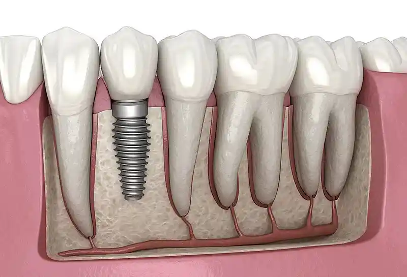
The most revolutionary contribution this advanced-technology micro-endodontic approach makes to the biology of tooth preservation is protecting the tooth’s “organic presence in the mouth” by saving it from being condemned to surgical extraction. No matter how advanced they become, artificial titanium implants can never possess the same periodontal ligament fiber networks and natural nerve receptors — which sense chewing pressure — that a person’s own natural tooth has. By cleaning away only the microscopically infected inner core of the tooth while preserving the outer bone and root integrity exactly as it was, root canal treatment under a microscope offers an invaluable contribution to the patient sustaining natural, comfortable chewing reflexes with their own tooth for a lifetime.
- Natural Chewing Sensation (Proprioception): The permanence of the ability to sense the hardness of food, thanks to the preservation of the living fibers around the tooth root.
- Maximum Dentin Conservation: Preventing unnecessary deep carving that would weaken the tooth, and preserving its fracture resistance, in work carried out while monitoring through the microscope eyepiece.
Frequently Asked Questions About Root Canal Treatment Under a Microscope
Below are the most common practical concerns on the minds of patients planning to undergo microscopic root canal treatment in order to prevent tooth loss and get rid of stubborn root abscesses, along with their clear, straightforward medical answers:
Will I feel a lot of pain or ache during a root canal treatment session under a microscope?
No, this is definitely not a procedure that causes pain or ache. Before the procedure, the tooth and surrounding connective tissue are completely numbed using advanced local anesthetic solutions. Since the nerve pathway is blocked throughout the session, all you will perceive, in a numbed state, is the gentle vibration of the device head and the coolness of the water rinse; the process is completed in total comfort, as if you were reading a newspaper in the chair.
Does a tooth that has had root canal treatment turn yellow or black because it has lost its vitality?
In old-style conventional treatments, tissue remnants left in the canal or low-quality canal filling paste could stain the tooth from the inside over time, causing gray-black discoloration. In modern microscopic endodontics, however, since the pulp chamber is cleaned under the microscope so that not a single cell remnant remains, and new-generation, non-staining bio-ceramic gels are used, permanent pathological discoloration in the tooth after the procedure absolutely does not occur.
What happens if that broken metal instrument fragment inside the root canal isn’t removed?
If a metal fragment (the tip of a file) that breaks inside the root canal is left there, it blocks the canal corridor like a mechanical plug, preventing the dentist from reaching the bacteria at the root tip and flushing it with disinfectant fluids; this leads to the inflammation (cyst) at the root tip chronically growing, eroding the bone, and ultimately causing the tooth to have to be extracted due to severe, painful abscesses — removing the fragment under a microscope is essential.
After root canal treatment is finished, should the tooth get a filling or a crown — which is correct?
This decision is made by the dentist based on how much of the tooth’s healthy crown (dentin) wall remains at the end of the microscope session. If the tooth’s side walls are largely intact, a standard composite filling is sufficient; however, if the decay was very extensive and only thin shell walls remain, then, to prevent the tooth from splitting in half under chewing pressure, placing a zirconium crown or a porcelain onlay filling that wraps around and protects the tooth like a cap is a medical rule for the tooth’s longevity.
Can I go back to my normal life and work right away on the day I have root canal treatment under a microscope?
Yes, microscopic root canal treatment is a completely local, non-invasive piece of surgical work that does not affect consciousness or motor skills; therefore, the moment the session ends, the patient can get up from the chair and continue their normal daily, professional, social, or academic life rhythm right where they left off, without interruption. There is no clinical obstacle to driving or attending work meetings immediately after the procedure.
Who We Are? & Panorama Ankara Oral and Dental Health
Panorama Ankara is a licensed oral and dental health center that brings together an academic team of specialists and the latest global bio-technological infrastructure under one roof — not only in oral, dental, and maxillofacial surgery, implantology, orthodontics, and cosmetic dentistry, but also in microscopic endodontics (root canal treatment specialization) — providing patients with institutional health assurance. Rather than condemning teeth with deep decay, stubborn root-tip cysts, and past failed fillings to surgical extraction, our center places vital importance on saving them through evidence-based medical disciplines, and uncompromisingly operates high-resolution, original Surgical Dental Microscope Root Canal Treatment ecosystems in its clinical operating workflow.
Across all radiological, screening, design, and microscopic surgical interventions carried out at Panorama Ankara, the principles of absolute sterilization, high patient safety, transparency, and honesty are enforced without compromise, in accordance with our founding charter. In our clinical practice, proprietary dental microscope units offering up to 25x optical magnification and shadow-free coaxial LED light power, along with micro-ultrasonic tips, are applied with millimetric precision under the supervision of our team of expert endodontists. Our aim is not to impose on our patients traditional treatment models carried out in the dark with cookie-cutter formulas — models that are risky and carry a high chance of failure — but to analyze, in clear detail on computer screens, the hair-thin tunnels within each tooth’s root canals; to provide maximum session comfort without harming surrounding soft tissue or blood vessels, the safety of broken-instrument removal, and watertight sealing quality that lets you smile with your own original teeth for a lifetime. If you’re tired of stubborn toothaches and root abscesses that won’t go away, yet keep postponing treatment out of fear of extraction or pain, and you want to begin your smart, microscopic treatment process from start to finish with scientific assurance at an expert center, you can get in touch with the expert team at Panorama Ankara and protect your health and the integrity of your smile through detailed 3D tomographic and microscopic analyses.