What is Digital Dentistry?
Digital dentistry is a modern dental discipline based on the integration of computer, software, and robotic manufacturing technologies into clinical dental practices. Workflows that conventionally relied on the clinician’s manual dexterity, rule-of-thumb adjustments, and chemical materials prone to shrinkage/expansion are replaced in the digital model by mathematical algorithms offering micron-level precision. This approach transfers the patient’s jaw structure onto the computer screen as a digital twin. The entire workflow, from the diagnostic phase to the final moment the prosthesis is manufactured, is managed over digital networks without data loss, zeroing out the margin of error with a perfection that surpasses human perception.
- CAD/CAM Integration: Introducing computer-aided design (CAD) and computer-aided manufacturing (CAM) directly into the operatory room.
- Data Standardization: Converting impression and radiological data into universal, non-deformable file formats such as STL, PLY, or DICOM.
- Objective Diagnosis: Verifying micro-pathologies that clinicians might overlook, with the assistance of smart artificial intelligence filters.
Which Technologies Are Used in Digital Dentistry?
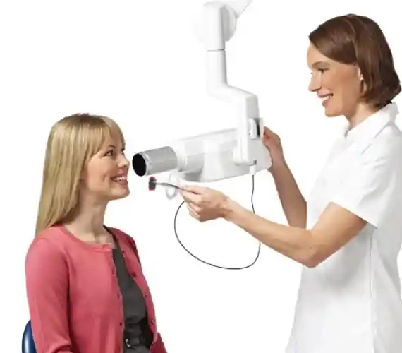
The highly efficient performance of digital dentistry within a clinical environment relies on the synchronized operation of a series of advanced technology hardware and software ecosystems. These systems convert light and X-ray reflections captured from intraoral tissues into processable engineering data within seconds. Each apparatus utilized in clinics is engineered to fortify accuracy at a distinct stage of treatment.
The primary advanced technologies forming the foundation of digital dental architecture include:
- Intraoral Scanners: Optical cameras that generate the 3D topographic map of the teeth,
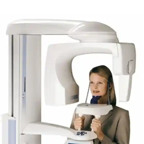
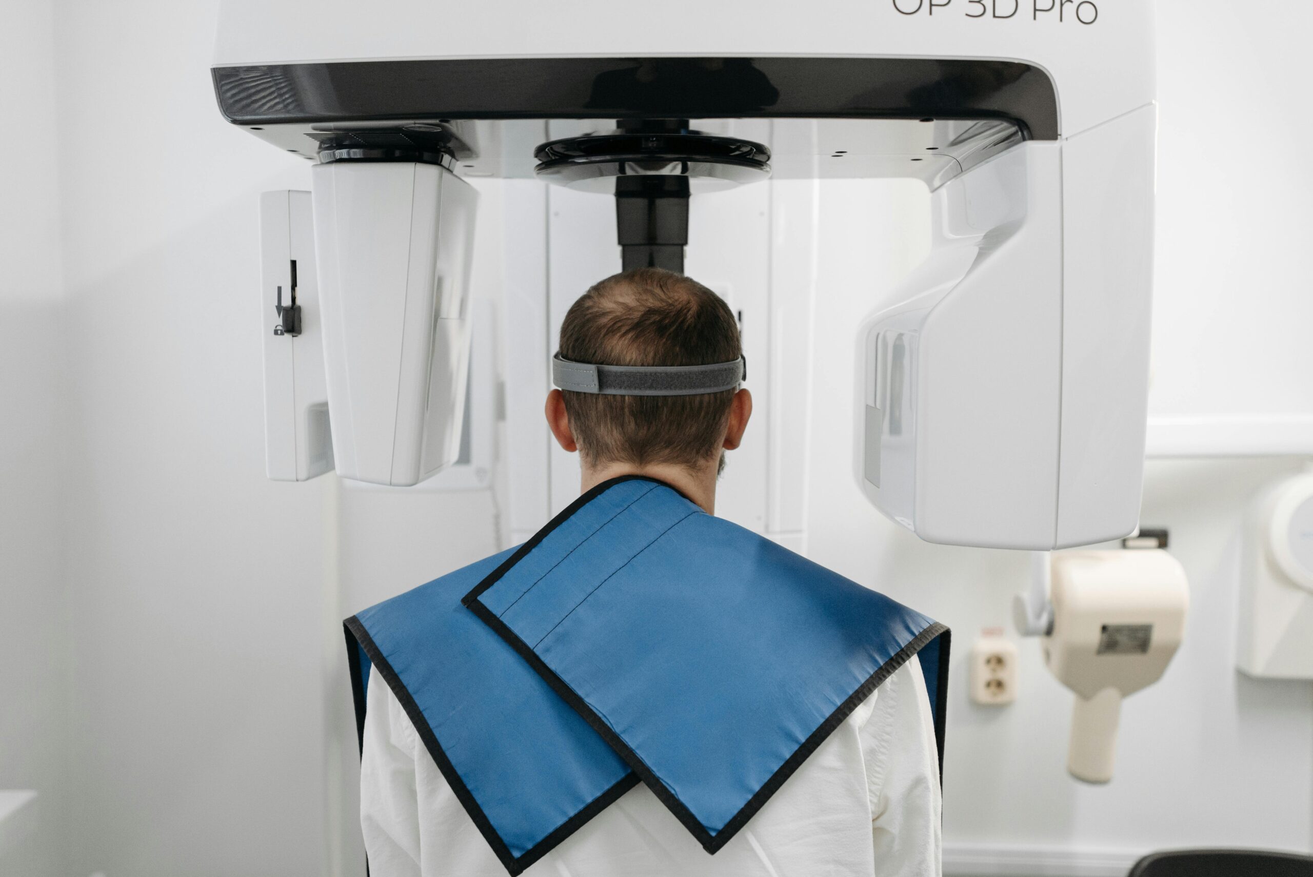
- CBCT 3D Dental Tomography: Cone-beam scanners displaying the jawbone and nerve canals in sections at a 1:1 scale,
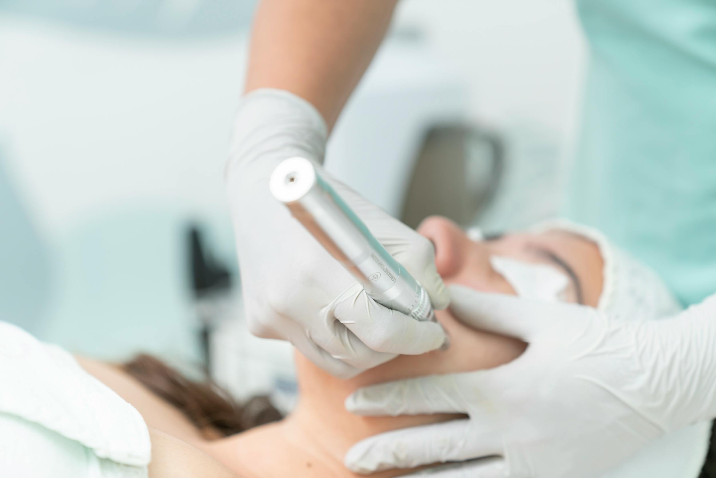
- Milling Machines: Robotic units that carve the designed prostheses out of zirconium blocks using diamond burs,
- 3D Dental Printers: Additive printers that fabricate surgical guide plates, temporary teeth, and model templates from liquid resin,
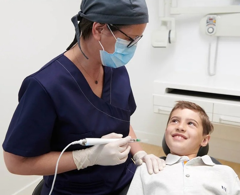
- Digital Anesthesia Devices: Smart systems providing painless numbing through computer-controlled infusion pumps.
In Which Treatments is Digital Dentistry Used?
Modern digital dentistry applications broaden their horizons daily, serving as the primary operational infrastructure across nearly all major dental specialties. It plays an active role at every step, from enhancing diagnostic accuracy to perfecting prosthetic adaptation. Eliminating speculative clinical estimates, this integrated workflow facilitates clinicians in achieving rational outcomes even in the most complex jaw reconstructions.
| Dental Specialty Branch | Utilized Digital Technology Component | Clinical Contribution Provided to the Treatment Process |
|---|---|---|
| Prosthodontics | Intraoral scanner and CAD/CAM milling units | Zero-error production of zirconium crowns, porcelain bridges, and laminate veneers within the same day. |
| Oral and Maxillofacial Surgery | CBCT 3D Tomography and Surgical Guide Plates | Placing implants into the bone through the safest angles that prevent nerve damage, flapless and sutureless. |
| Orthodontics / Pediatric Dentistry | 3D Scanners and Clear Aligner Software | Simulating tooth movements on the computer during wireless orthodontics workflows to robotically print successive aligners. |
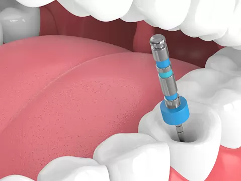
| Endodontics (Root Canal Treatment) | Microscopic RVG and Axial CBCT Sections | Detecting overlooked extra root canals and vertical root fractures at an incipient stage. |
Who is Digital Dentistry Suitable For?
Digital dentistry methods are suitable for any adult individual over the age of 18 who wishes to enhance their oral and dental health quality, replace missing teeth, or achieve an aesthetic smile. Because the system incorporates protective equipment that minimizes ionizing radiation doses and rejects the use of dough materials during the impression stage, it demonstrates high compatibility in sensitive patient cohorts (the elderly, pregnant patients, and individuals with disabilities). It is the most ideal treatment philosophy for active business professionals bound by intense time constraints or individuals harboring a fear of sitting in the dental chair.
- Those with a Severe Gag Reflex: Sensitive constitutions experiencing a vomiting sensation when an impression tray enters the mouth,
- Those Experiencing Dentophobia (Fear of the Dentist): Individuals wishing to minimize chairside stress thanks to digital anesthesia and expedited workflows,
- Those with Time Constraints: Patients traveling from abroad or those who, due to a busy work schedule, request veneer treatments to be finalized within 1 day.
What Are the Advantages of Digital Dentistry?
The verified clinical advantages of digital dentistry compared to traditional methods are not confined to intra-operative practicality; they also directly elevate the structural lifespan of prostheses. Human-induced impression, plaster, and casting errors are entirely eliminated from this chain. Designs executed at a micron level on a computer screen push the physical boundaries of materials, ensuring maximum biomechanical resistance and passive fit.
- Maximum Adaptation Quality: Preventing micro-leakage decay because no microscopic gaps (marginal gaps) remain between the prosthesis boundary and the gum margin.
- Same-Day Dentistry: Completing the scan and final prosthetic assembly within hours, instead of waiting days for laboratory shipping.
- Reversible Archive: Oral impression data is preserved indefinitely on cloud servers; if a tooth fractures years later, a replacement can be milled within seconds from the historic data.
- High Aesthetic Naturalness: Designing contours in perfect golden ratio harmony with the individual’s facial anatomy, thanks to digital smile design software.
How is Diagnosis and Treatment Planning Formulated in Digital Dentistry?
The diagnostic and planning phase in digital dentistry relies on the principle of superimposing and merging all anatomical data gathered from the patient’s mouth within a computer environment. The process initiates by pairing bone data (DICOM) derived from the CBCT device with the soft tissue map (STL) captured by the intraoral scanner inside specialized planning software. The clinician monitors the 3D simulation of the patient’s jawbone and gums on the screen. Within this interface, which tooth will be intervened with at what exact angle, and the capacity of the bone density to bear the implant screw are evaluated via rational graphics, finalizing every step of the operation in the virtual world before touching the skin.
- Multidimensional Registration: Consolidating bone, gum, and facial photograph datasets over a single digital model.
- Algorithmic Risk Analysis: The software automatically warning the clinician whenever anatomical hazard boundaries (nerve canals, sinus cavities) are approached.
- Predictable Outcomes: Outlining the treatment timeline and budget analysis clearly from the absolute beginning based on faultless gathered data.
How Do 3D Imaging Technologies Support Digital Dentistry?
Three-dimensional imaging technologies fortify the diagnostic power of digital dentistry by completely eradicating the illusion of “overlapping tissues obscuring structures,” which is the greatest defect of traditional two-dimensional X-rays. CBCT (Cone Beam Computed Tomography) devices scan the jawbone with millimetric horizontal, vertical, and transverse sections to deliver the true thickness and anatomical quality of the bone at a 1:1 scale. This support grants the surgeon total visual command, particularly when preparing implant sockets and outlining intraosseous cyst boundaries.
- Depth Information Cataloging: The capability to measure not only the height of the bone but also its width (thickness) at a micron level.
- Nerve Line Isolation: Eliminating paresthesia risks by 3D mapping the trajectory navigated by the main lower jaw nerve canal.
- Sinus Floor Analysis: Assessing the volumetric status and degree of sagging of the air spaces located in the upper jaw molar region.
How Are Intraoral Scanners Used in Digital Dentistry?
Intraoral scanners are deployed as the most critical data collection tool in digital dentistry to transfer the patient’s dental impression into a digital laboratory environment with zero distress and maximum comfort. While the scanner tip, resembling a camera, is guided over the teeth, the device projects thousands of structured LED light beams onto the tissue per second. The beams reflecting back from dental surfaces are processed via triangulation algorithms to generate a flawless 3D dental model within seconds; this model is accepted as the baseline data for verifying prosthetic preparation boundaries.
- Precise Margin Detection: Capturing the dental preparation lines (finish lines) where the crown will seat with micron-level clarity.
- Dynamic Occlusion Record: A phase where the right-left bite relationship is scanned instantaneously as the patient brings their jaws into a normal biting position.
- Instant Error Rectification: If a missing point is identified during scanning, that specific zone can be updated locally within seconds without remaking the entire impression from scratch.
How is CAD/CAM Technology Applied in Digital Dentistry?
CAD/CAM (Computer-Aided Design / Computer-Aided Manufacturing) technology is the major industrial application methodology forming both the design and robotic manufacturing muscle of digital dentistry. The workflow operates in two phases: In the first phase (CAD), the anatomical form, chewing cusps, and gingival transitions of the prosthesis are millimetrically tailored with a computer mouse over the 3D model derived from the intraoral scanner, utilizing facial aesthetic software. In the second phase (CAM), this approved digital design dataset is dispatched to multi-axis robotic milling machines; diamond burs within the device mill monolithic zirconium or ceramic blocks untouched, physically manifesting a faultless prosthesis within minutes.
- Computerized Modeling (CAD): Virtually drafting dental dimensions millimetrically in accordance with the golden ratio guidelines of the patient’s facial contours.
- Robotic Milling (CAM): Carving block materials via computerized commands, zeroing out human-induced casting and finishing errors.
- Strength of Block Materials: Utilizing homogenous, ultra-durable zirconium blocks manufactured under high pressure in industrial settings, completely free of air bubbles.
How is Digital Dentistry Used in Implant Treatment?
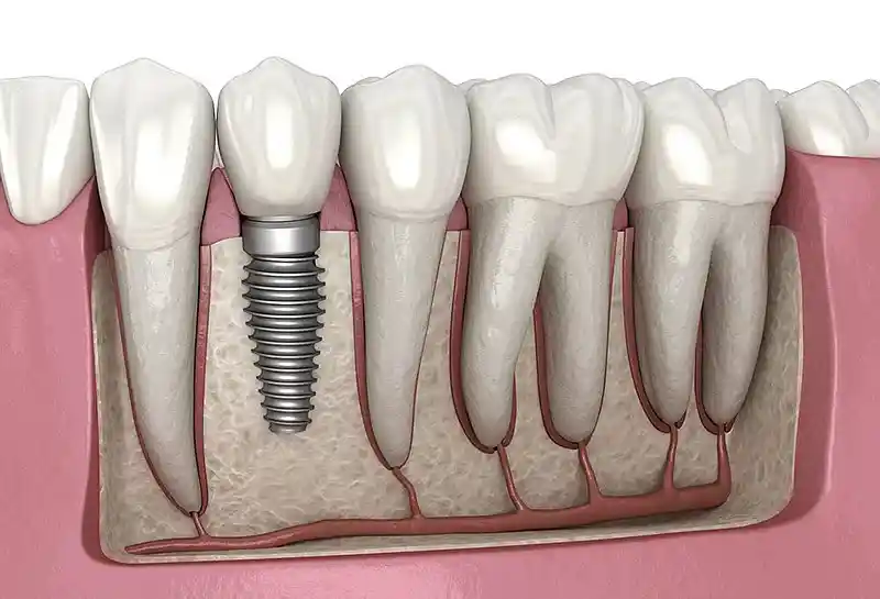
In implantology practices, digital dentistry elevates success rates to the pinnacle by imbuing surgical workflows with a completely “predictable and navigated” identity. Once CBCT tomography data and gingival records from the intraoral scanner are superimposed on the computer, the exact millimetric coordinates and precise angles where implant screws will be anchored into the jawbone are virtually simulated. Leveraging this data, a customized “Surgical Guide” plate is printed via 3D dental printers; during the operation, the surgeon avoids cutting open the gums (flapless/sutureless implant technique), anchoring the implant flawlessly into the bone bed within milliseconds by strictly navigating through the guide channels on this plate.
- Flapless Surgical Comfort: Because the gums are not incised with a scalpel, intra-operative bleeding and post-operative swelling and pain remain minimal.
- Scan Body Technology: Flawlessly capturing implant-supported prosthetic records with the aid of digital pins fitted onto the implant post-placement.
- Immediate Loading (Same-Day Teeth): Measuring the structural anchor strength (torque) of the implant within the bone via software, extending the luxury of fitting temporary fixed teeth during the very same appointment session.
How Does Digital Dentistry Support Orthodontic Treatment?
In orthodontic practices, digital dentistry establishes the sole technical infrastructure for clear aligner workflows (e.g., Invisalign), focusing on transparency to eradicate the aesthetic anxieties triggered by traditional metal brackets and wires. The 3D model acquired via the intraoral scanner is imported into orthodontic planning software (e.g., ClinCheck); artificial intelligence algorithms mathematically calculate how many microns the teeth will move with each sequential clear aligner exchange. Thanks to this data chain, patients can monitor through live simulations on the computer screen how their teeth will align months later and what their final occlusal profile will look like during the very first week of their treatment.
- Digital Setup Production: Virtually charting the trajectories for teeth to navigate toward their ideal coordinates within the jawbone matrix.
- Cumulative Aligner Manufacturing: High-precision printing of 20–40 sequential clear aligners via 3D printers to be delivered to the patient.
- Angular Root Control: Preventing the risk of tooth roots breaching the bone boundaries and generating fenestrations (perforations) during active movement.
How is Digital Dentistry Used in Aesthetic Dental Treatments?
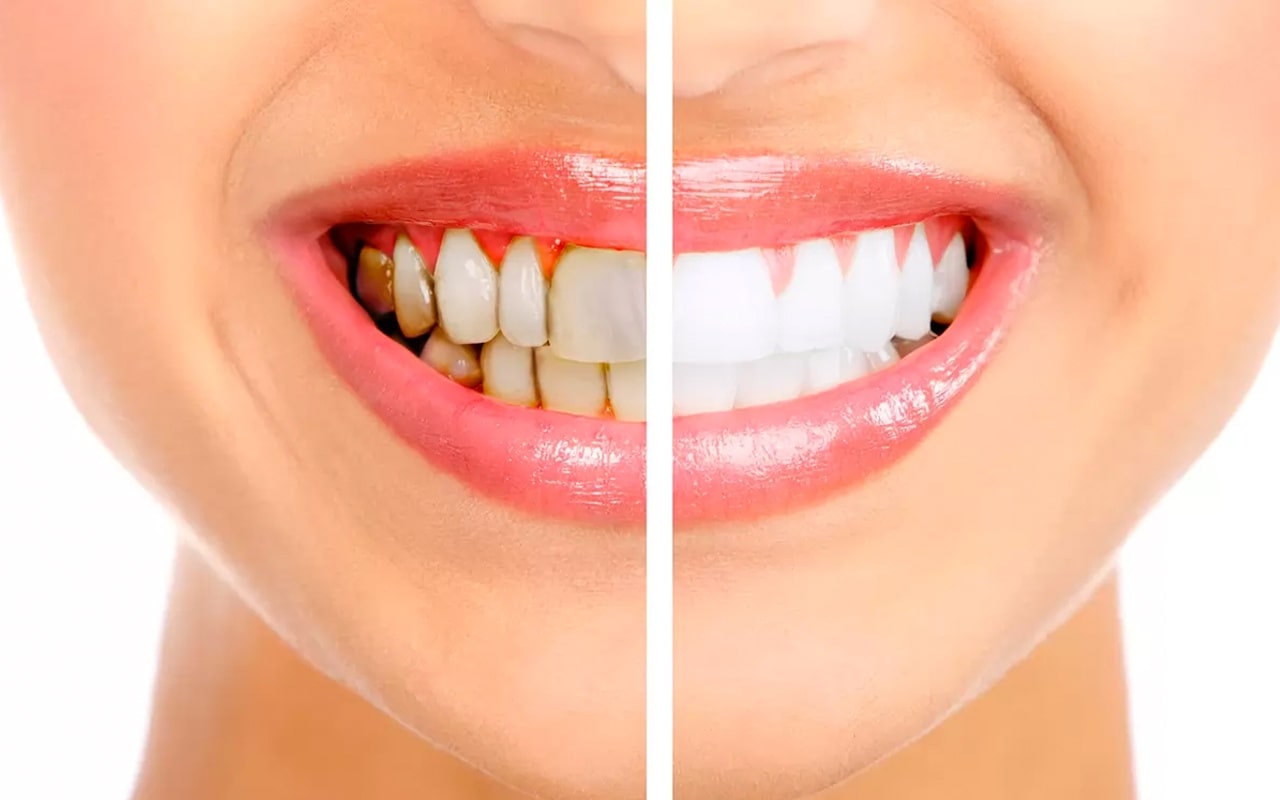
In aesthetic dentistry and smile design applications, digital scanners and design software are selected to establish a seamless visual harmony between the patient’s facial morphology and dental anatomy. The patient’s facial features, lip smile lines, and interpupillary axis are captured with high-resolution cameras and merged with the 3D oral model within the software. The clinician millimetrically tailors the length, width, gingival margins, and curve of the smile line using screen golden ratio calipers to construct an authentic “smile architecture” that best complements the individual.
- Mock-Up Live Trial: Printing the newly designed teeth as a temporary plastic model via a 3D printer to execute a live trial inside the patient’s mouth before any tooth reduction takes place.
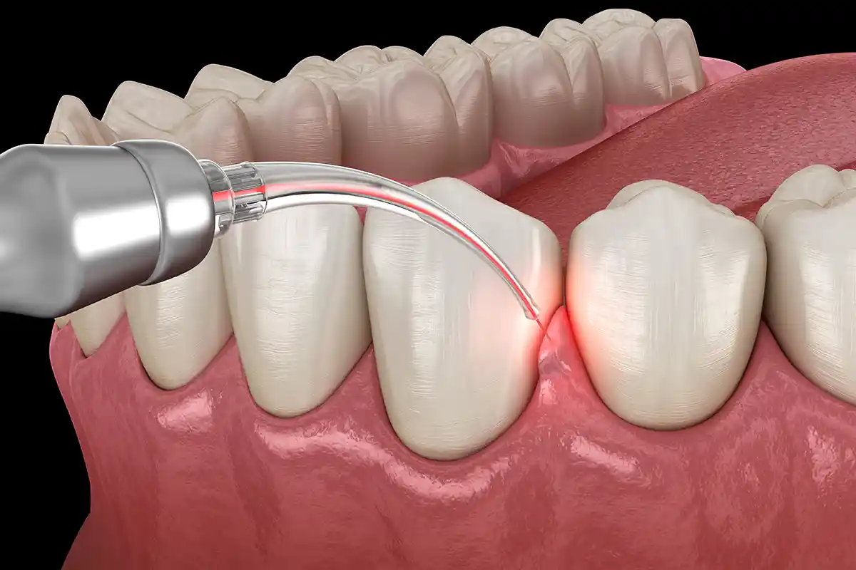
- Pink Aesthetic Calibration: Millimetrically mapping asymmetries along the gingival margins on a digital screen prior to performing laser surgery.
- Natural Enamel Optics: Fabricating translucent restorations that transmit light like natural teeth, thanks to premium glass-ceramic blocks utilized in CAD/CAM milling units.
How is Digital Dentistry Used in Prosthetic Treatments?
The digital workflow in prosthetic dental treatments (zirconium crowns, bridges, and porcelain restorations) interrupts the entire chain of human and material-induced error variables common in traditional laboratory stages. Dimensional deviations encountered in old-school casting methodologies, such as metal shrinkage or plaster expansion, are entirely filtered out in the digital matrix. Emerging from CAD/CAM milling units with micron precision, the restorations seat against the prepared tooth surface with absolute passive fit, avoiding high spots or binding on the first attempt; this radically downscales the number of clinical trial appointments.
- Inlay / Onley Smart Restorations: Scanning strictly the decayed block of the tooth and carving the missing fragment out of porcelain to be bonded into the socket like a puzzle piece.
- Marginal Adaptation Power: Eradicating gingival discoloration and bruising because the restoration margin seats against the gum line with zero error tolerance.
- Rapid Infrastructure Validation: Cross-sectionally testing the skeletal fit of prosthetic infrastructures (zirconium or titanium) on the computer screen.
How Does Digital Dentistry Enhance Patient Comfort?
The greatest comfort digital dentistry extends to the patient is completely eliminating the most disliked and anxiety-inducing steps of dental treatments, such as dough impression tracking, drawn-out trial sessions, and needle stings. The stress of large metal trays pressing against the palate and the fear of suffocation triggered by fluid chemical pastes seeping down the throat belong entirely to the past, thanks to intraoral scanners. The patient can swallow, signal the clinician to halt the process, rest, and comfortably resume scanning right where they left off with zero data loss.
- Zeroing the Gag Reflex: Because capture is executed purely through light waves near the outer architecture of the mouth, a sensation of nausea is never triggered.
- Digital Anesthesia Support: Preventing injection-induced needle stings through computer-controlled painless numbing delivery.
- Visual Participation Comfort: The comfort of patients auditing the 3D model of their own oral cavity on a giant screen alongside their clinician, transparently partnering in the treatment steps.
How Does Digital Dentistry Optimize Treatment Time?
The digital dental workflow radically optimizes and shortens the distressing time losses that consume weeks in traditional dentistry, which historically forced the patient to travel back and forth repeatedly between the clinic and the laboratory. Analog steps such as pouring plaster models, dispatching them via couriers, and executing casting trials are equated to internet speeds within the digital framework; impression datasets land before the technician online within seconds. This technological velocity delivers priceless treatment practicality for individuals managing busy modern schedules.
- Same-Day Delivery: The convenience of fitting a zirconium crown milled in the afternoon to a patient scanned in the morning, thanks to in-office CAD/CAM units.
- Reduction in the Number of Trials: Eliminating alteration and refinement appointments because the primary record is gathered digitally with micron precision.
- Rapid Transit Cycle: Physical courier stages making way for instantaneous email or cloud-based data transfers.
How Does Digital Dentistry Facilitate Impression Processes?
Uncontrollable physical risks encountered in conventional tray impressions—such as dimensional instability from dough shrinking during setting or plaster expanding during drying—are completely eradicated in digital impression processes. A digital impression captured via an intraoral scanner is a fixed “mathematical point cloud” dataset unaffected by elapsed time or ambient temperatures, rendering physical deformation impossible. If a lack of clarity is identified along a margin during scanning, rather than throwing the record away, the clinician simply deletes that small faulty field from the computer to update it locally within seconds; this streamlines the process tremendously.
- Zero Material Waste: Completely filtering out chemically scented, sticky alginate or silicone paste waste from the operatory.
- Luxury of Localized Refinement: Finalizing incomplete fragments of the impression within seconds by focusing strictly on that specific tooth, without distressing the entire mouth.
- Flawless Digital Archiving: Preserving impression data as gigabyte-sized files to serve as a reference template for potential prosthetic fractures in the future.
How Does Digital Dentistry Support Treatment Accuracy?
The greatest support digital dentistry delivers to treatment accuracy (precision coefficient) is auditing micro-boundary discrepancies brought on by human vision limitations or hand tremors during diagnostic and manufacturing phases through artificial intelligence algorithms. The 3D model on the computer screen empowers the clinician to audit the topography magnified hundreds of times; consequently, millimetric preparation margin errors or undercuts invisible to the naked eye are flagged by the software with red alert indicators before the prosthesis is ever manufactured. The robotics of milling devices carve the drafted design into the material with micron-level fidelity to guarantee flawless marginal adaptation.
- Minimizing Marginal Gap: Scaling down the space between the crown margin and the tooth surface below 20 microns to guarantee a leak-proof seal.
- Occlusal Contact Mapping: Perfectly balancing the pressure intensities of points where opposing teeth meet via color graphics.
- Predictable Surgical Precision: Guaranteeing that osteotomy sockets drilled into the bone via implant surgical guides do not deviate by even 0.1 mm from the pre-planned axis.
What Are the Imaging Systems Used in Digital Dentistry?
Advanced optical and radiological imaging ecosystems that execute high-resolution scans from both intraoral and extraoral vantage points constitute the data acquisition and diagnostic sharpness muscle of digital dentistry. While keeping the patient’s ionizing radiation exposure coefficient at the safest levels (under the ALARA principle), these systems project the internal architecture of tissues layer by layer onto the screen. Every imaging component stands as a radiological pillar ensuring the digital workflow advances without error.
The main imaging systems actively deployed in digital dental architecture include:
- CBCT (Cone Beam Computed Tomography): The leading radiological unit displaying hard bone tissues through 3D sections,
- Digital Panoramic Radiography: A system that screens the entire upper-lower jaw and dental alignment over a single, broad two-dimensional plane,
- RVG Sensors (Radiovisiography): Micro-sensors capturing high-resolution periapical sections chairside within seconds during active root canal treatment,
- Intraoral Optical Scanners: Radiation-free cameras outlining gingival and enamel topographies purely through LED light reflections.
Why is Digital Dentistry Addressed as the Treatment Approach of the Future?
Digital dentistry is accepted as the sole inevitable treatment standard of the future because it is not merely a practical hardware application, but the complete integration of artificial intelligence (AI), robotic automation, and cloud data transfer revolutions of the medical world into the dental sector. In subsequent years, artificial intelligence algorithms are reaching a level where they scan the patient’s radiograph and 3D record to identify decay, cysts, or orthodontic anomalies before the clinician, automatically drafting treatment alternatives; this bio-technological velocity and faultlessness render analog dentistry patterns entirely obsolete.
- Artificial Intelligence-Backed Diagnosis: The power of smart software to automatically analyze bone resorption and decay foci at a micron level,
- Teledentistry and the Global Laboratory: The luxury of dispatching oral data to an expert technician at the other end of the globe within a second to execute collaborative designs,
- Personalized Regenerative Medicine: The potential for 3D printers to print biological tissues and living cells in the future to manufacture bio-compatible jawbones.
What Are the Frequently Asked Questions About Digital Dentistry?
The most common practical anxieties arising in the minds of patients planning to benefit from digital workflow comfort during dental treatments and examinations, along with their uncensored, bare medical answers, are presented below:
Do intraoral digital scanner cameras generate radiation damage along the skin or gums?
No, they absolutely do not. Intraoral digital scanners are not X-ray devices; they harbor absolutely no ionizing radiation or X-rays within their configuration. These devices simply capture thousands of photographs per second utilizing harmless medical LED light sources or structured laser light waves, merging them into a three-dimensional model; therefore, they are 100% secure optical systems that can be utilized without limitations even on pregnant women or infants.
Is the lifespan of crowns and zirconium fabrications executed with digital impressions longer compared to dough impressions?
Yes, this status has been clinically verified. Millimetric shrinkages encountered during the setting/drying of traditional dough impressions and plaster models generate invisible microscopic voids (preparation margin discrepancies) between the crown edge and the gum; saliva and bacteria leaking through these voids insidiously decay the tooth underneath the crown over time. In digital impressions, conversely, the margin fit is milled faultlessly at a micron level, maximizing the seal and substantially extending the lifespan of the tooth.
If I do not like the outcomes after the digital dental design is finalized, is a reversal or alteration possible?
This scenario represents the greatest flexibility advantage digital dentistry extends to the patient. The 3D model drafted over the design software is presented for the patient’s approval, allowing the individual to evaluate the length, contour, and curves of the teeth on the screen before they are ever manufactured. Disliked attributes can be altered with a mouse within seconds; furthermore, this design can be printed as a plastic model via the “mock-up” technique for a live intraoral trial. Physical milling is initiated exclusively when the patient grants full approval.
Are digital dentistry applications vastly more expensive compared to traditional treatments?
The technological infrastructure and consumable material costs of the hardware used in digital dentistry (scanners, tomographies, CAD/CAM robots) may initially appear elevated; however, these systems downscale treatment session counts, eliminate material waste (dough, plaster, shipping logistics), and most importantly, eradicate the remaking costs of items bound by impression discrepancies. When evaluated against the time savings and dental longevity quality it yields in the long run, it is rationally far more economical.
I have old-school implants or porcelain bridges in my mouth; can an intraoral scanner record them?
Yes, intraoral scanners can capture all existing restorations, old porcelain bridges, crowns, or implant-supported frameworks within your mouth with high resolution. The software of next-generation scanners features optical filters capable of filtering out the glare reflecting from metal or ceramic surfaces. If those old prostheses are scheduled for replacement, historic attributes can be utilized as a baseline reference point to build new designs seamlessly over the existing prosthetic guidelines.
Who Are We? & Panorama Ankara Oral and Dental Health
Panorama Ankara is a licensed oral and dental health center extending institutional health assurances to its patients by uniting an academic expert staff and the latest global technological equipment under a single roof, particularly in oral, dental, and maxillofacial surgery, implantology, orthodontics, and aesthetic dentistry. Attaching vital importance to completely eliminating speculative estimates, marginal preparation margin errors induced by human hands, and those old dough-loaded impression trays that subject the patient to gag stress during treatment practices, our center operates seamless Digital Dentistry (CAD/CAM) ecosystems from the very inception to the conclusion of the clinical operational workflow.
Across all radiological, scanning, design, and robotic manufacturing interventions executed under the roof of Panorama Ankara, absolute sterilization, high patient safety, transparency, and honesty principles are operated with uncompromising determination as per our charter. In our clinical practices, proprietary intraoral scanners possessing micron-level point cloud processing capabilities and smart artificial intelligence integrations, along with high-axis milling devices, are meticulously applied under the supervision of our expert medical staff and technician cohort. Our goal is not to impose old-school, slow, and non-compliant restoration patterns on our patients; it is to 3D analyze every individual’s jawbone quality, occlusion mechanics, and facial aesthetic golden ratio boundaries on a digital screen prior to the operation, achieving maximum session comfort, same-day dentistry standards, and smooth smile designs that will be utilized with lifelong sealing integrity without damaging surrounding soft tissues and vascular pathways. If you are tired of your missing teeth, old worn-out crowns, or crowding when looking in the mirror, yet postpone your treatments due to fear of the dental chair or time constraints, and wish to initiate your end-to-end digital smart treatment workflow with a scientific guarantee in an expert center environment, you can safely protect your health and smile integrity by contacting the expert staff of Panorama Ankara for your detailed 3D intraoral scanning and digital planning analyses.