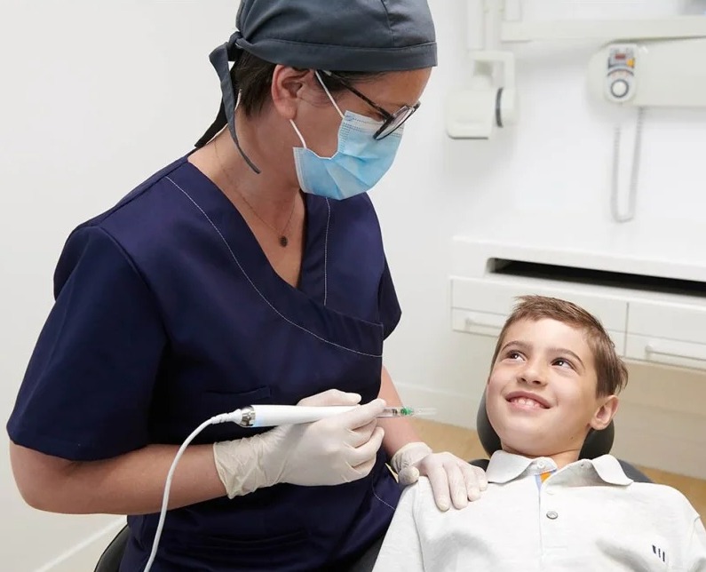
What is an Intraoral Scanner?
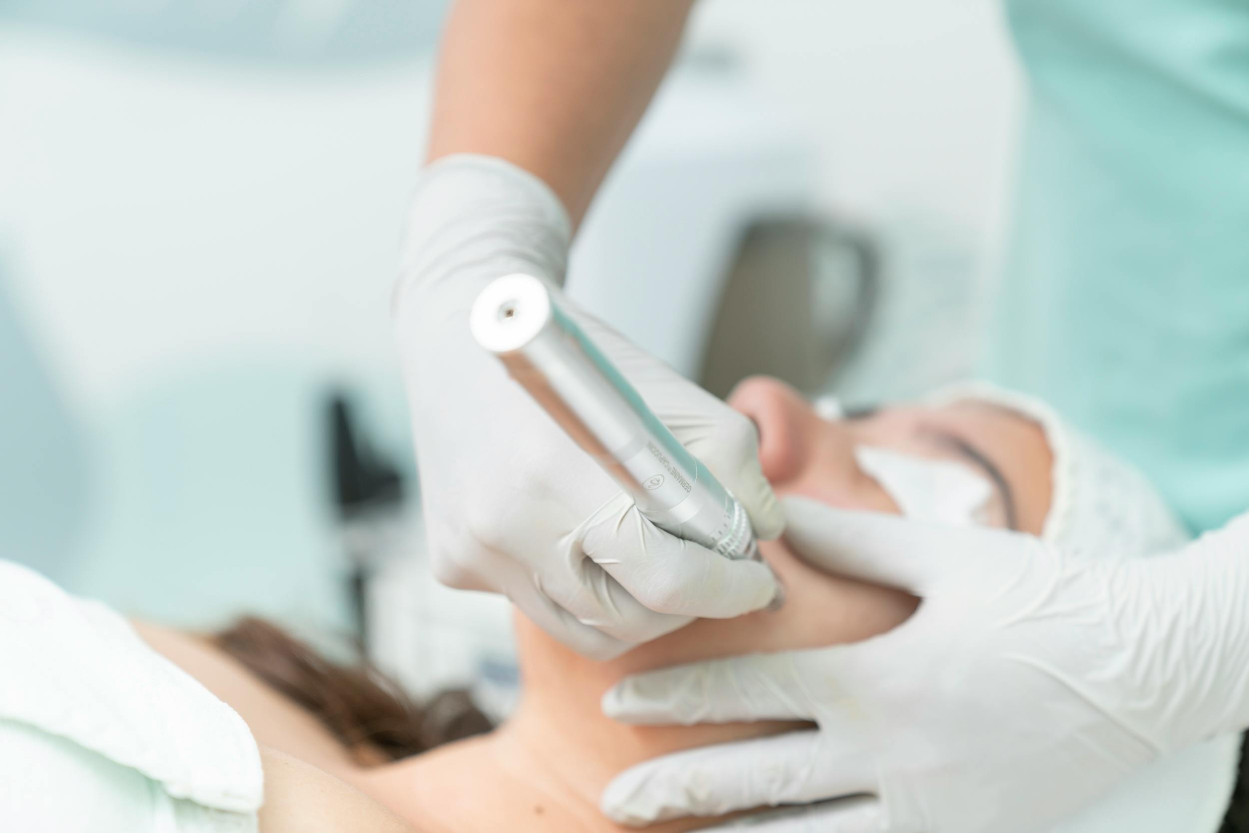
An intraoral scanner is a handheld medical scanning device equipped with high-resolution optical video capture technology, developed in dentistry to directly transfer a patient’s intraoral hard and soft tissue topography into a digital environment. These devices process and record thousands of three-dimensional point cloud data structures per second, capturing the geometric shape of teeth, occlusion (bite) relationships, and marginal gingival boundaries onto a computer within seconds. Completely eliminating traditional impression phases such as trays, plaster models, and shipping logistics to laboratories, intraoral scanners stand as the most fundamental starting and data collection pillar of modern digital dentistry (digital workflow).
- Optical Triangulation: Computes the time it takes for light waves to strike and bounce back from the tooth surface to map out a millimetric depth guide.
- True-Color 3D Model: Projects dental integrity onto the screen not merely geometrically, but also displaying the exact color shades of enamel and gingival tissues.
- Open Data System (STL/PLY): Enables the seamless online transfer of acquired digital data to dental laboratories worldwide within seconds.
How Do Intraoral Scanners Work?
The operating principle of intraoral scanners relies on “continuous video radiation” or “structured light projection” systems. While the scanner’s camera-like handpiece (wand) is guided over the teeth, thousands of structured light or laser waves are projected onto the tissue from its tip per second. Striking the indented and complex anatomical surfaces of the teeth, these beams reflect back into the sensor core of the device via angled mirrors. Advanced triangulation algorithms calculate millimetric depth, width, and height coordinates from the angles and timing of the reflected light; these coordinates are superimposed instantaneously to simulate a flawless 3D oral model within the cranial anatomy.
- Light Distribution: Calibrating laser or blue LED light waves according to the reflection coefficient of the enamel layer,
- Software Stitching: Leveraging artificial intelligence software to merge consecutive micro-photographs captured as the camera moves, much like assembling a puzzle,
- Instant Occlusion Analysis: A phase where, after scanning the upper and lower jaws independently, the patient is asked to close their jaws to dynamically record the bite relationship.
In Which Dental Treatments Are Intraoral Scanners Used?
Digital intraoral scanning systems are actively deployed in contemporary clinical dentistry for nearly all major and minor dental restoration processes that require replicating the patient’s dental impression and jaw anatomy within a laboratory environment. The high precision and error-eliminating digital intelligence delivered by these scanners radically enhance the adaptation quality of prostheses while shortening treatment times. The clinician can electronically inspect impression-related discrepancy risks before the data even leaves the clinic.
The primary dental treatments where intraoral scanners are most heavily and efficiently utilized include:
- Fixed Prosthodontic Treatments: Single-tooth porcelain crowns, zirconium bridges, inlay and onlay restorations,
- Aesthetic Restorations: Porcelain laminates (porcelain veneers) and all impression cut measurements for smile designs,
- Orthodontic Treatments: Formulating treatment plans and digital analysis models for clear aligner (wireless orthodontics) processes,
- Implantology Applications: Implant-supported prosthetic impressions and guide plate manufacturing data for navigated surgery.
Who Are Intraoral Scanners Suitable For?
Intraoral digital scanning technology is medically suitable for children and adult individuals of any age group who require dental impressions and can hold their mouth open for a specific duration. Because the system involves no ionizing radiation (X-rays) and operates strictly via harmless light waves, it can be safely utilized on any client regardless of their general health status (including pregnant patients and individuals with cardiac conditions). It serves as a clinical lifesaver, particularly for sensitive constitutions that demonstrate extreme hypersensitivity reactions to classic dough impression materials.
- Those with a Severe Gag Reflex: Individuals who experience nausea or an urge to vomit when trays and paste enter the mouth,
- Pediatric Patients: Children who grow restless while waiting for impression dough to set or struggle to stay cooperative in the chair,
- Those with Temporomandibular Joint (TMJ) Disorders: Joint patients who cannot open their mouth very wide or maintain a stationary position for extended periods.
What Are the Advantages of Intraoral Scanners?
The advantages brought by digital intraoral scanners to clinical dental practices maximize the treatment comfort of the patient while minimizing the margin of error between the clinician and the laboratory. Dimensional stability losses common in traditional plaster models—such as fracturing, shrinkage, or expansion—are conclusively prevented in digital datasets; STL data retains its first-day millimetric accuracy even after years pass. The comparison table below presents the differences between digital scanning and old-school dough impression methods:
| Clinical Evaluation Parameter | Intraoral Scanner (Digital Impression) | Traditional Tray Impression (Alginate/Silicone) |
|---|---|---|
| Patient Comfort and Nausea Risk | Extremely high; scanned solely with light, generates no sensation of nausea. | Low; large metal trays and fluid pastes leaking into the throat strain the patient. |
| Margin of Error and Dimensional Accuracy | At micron level; software instantaneously flags non-compliant areas. | High; risks exist due to dough shrinkage during setting or plaster expansion. |
| Delivery Time to Laboratory | Takes seconds; online data transfer (e-mail/cloud) is executed over the internet. | Takes days; transportation of the plaster model via courier or shipping is mandatory. |
| Need for Remaking Impressions | Virtually non-existent; missing zones are scanned locally within seconds. | Moderate-high; if air bubbles remain trapped in the dough, the impression must be remade from scratch. |
How Do Intraoral Scanners Differ From Traditional Impression Methods?
In traditional impression methods, hydrocolloid (alginate) or silicone-based chemical doughs are loaded into large metal or plastic trays, placed inside the patient’s mouth, and held for an average of 3–5 minutes for the material to set; during this window, breathing and swallowing become difficult for the patient. The digital scanning method completely eradicates this distressing physical contact. Without touching the scanner tip to the gums, the clinician guides it over the teeth much like filming a video. The most radical difference is that while an error in a traditional impression requires the entire process to be repeated from the beginning, digital workflows allow the clinician to simply erase the faulty minor area from the computer and rescan it in seconds.
- Elimination of Physical Materials: Discarding chemically scented, fluid pastes that can seep down the throat, along with sticky consistencies,
- Visual Feedback: The luxury of instantly auditing the quality of the impression on a 3D giant screen while the patient is still in the chair,
- Archiving Convenience: Storing data indefinitely on gigabyte-sized cloud servers instead of occupying physical storage space with plaster models in the clinic.
How is the Intraoral Scanning Procedure Executed?
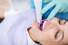
The intraoral scanning procedure is a highly technological session phase that requires no anesthesia or surgical intervention, allowing the patient to lean back comfortably in the clinical chair. The process initiates with the clinician introducing the handpiece wand of the scanner into the oral cavity. A specific geometric route is followed during the scan; typically starting from the chewing surfaces (occlusal) of the lower jaw, followed by the cheek (buccal) and tongue (lingual) surfaces. The identical protocol is repeated for the upper jaw. In the final stage, the patient is asked to close their teeth normally, and the device captures the right-left bite relationship, locking the digital jaw model.
- Rotational Movement: Guiding the device tip at flexible angles to align perfectly with the curved anatomical contours of the teeth,
- Artificial Intelligence Filtering: Automatically purging tissues such as the tongue, cheek, or clinician’s finger that accidentally enter the frame during scanning,
- Occlusal Contact Mapping: Projecting full-color graphics onto the screen to monitor exactly at which points and with how much force the upper and lower teeth meet.
Is Preparation Required Prior to Intraoral Scanning?
The 3D intraoral scanning application is a practical workflow that requires no prior fasting or ingestion of chemical medications by the patient. The sole and most critical clinical preparation before the session is optimizing the dental surfaces so that they do not deflect the scanning beams. A thick layer of sebum (oil), heavy accumulation of saliva, or bleeding on the teeth can obstruct optical beams from penetrating the enamel layer, leading to deviations (artifacts) in the image; therefore, keeping the tissues dry and clean directly dictates scanning quality.
The fundamental preparation steps executed in the clinical environment prior to the scan include:
- Purging plaque and food debris from dental surfaces using dental cleaning brushes,
- Drying the dense saliva layer on the teeth with the aid of an air-water syringe,
- Spraying a very thin, harmless medical titanium dioxide powder onto the tooth surface in certain older or specialized scanner models to prevent reflections.
How Are Intraoral Scanners Used in Implant Treatment Planning?
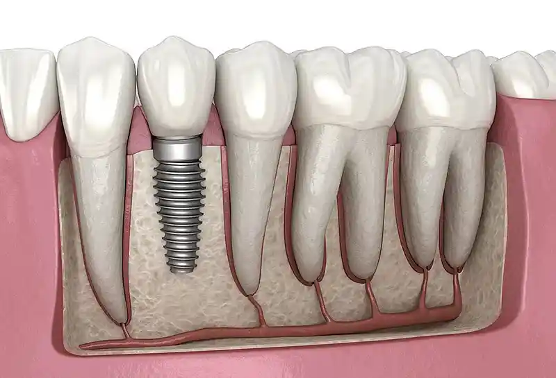
Intraoral scanners function as a digital bridge that zeroes out the margin of error in the fabrication of implant-supported prostheses and surgical planning. Following implant surgery, it is standard protocol to fit a specially coded digital pin called a “scan body” inside the implant to transfer the exact intraoral angle and position of the artificial roots embedded in the bone to the laboratory. Scanning this pin, the device records the three-dimensional x, y, and z coordinates of the implant within the jawbone into the computer at a micron level; this ensures that implant-supported screw-retained teeth or zirconium porcelains seat with absolute passive fit without imposing even the slightest strain or tension onto the jawbone.
- Scan Body Integration: Pairing the implant orientation identically with dental library configurations within the digital software,
- CBCT Superimposition: Merging 3D dental tomography bone data with soft tissue data derived from the intraoral scanner,
- Flapless Implant Infrastructure: Leveraging this dual dataset to design navigated surgical templates that empower the surgeon to place implants without making structural incisions in the gums.
How Do Intraoral Scanners Support Orthodontic Treatment?
Intraoral scanners establish the most vital technical infrastructure for modern clear aligner (wireless orthodontics—Invisalign, etc.) treatments because clear aligner manufacturing is entirely a digital engineering workflow. The STL data acquired from the scanner is imported into orthodontic software, where the micro-movement directions of the teeth for each phase are calculated through computer algorithms. Furthermore, thanks to “Orthodontic Simulator” software provided by the system, patients can rationally observe on the screen how their teeth will align weeks later and what kind of smile they will possess upon treatment completion before even starting their procedures.
- Digital Setup Preparation: Virtually simulating the movement boundaries of teeth within the jawbone inside a computerized matrix,
- Staged Aligner Manufacturing: Sending error-free data gathered from the scan to 3D printers to print successive batches of 20–30 clear aligners all at once,
- Progress Monitoring: Conducting mid-treatment scans to evaluate how closely the teeth are adhering to the pre-planned trajectory.
Why Are Intraoral Scanners Preferred in Aesthetic Dental Treatments?
In aesthetic dentistry—particularly in porcelain laminate veneer or full-ceramic (E-max) crown applications—micron-level marginal adaptation is the most uncompromising criterion dictating both the lifespan and natural appearance of the tooth. Intraoral scanners capture the dental preparation margins sculpted by the dentist with the sharpest resolution and transmit them to CAD/CAM milling machines in the laboratory. This technological precision ensures the crown seats against the gingival margin with a zero-error line, preventing artificial thicknesses, preserving the natural pink form of the gums, and completely eradicating the risk of insidious micro-leakage decay developing underneath the crown.
- Sharp Margin Detection: Automated recognition of tooth preparation boundaries (finish lines) by the software, eliminating human error margins,
- Superficial Smoothness: Constructing a micro-design infrastructure that enables light to reflect homogeneously from porcelain surfaces just like natural enamel,
- Minimum Preparation: Preventing unnecessary deep reduction of tooth structure due to the clarity and accuracy of the digital impression.
How Are Intraoral Scanners Used in Prosthetic Treatments?
Utilizing intraoral scanners in prosthetic dental treatments (zirconium crowns, bridges, or implant-supported hybrid prostheses) interrupts the entire chain of human-error variables common in conventional laboratory phases. The 3D oral model acquired from the scan lands directly in front of the digital laboratory technician; using a computer mouse, the technician virtually contours and designs the anatomical form and chewing cusps of the prosthesis (CAD). The moment the design is finalized, data is dispatched to computerized milling machines operating with diamond burs, and zirconium blocks are milled untouched with zero-error tolerance to produce prostheses in perfect harmony with the patient’s mouth.
- Digital Modeling (CAD): Virtual drafting of dental anatomy on a computer screen tailored to proportions corresponding to the patient’s facial features,
- Robotic Milling (CAM): The phase where the designed tooth is carved with micron precision out of zirconium or porcelain blocks,
- Rapid Trial Cycle: Near-total elimination of bite high spots and soreness issues during seating trials caused by impression discrepancies.
How Do Intraoral Scanners Contribute to Digital Smile Design?
Digital Smile Design is the process of synthesizing the aesthetic proportions between a patient’s facial morphology, lip movements, and dental architecture within a computer environment; the intraoral scanner is the most vital data supplier for this synthesis. The 3D dental model acquired from the scanner and high-resolution facial photographs captured in a studio setting are superimposed within specialized aesthetic software through three-dimensional registration methods. Consequently, rather than evaluating the tooth in isolation, the clinician visualizes on-screen how the newly engineered dental forms align with the individual’s interpupillary axis, lip smile line, and skin tone to construct a flawless “smile architecture.”
- Holistic Golden Ratio: Computerized calculation of dental lengths and widths relative to the vertical dimensions of the individual’s facial features,
- Mock-Up Experience: Printing the newly engineered smile designed on the computer as a temporary plastic model via a 3D printer for a live intraoral trial,
- Predictable Success: The comfort of patients embarking on the workflow knowing exactly what kind of aesthetic smile they will achieve at the end of the sessions.
How Long Does an Intraoral Scanning Process Take?
The intraoral scanning application is an exceptionally rapid procedure that fits perfectly into the intense time constraints of modern metropolitan life, consuming almost none of the patient’s time in the clinical calendar. Thanks to next-generation high-speed scanning heads, completely capturing a single dental arch (either upper or lower jaw) is completed in an average of 45 to 60 seconds. Based on the patient’s oral condition, scanning the lower jaw, upper jaw, and bite relationship as a whole and locking the 3D model on-screen encompasses a comfortable timeline of only 3 to 5 minutes in total.
- Single Arch Scanning Duration: 45–60 seconds on average (The period during which the high-speed optical wand is guided over the teeth).
- Bite (Occlusion) Scan: Approximately 15–20 seconds (Locking the static relationship of the jaws to one another).
- Instant Online Dispatch: The moment the scan is completed, data is transmitted to the laboratory at email speeds within seconds, completely eliminating shipping transit times.
How Does Utilizing an Intraoral Scanner Enhance Patient Comfort?
The greatest comfort computerized intraoral scanners extend to the patient is completely eradicating the “impression nightmare,” which is historically the least liked step of dental treatments. The pressure of large metal trays against the palate and the fear of suffocation or breathlessness caused by sticky, muddy pastes leaking down the throat are completely non-existent in digital configurations. The patient can signal the clinician at any given moment during scanning to pause the process, swallow, rest, and resume scanning right where they left off with zero data loss; this flexibility elevates the session into a genuine wellness experience.
- Zero Gag Stress: Optical capture is executed purely through light, failing to stimulate nerve endings and zeroing out the gag reflex.
- Chair Time Savings: Eliminating the obligation to wait uncomfortably for minutes in the chair for impression materials to set.
- Transparent Communication: The comfort of patients auditing fractures, decay, or occlusal misalignments in their own teeth on a 3D screen alongside their clinician.
How Are Digital Impressions Evaluated Post-Intraoral Scanning?
The moment the scanning workflow concludes, evaluating the generated 3D digital model is an analytical process where the clinician merges occlusion mechanics and dental anatomy expertise with on-screen software tools. Rotating the model 360 degrees on the screen, the clinician evaluates the sharpness of dental preparations, subgingival preparation lines, and the margin depths where the porcelain crown will seat. Integrated artificial intelligence analysis filters within the software report millimetrically to the clinician via color scales whether adequate vertical clearance remains between the upper and lower teeth for the prosthesis.
- Bite Contact Map (Color Map): Monitoring the force intensities of points where teeth meet, color-coded in red, green, and blue.
- Undercut Audit: Immediate on-screen detection during the session of reverse angles (undercuts) that could cause the prosthesis to bind during insertion.
How Do Intraoral Scanners Facilitate Treatment Planning?
Intraoral scanners facilitate the planning of evidence-based and fully predictable treatment algorithms, eliminating speculative clinical estimates. Because the clinician can model the patient’s intraoral status as a whole on a computer screen, they can smoothly chart the geometric axes of prosthetic bridges or implants destined for edentulous spans before even initiating the operation. This digital ease accelerates workflow management by enabling specialist clinicians to make collaborative decisions on the identical 3D dataset in complex, multidisciplinary cases involving orthodontics, surgery, and prosthodontics.
- Virtual Preparation Rehearsal: Digitally testing how much crown clearance will be reduced from the teeth on the computer,
- Rapid Laboratory Synchronization: The ease of the technician transmitting the engineered dental form to the clinician online to acquire digital approval prior to the appointment session.
Are Intraoral Scanners a Safe Imaging Method?
Yes, intraoral scanners are a completely harmless and 100% safe imaging method harboring no side effects, risks, or complication coefficients in medical and dental radiology standards. The devices emit absolutely no ionizing radiation (X-rays); their operating mechanisms rely strictly on the visible light spectrum emitted by standard digital cameras or LED projection units. Due to this biological safety, the procedure can be applied without limitations or the obligation of protective lead aprons to pregnant women, infants, or sensitive individuals with severe systemic illnesses.
- Radiation-Free Scanning: The security of executing repeat scans as many times as desired throughout the day since it involves no X-rays.
- Absolute Sterilization: The strict obligation to sterilize the intraoral scanner tips in high-temperature autoclave units after every single session or operating with single-use protective sleeves to prevent cross-contamination.
What Are the Advantages of Digital Dentistry With Intraoral Scanners?
The era of digital dentistry initiated by intraoral scanners translates into accelerated clinical workflows, an end to material waste, and patient satisfaction reaching its peak. Thanks to the digital flow, porcelain or zirconium crown processes—which conventionally span at least 3–4 sessions and consume weeks—can today be completed comfortably within a single day or even a few hours under the “same-day dentistry” concept, encompassing scanning, design, and robotic production.
- Maximum Time Savings: Decreasing treatment days due to the elimination of shipping turnarounds and the velocity of robotic manufacturing,
- Flawless Marginal Adaptation: Long-lasting, leakage-free prostheses uniting with the gums at a zero-error line because human manipulation and plaster expansion errors are eliminated,
- Ecological and Clean Clinic: An environmentally friendly digital operation infrastructure purged of chemical impression waste and plaster dust.
What Are the Frequently Asked Questions About Intraoral Scanners?
The most common practical anxieties arising in the minds of patients scheduled to undergo digital intraoral scanning prior to prosthetic or orthodontic treatments, along with their uncensored, bare medical answers, are presented below:
Will the intraoral scanning process hurt during the session or give me an electric shock?
No, you will absolutely feel no pain, ache, soreness, or electrical sensation. The procedure is entirely non-invasive; the smooth plastic tip at the head of the device is simply guided over the surfaces of your teeth with gentle contact. Since the light waves emitted by the scanner generate no heat or stimulation in the tissues, the session is completed in a highly comfortable and pleasant manner.
Is it guaranteed that teeth made with digital impressions fit better than traditional dough impressions?
Yes, this is a clinically verified fact. Traditional dough materials can flex while being withdrawn from the mouth, shrink while setting, and the plaster poured over them can expand millimetrically as it dries; this induces micro-discrepancies in the prosthesis. The digital scanner, conversely, captures dental anatomy as a computer dataset immune to risk of distortion, so teeth emerging from robotic milling machines seat in the mouth with absolute precision without creating sore spots.
What happens if I experience nausea or need to swallow during scanning?
This scenario highlights the greatest flexibility advantage digital scanning extends to the patient. When a traditional dough impression tray enters your mouth, it cannot be removed until it sets, forcing you to wait regardless of nausea. In digital scanning, the moment you feel nauseous or wish to swallow, you simply signal your clinician; the clinician pauses the device. After you rest and rinse your mouth, the device automatically recognizes the tooth patterns right where it left off, resuming the scan with zero data loss.
Does the light emitted by intraoral scanner devices damage my eyes or my health?
No, it poses absolutely no harm. The rays emitted by the devices contain no X-rays (radyasyon); they reside strictly within the safe visible light spectrum emitted by standard LED lamps or smartphone screens in our homes. Unless held directly inside the eye for an extended duration, it has zero negative impact on ocular health or general body biology; it can be used infinitely with this safety even on pregnant patients.
Will I wait for days for my teeth to be fitted after an impression is taken with digital scanning?
No, the waiting turnaround is radically compressed. While traditional impressions sacrifice at least 1–2 days for plaster to set and transit to the lab via courier, transferring digital scanning data to the laboratory consumes a mere 5 seconds. If the clinic houses an integrated dental CAD/CAM milling unit, your tooth can be designed and robotically manufactured within a few hours following the scan, allowing it to be permanently fitted into your mouth on the same day.
Who Are We? & Panorama Ankara Oral and Dental Health
Panorama Ankara is a licensed oral and dental health center extending institutional health assurances to its patients by uniting an academic expert staff and the latest technological digital infrastructure under a single roof, particularly in oral, dental, and maxillofacial surgery, implantology, orthodontics, and aesthetic dentistry. Attaching vital importance to gathering data with millimetric accuracy at the very inception of treatment tracks, and shielding its patients from the suffocation and gagging stress induced by dough-loaded trays, our center welcomes its clients with globally certified, high-speed premium Digital Intraoral Scanner systems within its clinical assets.
Across all radiological, scanning, and robotic manufacturing interventions executed under the roof of Panorama Ankara, absolute sterilization, high patient safety, transparency, and honesty principles are operated with uncompromising determination as per our charter. In our clinical practices, proprietary intraoral scanners possessing micron-level point cloud processing resolutions and smart tissue filtering software are meticulously applied by our expert medical staff to maximize the patient’s intraoral comfort and prosthetic adaptation quality. Our goal is not to exhaust our patients in week-long trial sessions with arbitrary frameworks or non-compliant impressions; it is to analyze every individual’s jaw occlusion mechanics and gingival boundaries bare to the eye on a computer screen, completing virtual smile designs before the operation to achieve maximum session comfort, same-day dentistry standards, and the highest treatment success rates without damaging surrounding tissues and vascular pathways. If you are tired of your missing teeth, old worn-out crowns, or crowding when looking in the mirror, yet postpone your treatments out of fear of dough impressions, and wish to initiate your digital treatment workflow with a scientific guarantee in an expert center environment, you can safely protect your health and smile integrity by contacting the expert staff of Panorama Ankara for your detailed 3D intraoral scanning analyses.