What is CBCT Dental Tomography? (What is CBCT?)
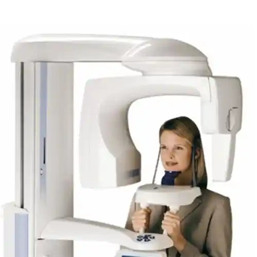
CBCT (Cone Beam Computed Tomography) is a specialized X-ray technology developed to obtain three-dimensional anatomical images of the maxillofacial (jaw and face) region. Compared to medical tomography, it uses a much smaller, narrower, and cone-shaped X-ray beam. This system captures hundreds of micro-sections from different angles by making a single 360-degree rotation around the patient’s head. Unlike traditional panoramic or periapical X-rays, it displays the depth and width of bone tissues, the route of nerve canals, and anatomical spaces in their true dimensions (1:1 scale), reducing the margin of error to zero in diagnosing intraoral pathologies.
- Three-Dimensional Resolution: Provides complete volumetric data of tissues in x, y, and z axes.
- Low Radiation Profile: Thanks to its cone beam geometry, it emits up to 90% less radiation compared to medical tomography.
- Isotropic Voxel Structure: Produces digital image pixels that ensure millimetric accuracy (up to 0.1 mm) in measurements in all directions.
How Does CBCT 3D Dental Tomography Work?
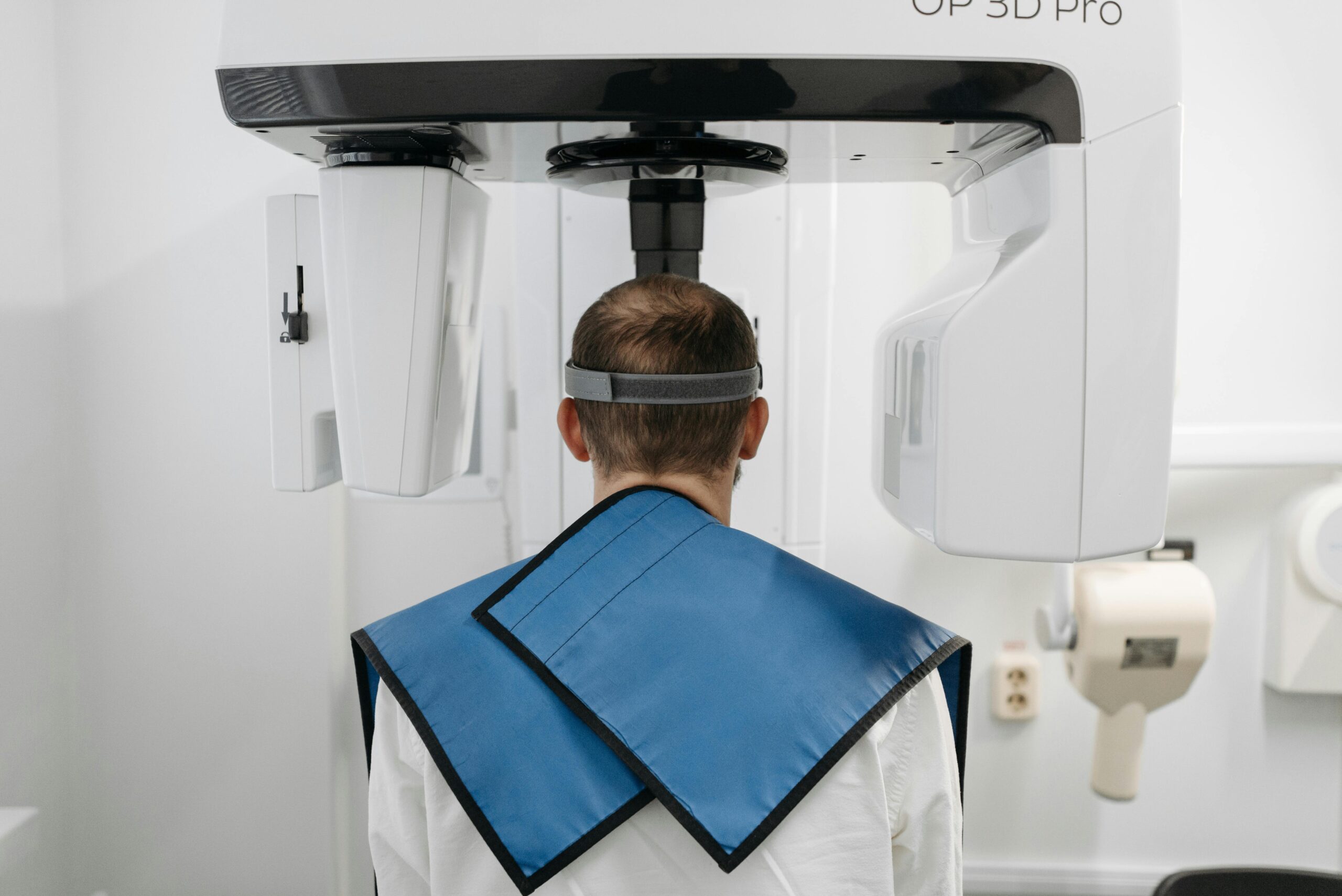
The operating mechanism of CBCT devices is based on the synchronized movement of an X-ray source mounted on a rotating gantry and a digital flat panel detector (FPD) positioned directly opposite to it. While the patient stands stationary in the center of the device, the beam source passes cone-shaped scanning waves through the scalp and bone structures. The beams that penetrate the hard tissues and reach the detector are transmitted to the computer software as “raw data” within seconds. Using advanced mathematical reconstruction software such as the “Feldkamp algorithm,” the system converts this raw data into thousands of stacked axial, sagittal, and coronal sections, creating a three-dimensional volumetric model for the clinician to examine.
- Single-Rotation Scan: The entire dataset is collected with just one single rotation of the device head around the patient.
- Field of View (FOV) / Volumetric Capture: Minimizes unnecessary radiation exposure by focusing only on the targeted jaw region (e.g., a single tooth or the entire jaw).
- Digital Remodeling: The process where raw data is processed by the software within seconds and converted into a 3D bone density map.
When is CBCT 3D Dental Tomography Used?
Three-dimensional dental tomography is utilized in all complex oral and maxillofacial pathologies where standard two-dimensional panoramic X-rays fall short due to their inability to provide depth information and their tendency to hide anatomical structures by overlapping them. CBCT scanning becomes a clinical necessity in every case where the anatomical boundaries, volume, and quality of the hard tissue must be known prior to a surgical procedure. This data is leveraged not only for diagnosis but also for preparing treatment simulations and manufacturing surgical guides.
The primary situations where CBCT technology is actively deployed in dentistry include:
- Measuring the width and height of the jawbone prior to implant placement in borderline cases with insufficient bone volume,
- Scanning the millimetric distance of impacted wisdom teeth or canines from the main lower jaw nerve canal (inferior alveolar nerve),
- Detecting the boundaries of cysts, tumors, or abscesses developing within the jawbone and the destruction they cause in the bone,
- Identifying overlooked extra root canals or root fractures during root canal retreatment procedures.
Who is CBCT 3D Dental Tomography Suitable For?
CBCT 3D dental tomography is suitable for any adult dental patient requiring advanced diagnosis and planning in the oral and maxillofacial region. Specifically, dental implant candidates, individuals experiencing temporomandibular joint (TMJ) disorders, or patients scheduled for orthognathic surgery are the primary target audience for this technology. It can also be safely applied to pediatric patients by selecting low-dose pediatric protocols to determine the position of impacted teeth or examine cleft palates. However, except for emergency medical indications, tomography scans should be postponed for patients with suspected pregnancy or those in their first trimester of pregnancy.
| Patient Profile | Clinical Indication Status | Diagnostic Contribution of Tomography |
|---|---|---|
| Dental Implant Candidates | Fully Suitable / Mandatory | Measurement of bone height-width, detection of the sinus floor, and bone density analysis. |
| Patients with Impacted Teeth Complaints | Fully Suitable | Three-dimensional determination of the proximity relationship of roots to the mandibular nerve or sinus cavity. |
| Chronic TMJ Patients | Fully Suitable | Analysis of condylar abrasions, joint space, and bony defects. |
| Pregnant Patients | Not Suitable (Except emergencies) | It is standard protocol to postpone the scan until post-delivery to prevent fetal radiation risks. |
What Are the Advantages of CBCT 3D Dental Tomography?
The greatest advantage CBCT technology brings to modern dentistry is that it completely eliminates the margin of error in diagnostic processes and offers the clinician the opportunity to visualize the sub-jaw anatomy naked to the eye prior to the operation without making an incision. Compared to conventional computed tomography (CT), it offers radical advantages regarding cost, scanning speed, and patient safety. The entirely digital execution of the workflow also facilitates patient-clinician communication and treatment approval processes.
- 1:1 True Scale: Precise anatomical measurements without magnification, reduction, or distortion in the image,
- Minimal Radiation Dose: Up to 90% reduction in the radiation load exposed to the patient compared to medical fan-beam tomography,
- Fast Scanning Time: Collection of all head and jaw data in a very short duration, averaging 10 to 20 seconds,
- High Image Quality: Perfect visualization of hard bone tissue details with micron-level voxel resolution.
Which Structures Can Be Visualized with CBCT 3D Dental Tomography?
Cone-beam computed tomography visualizes all hard tissue structures in the craniofacial and jaw anatomy along with their spatial relationships with high resolution. Although soft tissue details (muscle, fat, lymph nodes) cannot be differentiated as deeply as in medical MRI or CT, air spaces, nerve canals, and intraosseous pathologies can be clearly scanned. This allows for a complete topographic mapping of the maxillofacial region.
- Maxilla and Mandibula: The entire internal and external bone architecture, both cortical and trabecular, of the upper and lower jawbones.
- Dental Structures: Anatomy of tooth roots, number of root canals, pulp chamber, and periodontal spaces.
- Anatomical Spaces: Maxillary sinus cavities, nasal cavity, and respiratory tract obstructions (airway analysis).
- Nerve Networks: Neurovascular structures critical to protect during implant surgery, such as the mandibular canal and mental foramen.
Why is CBCT 3D Dental Tomography Critical in Implant Planning?
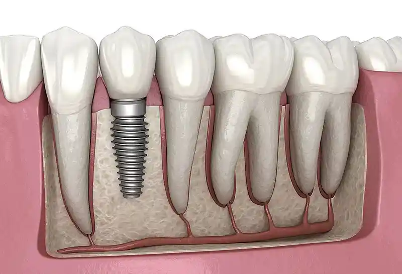
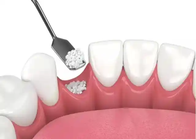
Success in implant surgery directly depends on placing the artificial titanium root into the jawbone at the correct angle, depth, and position (osseointegration); CBCT is the safest scientific guide for this planning. While panoramic X-rays only show the height of the bone but not its width (thickness), 3D tomography allows the sectional thickness of the bone to be clearly measured. The clinician can see millimetrically whether there is a need for bone grafting (bone powder) in the implant site, the sagging of the sinus floor, or the location of anatomical nerve canals, thereby definitively eliminating surgical complication risks such as vessel/nerve injury or sinus perforation during the operation.
- Bone Quality Assessment: Predicting the primary stability of the implant by measuring bone density (D1, D2, D3, D4 classification).
- Virtual Simulation: Pre-operative testing of virtual placement of the appropriate size implant onto the digital bone model within specialized software.
- Surgical Guide Production: Transferring tomography data to 3D printers to prepare “navigated surgical guides” that enable flapless, sutureless implant placement.
How is CBCT 3D Dental Tomography Used in Jaw Surgery?
In oral and maxillofacial surgery practices, clearly outlining the boundaries of pathological lesions and pre-mapping surgical margins are vital to the success of the operation; CBCT provides the surgeon with this tactical edge. Cysts or tumors can erode the jawbone from within and perforate the cortical bone; tomography displays the extent of this destruction and how close the lesion is to healthy tooth roots. Additionally, detecting complex post-traumatic jaw fractures and determining the direction of fracture lines directly shapes the surgeon’s operative plan.
- Cyst and Tumor Boundaries: Calculating the volume of the lesion in cm³ to determine the margins for enucleation (curettage).
- Impacted Tooth Operations: Minimizing bone removal by determining the exact approach direction (lingual or palatal) for teeth embedded under the bone.
- Orthognathic Surgery Foundation: Digital simulation of bone incisions (osteotomy lines) to be performed for correcting jaw misalignments.
How Does CBCT 3D Dental Tomography Support Orthodontic Treatment?
In orthodontic and pediatric dentistry applications, the impacted positions of teeth within the jawbone, the proximity of roots to one another, and the thickness of bone barriers directly affect treatment planning; CBCT supports these workflows with rational parameters. Especially when planning to orthodontically erupt and align canine teeth impacted in the palate, the exact position of the tooth is located via tomography. Furthermore, during mini-screw (skeletal anchorage) applications, the thickness of the bone site where the screw will be placed is measured from tomography sections to prevent nerve or root injuries.
- Root Resorption Analysis: Micron-level evaluation of whether impacted teeth are resorbing the roots of adjacent healthy teeth.
- Airway and Sleep Apnea: Calculating the respiratory tract volume in patients with maxillary constriction to measure the impact of orthodontic expansion on breathing.
- Cortical Bone Boundary: Preventing the risk of the tooth root breaching the jawbone boundary during orthodontic movements, which could cause fenestration (perforation).
Why is CBCT 3D Dental Tomography Preferred in Endodontic Applications?
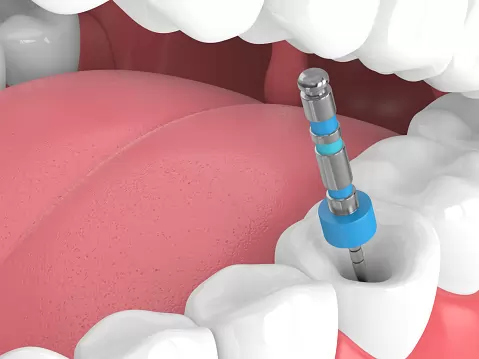
The specialty of endodontics (root canal treatment) relies on cleaning the microscopic nerve canals inside the tooth, and the anatomy of these canals can sometimes be too complex (C-shaped canals, extra lateral canals) for traditional X-rays to display; CBCT resolves these obstruction points with cellular clarity. A tooth that appears to have two roots on standard X-rays might hide a third or fourth canal behind it, and overlooking this can result in root canal treatment failure. 3D tomography scans the tooth in horizontal sections (axial plan), revealing all canal orifices in their bare reality.
- Extra Canal Detection: Pinpointing the locations of hidden canals, such as the MB2 in upper molars.
- Diagnosis of Root Fractures: Detecting microscopic vertical root fractures that cannot be distinguished on conventional X-rays, thereby preventing unnecessary treatments.
- Periapical Lesion Analysis: Deep evaluation of the propagation area of inflammation at the root apex within the bone, as well as perforations.
Is Preparation Required Before CBCT 3D Dental Tomography?
A 3D dental tomography scan is a highly practical, non-invasive radiological procedure that requires no prior fasting, medication use, or administration of contrast media (medicated liquid). The sole and most critical medical preparation before entering the scanning room is the removal of all metal objects in the head and neck region that could block X-rays and cause “artifacts” (metal flare/shadowing) on the image. Since these flares degrade image clarity and reduce diagnostic quality, strict adherence to this rule is necessary.
The basic items that the patient must remove prior to the scan include:
- Removable partial dentures or false teeth containing metal components inside the mouth,
- Earrings, nose studs, piercings in the ears/nose, and metal necklaces around the neck,
- Glasses, metal hairpins, hearing aids, and any metal hair extensions or clips.
How Long Does a CBCT 3D Dental Tomography Scan Take?
The 3D dental tomography application is an incredibly fast and practical procedure that does not consume the patient’s time in modern clinical scheduling. The active rotation and radiation emission time of the device around the patient’s head usually takes only 10 to 20 seconds, depending on the size of the selected field of view (FOV). When including the phase where the process is computed by the software and reconstructed into a 3D model, the total time the patient spends in the tomography room does not exceed a few minutes; images hit the clinician’s screen immediately after the session.
- Active Irradiation Time: 10 – 20 Seconds (The 360-degree rotation period of the device head).
- Digital Reconstruction: 1 – 2 Minutes (The duration for the software to process raw data and generate sections).
- Total Process: The patient completes all steps and leaves the room in approximately 3–5 minutes.
What Should Be Considered During a CBCT 3D Dental Tomography Scan?
Critical rules must be followed during the tomography scan to both safeguard the patient’s radiation safety and maximize the resolution quality of the acquired 3D image. The most vital rule is that the patient must remain absolutely still, avoid swallowing, and maintain a stable position throughout the 10-20 second active scanning window while the device rotates. Even the smallest micro-movement of the head can cause “motion artifacts” (blurring) in the digital sections, rendering the tomography invalid and forcing a rescanning process.
- Absolute Immobility: Holding the breath slightly during the scan and standing still like a statue until the device exposures are finished.
- Protective Equipment: Vesting the patient with a lead apron and a lead thyroid shield collar to minimize radiation exposure.
- Correct Positioning: Placing the jaw accurately onto the bite block indicated by the clinician, guided by the laser alignment lines of the device.
Is CBCT 3D Dental Tomography a Safe Imaging Method?
Yes, although it involves ionizing radiation, CBCT 3D dental tomography is an exceptionally safe imaging method developed in accordance with the “ALARA” (As Low As Reasonably Achievable) principle in modern radiology standards. Because the system utilizes cone-beam technology, it delivers up to 90% less radiation dose to the patient compared to conventional medical computed tomography (CT) scans performed in hospitals. When compared to the background radiation doses we naturally receive from nature, cosmic rays, or airplane travel in daily life, the risk coefficient emitted by a single dental tomography is negligible.
- Radiation Comparison: While a single medical chest CT scan involves high doses at the micro-sievert level, the dental CBCT dose is significantly lower.
- Target-Oriented Scanning: Thanks to FOV limitations, beams are directed only to the specific dental region to be operated on, protecting sensitive organs like the brain or eyes.
Can One Return to Daily Life Immediately After CBCT 3D Dental Tomography?
Yes, a 3D dental tomography scan is a completely non-invasive procedure that induces no side effects, fatigue, or numbness on the patient’s nervous system, motor skills, or general functionality. No injections are made to the skin nor is any medicated contrast liquid introduced into the body during the procedure; therefore, the exact minute the session concludes, the patient can seamlessly resume their normal daily, social, professional, or academic life. There are no clinical barriers to driving, returning to work, or eating after the procedure.
- Zero Recovery Time: There is no requirement for resting or keeping the patient under observation after the scan.
- Flexible Scheduling: The scan can be performed quickly by taking a brief break even during midday working hours.
Which Dental Treatments Does CBCT 3D Dental Tomography Facilitate Planning For?
Three-dimensional dental tomography data facilitates the accurate and rational pre-treatment planning of roadmaps in almost all specialties of dentistry. Displaying tissues layer by layer gives the clinician the opportunity to prepare evidence-based and predictable treatment protocols instead of taking arbitrary steps. The table below summarizes the primary dental treatments whose planning is directly streamlined by CBCT along with software integration processes:
| Facilitated Dental Treatment | Clinical Convenience Provided by Tomography | Achieved Treatment Success Outcome |
|---|---|---|
| All-on-4 / All-on-6 Implants | 3D identification of the densest bone regions in fully edentulous jaws and adjustment of implant angles. | Enables same-day fixed teeth fabrication (immediate loading) by reducing the need for bone grafts. |
| Sinus Lifting (Sinus Elevation) | Measurement of anatomical septa inside the maxillary sinus and the thickness of the sinus membrane. | Ensures safe placement of bone powder by reducing membrane perforation risks to zero during operation. |
| Impacted Tooth Excision | Presenting the exact orientation of the tooth (palatal or labial side) to the surgeon with clear sections. | Reduces tissue trauma and swelling by allowing the selection of the correct surgical flap direction. |
How is Digital Treatment Planning Performed with CBCT 3D Dental Tomography?
Digital treatment planning is a high-tech process that begins with transferring 3D datasets in “DICOM” format obtained from the CBCT device into advanced computer-aided design (CAD/CAM) software. On the computer screen, the clinician virtually simulates the length, diameter, and exact coordinates where implants will be placed using a mouse on a digital model that is an exact replica of the patient’s jawbone. If guided surgery is to be performed, this tomography data is superimposed with impressions taken from intraoral digital scanners (stent matching) to enable the fabrication of millimetric surgical guide plates from 3D printers; thus, the margin of error is zeroed out before the operation.
- DICOM Integration: The phase of transferring radiological data to digital design programs without any data loss.
- Virtual Bone Density Analysis: Observing on the screen where the bone contains harder or softer tissue with the help of software color scales.
- Navigated Surgery Infrastructure: Drafting guide plate templates on the computer, which prevents unexpected incisions during the actual operation.
How Does CBCT 3D Dental Tomography Contribute to the Diagnostic Process?
The most revolutionary contribution of CBCT to the diagnostic process is making hidden intraoral foci visible—foci that remain hidden behind two-dimensional X-rays and cause chronic pain because they cannot be diagnosed. While conventional X-rays can only resolve damage on the anterior and posterior surfaces of a tooth, fine cracks on the lateral (proximal) walls of the tooth or fractures inside root canals can be missed due to overlapping shadows. Because 3D tomography scans and slices the tooth with axial slices at the micron level, it allows for the pinpoint detection of undiagnosed stubborn toothaches, hidden root-tip abscesses, and early-stage bone resorption foci, granting an opportunity for early intervention.
- Shadow Elimination: Eradicating superposition (overlapping) illusions created by bones or neighboring teeth.
- Early Diagnosis Capability: Catching aggressive lesions like cysts or tumors at an incipient stage before they create large cavities in the bone.
How Are CBCT 3D Dental Tomography Results Evaluated?
Evaluating the acquired three-dimensional tomography sections is a systematic analysis process that requires running radiological anatomy expertise and clinical dentistry vision in tandem. On the digital screen interface, the clinician examines the image in panels across three primary planes: axial (top view), coronal (front view), and sagittal (side view). Using the software’s measurement tools, distances to nerve canals and bone thicknesses are millimetrically cataloged; the boundaries of pathological areas are scanned by adjusting contrast settings, and the final treatment report is generated by matching the acquired data with the patient’s clinical intraoral examination findings.
- Multi-Planar Reconstruction (MPR): Detecting hidden canals by scanning the image layer by layer from bottom to top.
- Density Measurement (Hounsfield Unit): The phase of deciding on the implant brand and type selection by measuring bone density.
What Are the Frequently Asked Questions About CBCT 3D Dental Tomography?
The most common practical anxieties arising in the minds of patients who are recommended to undergo a three-dimensional dental tomography scan prior to treatment planning, along with their uncensored, bare medical answers, are presented below:
Will I feel pain or sense the radiation during the CBCT 3D dental tomography scan?
No, you will absolutely feel no pain, ache, or soreness. The procedure is entirely non-invasive, meaning no needles are pricked into your skin or any contact made; only the device head rotates around your head. As for radiation waves, they possess a physical nature imperceptible to human senses (sight, hearing, smell, or touch), so you will feel no heat or electricity during the scan.
I already had a normal panoramic dental X-ray, why is a 3D tomography requested additionally?
Panoramic X-rays only show behind the skin and bones in two dimensions (like a flat photograph); in these images, the height of the bone can be selected, but the width (transverse volume) and depth of the bone cannot be perceived. Furthermore, because tissues overlap, hidden canals or fractures can be concealed. 3D tomography, however, gives depth information, allowing the bone to be viewed from every angle in sections; 3D data is mandatory to avoid mistakes in risky steps like implant surgery.
I have numerous metal fillings and implants in my mouth, does this prevent a tomography scan?
No, fixed bridges, metal fillings, or titanium implants in your mouth are medically absolutely no barrier to undergoing a tomography scan; since these structures do not contain a magnetic field, they bear no risk of dislodgement or heating like in MRI machines. However, when X-rays strike metal, they can create linear white flares called “artifacts” on the digital screen; thanks to “metal artifact reduction software” (MAR) in modern CBCT devices, these flares are filtered out and the image is clarified.
Can breastfeeding mothers undergo 3D dental tomography, and is it necessary to pump and discard milk afterward?
Yes, breastfeeding mothers can safely undergo 3D dental tomography scans. X-rays are not a type of radiation that accumulates, gets stored in the body or breast milk, or alters the structure of the milk; the beams pass through the body and dissipate within milliseconds. Therefore, there is absolutely no need to pump breast milk and throw it away or avoid breastfeeding the baby after the scan; the mother can comfortably continue breastfeeding her baby the moment she steps out of the room.
Do I have to wait for days to receive the results following a dental tomography scan?
No, you definitely do not need to wait. The moment the exposure sweeps of the device finish, raw data flows into the computer software within seconds, and the three-dimensional reconstruction is completed in an average of 1-2 minutes, making the sections ready. The images instantly hit your physician’s screen over the clinic’s internal shared network; thus, you can transition straight into the digital planning stage right that minute without losing any time.
Who Are We? & Panorama Ankara Oral and Dental Health
Panorama Ankara is an institutional oral and dental health center that combines advanced technological infrastructure and academic expertise under a single roof, offering services at international health standards, particularly in oral, dental, and maxillofacial surgery as well as all specialty branches of dentistry. Especially in protocols requiring millimetric precision, such as fully edentulous cases, implant surgeries involving advanced bone resorption, and impacted tooth excisions, it utilizes state-of-the-art CBCT 3D Dental Tomography systems within its structure to guarantee diagnostic flawlessness.
Across all radiological and surgical interventions performed under the roof of Panorama Ankara, absolute sterilization, high patient safety, transparency, and medical ethical rules are operated with uncompromising determination. In our clinical practices, proprietary CBCT devices working at ultra-low radiation doses, possessing micron-level voxel resolution and smart artifact filtering software, are meticulously calibrated and applied by our expert medical staff to map the anatomical landscape of the patient. Our goal is not to plan risky surgeries with arbitrary patterns, but to analyze every patient’s jawbone quality and the coordinates of nerve beds bare to the eye on the computer screen, completing virtual simulations before the operation to achieve maximum session comfort and the highest treatment success rates without damaging the skin and vascular pathways. If you are tired of your missing teeth when looking in the mirror, are concerned about surgical risks in implant treatments, and wish to initiate your diagnosis-treatment process with a scientific guarantee in an expert polyclinic environment, you can safely protect your health and smile integrity by contacting the expert staff of Panorama Ankara for detailed 3D tomographic analyses.