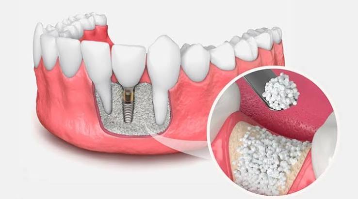
Ankara Bone Graft Application
The foundation of a healthy oral structure is a jawbone with sufficient volume and density that firmly supports the teeth and withstands chewing forces. However, tooth loss due to various reasons, advanced periodontal (gum) diseases, cystic formations, or physical trauma to the jaw can, over time, lead to bone resorption in the jaw—known in medical literature as “alveolar resorption.” In the absence of stimulation in the form of a tooth root, the body perceives the bone tissue in that area as unnecessary and gradually reabsorbs it. This condition not only causes facial collapse and visible signs of aging but also eliminates the biomechanical infrastructure required for future implant or prosthetic treatments. By adopting advanced surgical techniques and personalized treatment protocols in Ankara bone graft applications, Panorama Ankara aims to restore tissue integrity by providing internationally standardized medical solutions for the reconstruction of weakened or volume-deficient bone tissue.
Bone grafting, commonly known as “bone powder” treatment, involves the surgical placement of biologically compatible materials into areas with insufficient bone tissue to stimulate new bone formation. This treatment is not merely the placement of a physical filler; rather, it is a complex bioengineering process that activates the body’s natural healing and cellular regeneration mechanisms. In procedures performed by the experienced physicians at Panorama Ankara, the goal is for the added graft material to gradually become surrounded by the patient’s own living bone cells (osteoblasts), vascularized, and ultimately integrated into the patient’s native bone tissue as an inseparable whole. By enabling three-dimensional reconstruction of the jawbone, this method is considered one of the most critical surgical interventions influencing the success rate of future dental treatments.
Biological Dynamics of Bone Healing: Osteoconduction and Osteoinduction
The success of bone graft applications in oral and maxillofacial surgery is based on the fundamental principles of tissue engineering: osteoconduction, osteoinduction, and osteogenesis. Osteoconduction refers to the graft material serving as a porous physical scaffold (matrix) upon which new bone cells can grow. Blood vessels and cells migrate through the microchannels of this scaffold, transforming the area into living tissue. Osteoinduction, on the other hand, is a more active process; specific proteins and growth factors contained in the graft material stimulate surrounding stem cells to differentiate into bone-forming cells. In advanced grafting procedures performed at Panorama Ankara, high-quality, certified, and biocompatible medical materials are meticulously selected based on the patient’s anatomical needs and the size of the bone defect to maximize the efficiency of these biological mechanisms.
For this biological transformation process to proceed successfully, maintaining micro-level stability in the surgical area is of vital importance. To prevent soft tissues (gum cells) from invading the graft site, bone graft procedures are typically supported by special membranes known as “barrier membranes.” These membranes physically block rapidly proliferating gum cells from entering the graft area, providing slower-growing bone cells with the protected space and time they require. Thanks to the collagen-based, resorbable membranes frequently used in Panorama Ankara’s up-to-date surgical protocols, patients do not need to undergo a second surgical procedure to remove the membrane during later stages of healing.
Bone Graft Materials and Sources Used in Clinical Practice
Each patient’s physiological structure, the size of the jawbone defect, and the requirements of the planned prosthetic treatment differ. Therefore, there is no single type of bone graft material in medical practice; instead, various graft types obtained from different sources with distinct resorption rates and biological properties are available. When planning patient-specific treatment, Panorama Ankara determines the most suitable graft material based on radiological data. These materials are generally categorized into four main groups according to their source and structural characteristics. The table below presents a detailed analysis of the internationally recognized bone graft types evaluated in our clinic.
| Graft Type | Source | Clinical Features and Applications |
|---|---|---|
| Autogenous Graft (Autograft) | Bone tissue harvested from the patient’s own body (usually the chin, wisdom tooth area, or hip bone). | Considered the “gold standard” as it contains living bone cells. There is no risk of immune rejection, but it requires a second surgical site. |
| Allograft | Bone tissue obtained from human donors of the same species, processed and sterilized in medical laboratories. | Eliminates the need to harvest bone from the patient. Exhibits superior osteoconductive properties and is frequently preferred in large bone defect repairs. |
| Xenograft | Materials derived from a different species (commonly bovine, equine, or porcine), processed to remove all organic components, leaving a mineral scaffold. | Has a long resorption period, maintaining volume at the application site for an extended time and serving as an excellent scaffold. |
| Alloplastic Graft | Synthetic inorganic materials produced in laboratory settings, such as calcium phosphate, hydroxyapatite, or bioactive glass. | Free from disease transmission risk and provides an unlimited source. Designed to be gradually resorbed and replaced by natural bone tissue. |
3D Tomography-Supported Digital Diagnosis and Planning at Panorama Ankara
The most fundamental factor determining success in modern oral surgery is the accuracy of diagnosis and the precision of planning before intervention. In complex cases where bone deficiency cannot be clearly identified visually or with traditional two-dimensional panoramic X-rays, measuring bone width, height, and density with millimetric precision is essential. Panorama Ankara utilizes advanced 3D Volumetric Dental Tomography devices to create a three-dimensional digital map of the patient’s jawbone. Nerve pathways, maxillary sinus volumes, and adjacent tooth root positions are visualized in detail. Before surgery, the physician uses computer simulations to virtually plan where and in what volume the bone graft material will be placed, maximizing procedural predictability.
This digital infrastructure also plays a significant role in determining whether implant placement can be performed simultaneously with bone grafting. If the existing bone tissue provides sufficient volume to achieve primary stability for the implant, Panorama Ankara’s specialists may place the implant and fill surrounding gaps with bone graft material in a single surgical session. However, in cases of advanced bone loss, it is biologically more rational to first reconstruct the bone, wait approximately 4 to 6 months for cellular maturation, and then proceed with implant surgery once new bone has formed. In both scenarios, the objective is to establish a stable foundation capable of withstanding significant chewing forces for years.
Post-Surgical Tissue Healing Process and Patient Responsibilities
Bone graft procedures are high-comfort treatments performed under local anesthesia, during which patients do not experience pain. However, the postoperative period—when cellular integration and new blood vessel formation (angiogenesis) occur—is the most sensitive phase of the process. Panorama Ankara provides patients with detailed care and nutrition guidelines to follow after surgery. Mild swelling or minimal discoloration within the first 48 hours is a natural physiological response as the body increases blood flow to initiate healing. To manage this period comfortably and protect graft stability, patients should adhere to the following essential guidelines:
- Use prescribed antibiotics, anti-inflammatory medications, and analgesics exactly as directed,
- Avoid touching the surgical sutures with the tongue or fingers and refrain from activities creating negative pressure (such as using a straw or forceful spitting),
- Strictly avoid smoking and tobacco products, which impair oxygenation of healing tissues and significantly increase graft failure risk,
- Follow a lukewarm, soft diet (purees, soups) for the first few days and prevent hard foods from contacting the surgical area.
Building the Smile of the Future: A Multidisciplinary Approach
Jawbone loss not only leads to functional deficiencies but also disrupts facial proportions and causes deep wrinkles around the lips, directly affecting aesthetic perception. Providing Ankara bone graft services, Panorama Ankara goes beyond delivering medical treatment by adopting a multidisciplinary approach that restores both functional and aesthetic values. The clinic’s sterilization standards, vision aligned with the latest advances in tissue engineering, and transparent communication strategy prioritizing patient psychology create a reassuring atmosphere at every step of the treatment journey.
In conclusion, conditions such as “bone insufficiency,” “implant cannot be placed,” or “jawbone resorption” are no longer dead ends in modern dentistry. Thanks to biomaterials developed through scientific and technological advancements, the regenerative capacity of tissues can be effectively guided, and the jawbone can essentially be rebuilt. To achieve a long-lasting, healthy, and natural-looking oral structure, chew with confidence, and smile freely in social life, you may schedule a detailed tomographic analysis and consultation with the physicians at Panorama Ankara and establish the most appropriate structural foundation for your health under expert care.