Ankara Wisdom Tooth Extraction
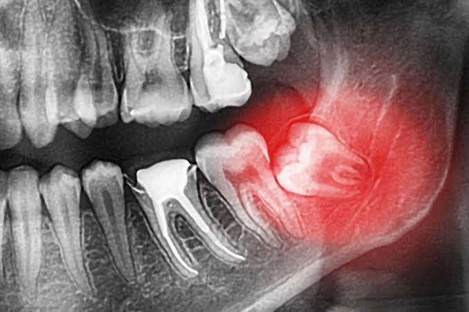
Over the thousands-year journey of human evolution, the dramatic shift in our eating habits—from raw meat and tough, fibrous foods to processed and softer diets—has led to notable changes in our skeletal structure, particularly in jaw anatomy. Third molars, which once fit comfortably into our ancestors’ broad jawbones and actively contributed to chewing, often cannot find enough space to erupt in the increasingly smaller jaws of modern humans. Commonly known as “wisdom teeth,” these teeth typically attempt to erupt into the oral cavity between the ages of 17 and 25; however, due to lack of anatomical space, they frequently become trapped within the jawbone or beneath the gum tissue and remain in an “impacted” or “partially impacted” position. With advanced surgical expertise in Ankara wisdom tooth extraction, Panorama Ankara aims to permanently eliminate acute pain episodes, chronic infections, and potential damage to surrounding tissues caused by this incompatibility through modern oral and maxillofacial surgery protocols.
The decision to keep or remove wisdom teeth depends not only on the tooth’s current condition, but also directly on its three-dimensional relationship with adjacent teeth, the jawbone, and nerve networks. A wisdom tooth that has fully erupted, establishes a correct occlusal relationship with its opposing tooth, and can be properly cleaned by the patient may be preserved. Unfortunately, in the vast majority of cases, these teeth develop in horizontal, tilted (angulated), or even completely inverted positions, exerting pressure on the roots of the healthy second molar in front of them. This mechanical pressure not only causes severe jaw pain that can radiate to the ear, but may also lead to root resorption in the adjacent healthy tooth or create deep carious pockets between the two teeth that are impossible to clean. Guided by detailed radiological examinations, Panorama Ankara’s expert maxillofacial surgeons detect these silent risks early and plan preventive and therapeutic interventions with meticulous care.
Anatomical Variations of Impacted and Partially Impacted Teeth and the Risk of Pericoronitis
In jaw surgery, wisdom teeth are subject to various medical classifications based on their position within the bone and their eruption angles. Fully impacted teeth are those completely covered by jawbone and thick mucosal tissue and cannot be visually detected inside the mouth. Although these teeth may remain silent, they require regular radiographic monitoring because the surrounding follicular (sac) tissue may carry a risk of transforming into cystic or tumoral structures over time. The group that causes the most significant and acute clinical problems is “partially impacted” wisdom teeth. In these cases, only a small cusp (tip) of the tooth breaks through the gum and opens to the oral environment, while the rest remains under a gingival flap (operculum). This partially open flap functions like a bacterial trap, allowing food debris and saliva to accumulate in a dark, warm, and moist area. Bacteria that rapidly multiply in this micro-pocket—where toothbrushes and floss cannot reach—trigger a severe localized infection known in medical literature as “pericoronitis.”
When pericoronitis develops, the patient may present with highly distressing symptoms such as difficulty swallowing (dysphagia), limited mouth opening (trismus), severe throbbing pain in the affected area, swelling of the submandibular lymph nodes, bad breath (halitosis), and sometimes systemic fever. Because performing surgical intervention during the acute infection phase can impair tissue healing and increase the risk of the infection spreading into the bloodstream, Panorama Ankara physicians first irrigate and cleanse the area with special solutions and, when necessary, initiate antibiotic therapy to suppress the acute episode. Once the infection is under control and the tissues have calmed, surgical removal of the partially impacted tooth—the source of the problem—is the only rational medical approach to prevent recurrence (relapse) of pericoronitis.
Wisdom Tooth Extraction Methods: Differences Between Simple Extraction and Surgical Extraction
Not every wisdom tooth procedure has the same degree of surgical difficulty. The anatomical form of the root structure, the density of the jawbone, and the eruption angle of the tooth can change all parameters—from the type of anesthesia to the duration of the procedure. At Panorama Ankara, transparent information is provided to our patients according to the nature of the intervention, and the process is structured accordingly. In dental practice, these procedures are broadly divided into two main categories: simple extractions and complex surgical extractions. The table below summarizes the clinical dynamics of these two procedures in a comparative manner to help patients understand which process they may fall under based on their radiological condition.
| Clinical Procedure Criterion | Simple Extraction of Erupted Teeth (Routine Extraction) | Surgical Extraction of Impacted Teeth (Flap Surgery) |
|---|---|---|
| Anatomical Position of the Tooth | The tooth is fully erupted, clearly visible in the mouth, and can be grasped with instruments. | The tooth is partially or completely under bone/gum tissue and is not aligned in a straight axis. |
| Type of Surgical Intervention | The gum is not opened. The tooth is luxated (loosened) and extracted using forceps and elevators. | The gum is surgically elevated (flap); if necessary, some surrounding bone is removed and the tooth is sectioned for removal. |
| Need for Stitches (Sutures) | In most cases, stitches are not needed; a clot forms naturally. | Stitches are required to reposition the elevated gum tissue and close the wound site. |
| Procedure Duration | Usually takes a few minutes after local anesthesia takes effect. | May vary between 20 and 45 minutes depending on anatomical difficulty and root structure. |
| Healing Period and Comfort | Minimal trauma; full return to normal life is typically possible the next day. | Mild edema, visible swelling, and chewing sensitivity are physiologically expected during the first 48 hours. |
Safe Surgical Planning with 3D Tomography Support at Panorama Ankara
In surgical extraction of impacted lower wisdom teeth, the most critical anatomical risk factor is the main nerve pathway called the “Inferior Alveolar Nerve” (Nervus Alveolaris Inferior), which provides sensation to the lower lip, chin, and lower teeth. The roots of tilted or fully impacted wisdom teeth often lie very close to this thick nerve canal, and in some specific cases, the nerve canal may even pass directly between the tooth roots. Traditional two-dimensional panoramic X-rays may be insufficient to reveal the true depth and three-dimensional distance of this proximity. Even minimal pressure or surgical trauma to the nerve can lead to postoperative “paresthesia” (numbness and sensory loss) in the lower lip that may last weeks or months, and rarely may become permanent. Prioritizing the neurological health of patients above all, Panorama Ankara routinely uses advanced 3D Volumetric Dental Tomography (CBCT) as part of its diagnostic protocol in all impacted-tooth cases considered high risk.
With 3D tomography data, the maxillofacial surgeon can evaluate—from every angle (sagittal, coronal, axial)—the tooth’s position, the number of roots and how curved they are, its millimetric distance to the nerve canal, and the surrounding bone density. This three-dimensional virtual mapping enables the surgical strategy to be built safely from start to finish. If the tooth root is interwoven with the nerve, instead of forcing extraction in one piece, the clinician may apply an advanced technique called “coronectomy,” removing the crown portion of the tooth while leaving the root portion entangled with the nerve inside the bone—thereby reducing the risk of nerve injury to near zero. The root fragment left in the bone generally does not cause infection and may integrate with bone tissue over time. In impacted upper wisdom tooth surgeries, tomography plays a critical role in preventing complications such as the tooth slipping into the maxillary sinus space during extraction or perforation of the sinus membrane (oroantral fistula) by revealing the relationship between the tooth roots and the maxillary sinus cavity.
Protecting Orthodontic Treatments and Preventing the Risk of Crowding
The potential harm of wisdom teeth is not limited to local pain and infections; these teeth can also be silent sources of force that threaten the aesthetic integrity of the entire dental alignment. For individuals who have undergone long, demanding, and costly orthodontic treatments (braces or clear aligners) during adolescence or young adulthood and achieved a perfectly aligned smile, impacted wisdom teeth represent a significant risk factor. As these teeth attempt to erupt from the very back of the jaw, they apply continuous and chronic pushing forces toward the front teeth. This can create a domino effect, transmitting pressure all the way to the incisors. As a result of this biomechanical load, previously corrected front teeth may rotate again, overlap, and develop crowding (orthodontic relapse).
Embracing not only surgical approaches but also multidisciplinary dentistry principles, Panorama Ankara safeguards patients’ orthodontic investments. Our orthodontics and maxillofacial surgery departments work in coordination to analyze radiographic development for patients who have completed or are about to begin orthodontic treatment. If the eruption path of wisdom teeth is angled in a way that threatens the front teeth, these teeth may be removed surgically for purely preventive (prophylactic) purposes—even before acute symptoms such as pain or inflammation appear—so that crowding is prevented before it begins. This proactive approach is one of the most critical medical decisions that helps preserve the perfection of a patient’s aesthetic smile for life.
Tissue Healing Dynamics After Surgical Extraction and the Golden Rules
The success of a surgical tooth extraction is measured not only by the clinician’s technical competence in the dental chair, but also directly by how well the patient follows postoperative care instructions during the recovery period. The first structure formed in the extraction socket (alveolus) is a blood clot. This clot functions like a natural bandage: it isolates exposed bone tissue from external bacteria, prevents pain, and establishes a biological scaffold for new bone cells to form. If the patient dislodges this clot by creating negative pressure or irritating the site, a healing complication called “alveolitis” (dry socket) may occur—leaving bone exposed and causing extremely severe pain. To prevent this, Panorama Ankara advises patients to meticulously follow these vital postoperative rules:
- After the procedure, bite firmly on the sterile gauze placed over the surgical area for at least 30–45 minutes to support clot formation,
- During the first 24 hours, apply intermittent cold compresses (ice packs) externally on the cheek to promote vasoconstriction and minimize swelling and bleeding,
- To avoid creating negative pressure that could dislodge the clot, never spit, do not use a straw, and on the first day switch to a lukewarm, smooth, puree-like, liquid-based diet instead of hot/hard foods,
- Completely avoid smoking and other tobacco products for at least 48–72 hours, as they dramatically reduce oxygenation of healing tissues and significantly increase dry socket risk.
A Painless, Fear-Free Surgical Experience with Panorama Ankara
Unfortunately, surgical interventions in oral and dental health are associated for many people with deep anxiety and fears known as “dental phobia.” When wisdom tooth extraction is mentioned, sounds of bone cutting, expectations of pain, or fear of needles may lead many patients to postpone treatment for years—allowing problems to progress into large cysts or jawbone loss. For patients seeking Ankara wisdom tooth extraction, Panorama Ankara employs modern anesthetic approaches to break this phobia and reframe the surgical experience as “a completely painless rest period.” The next-generation local anesthetic solutions used in the clinic block the target nerve within seconds, fully numbing the tissue; therefore, our patients do not feel even the slightest pain or discomfort during the procedure.
In addition, for patients with needle phobia, a very strong gag reflex, or those who experience panic attacks even at the thought of sitting in a maxillofacial surgery chair, “Conscious Sedation” protocols (laughing gas or intravenous relaxant medications) are safely administered under the supervision of our expert anesthesia teams. Under sedation, the patient enters a state of deep relaxation and does not perceive external sounds or surgical instruments, while still remaining conscious enough to follow the clinician’s instructions. After the procedure, the patient typically does not recall any traumatic memory of the operation and feels as if they have awakened from a short nap. If painful impacted teeth are reducing your quality of life and you are hesitant about surgical intervention, you can schedule a detailed radiological examination without delay to become acquainted with our safe treatment protocols—planned with state-of-the-art imaging systems and supported by fast, suture-free methods—in the high-comfort environment of Panorama Ankara.