Ankara Tooth Extraction
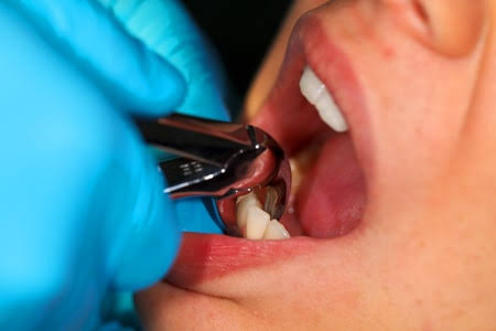
The fundamental philosophy—and foremost objective—of modern dentistry is to preserve a patient’s natural teeth in the mouth in a healthy, functional, and aesthetic manner for a lifetime. Thanks to today’s advances in endodontic (root canal) approaches, advanced periodontal (gum) surgeries, and high-level restorative materials, many teeth that were once thought to be lost can now be successfully saved and retained in the mouth. However, despite all these superior technological and medical efforts, in certain clinical situations keeping a tooth may reach a point where it compromises the overall health of the jawbone, the integrity of neighboring teeth, or the patient’s systemic health. In such unavoidable cases, “dental extraction,” as it is known in medical literature, is not merely the removal of a diseased organ; it is also a critically important medical step taken to prevent larger infections and to prepare a healthy foundation for future prosthetic or implant treatments. In Ankara tooth extraction applications, Panorama Ankara—guided by a preventive dentistry vision—transforms the process of separating a tooth from its tissues from a crude application of force into a microscopic engineering procedure carried out with maximum respect for the surrounding bone and gum tissues.
The most important factor that makes tooth extraction far from an ordinary procedure is that every patient—and even every tooth within the same patient’s mouth—has a completely unique anatomical structure. The number of roots a tooth has, the curvature of those roots within the jawbone, the density of the bone (cortical thickness), and the extent of destruction in the crown (upper) portion of the tooth directly determine the extraction strategy. While traditional approaches focus only on removing the tooth, the main focus in contemporary oral surgery protocols is the “quality of the remaining tissues.” Before beginning the extraction, Panorama Ankara’s experienced clinical team performs three-dimensional radiographic analyses and detailed clinical examinations to map the tooth’s biomechanical relationship with surrounding tissues. This meticulous preparation minimizes the risk of complications such as root fracture, jawbone damage, or injury to adjacent anatomical spaces (such as the maxillary sinus), while maximizing the time the patient spends in the chair and comfort during the healing period.
Advanced Clinical Indications and Biological Factors That Make Extraction Necessary
Deciding to extract a tooth is never a hasty choice; rather, it is the final option used when all boundaries of conservative treatments have been pushed and there is biologically no other alternative. Although tooth tissues are extremely hard structures composed of enamel and dentin layers, they can suffer irreversible damage in the face of neglected bacterial infections or severe physical trauma. Among the leading clinical indications that frequently make tooth extraction a medical necessity at Panorama Ankara are deep caries (decay) lesions that have progressed down to the roots and caused such extensive loss of tooth structure that no filling or porcelain crown (cap) can be placed. When decay extends far below the gum level and progresses into the jawbone, restoring the tooth becomes biologically impossible, and the tooth must be removed so it does not remain an infection source within the bone.
Not only the deterioration within the tooth itself, but also the destruction of the supporting tissues (the periodontium) that connect the tooth to the jawbone is a determining factor for extraction. In advanced periodontal disease (periodontitis), the jawbone surrounding the tooth resorbs due to chronic infection, and the tooth loses its bony support entirely and begins to loosen. Teeth with very high degrees of mobility not only fail to perform chewing function, but also continue to erode the surrounding healthy bone tissue. In addition, teeth that have fractured vertically or have broken roots, stubborn infections that do not respond to root canal treatment and form massive cysts at the root tip, or healthy teeth strategically sacrificed by the clinician to create space during orthodontic treatment (braces) also constitute a broad range of extraction indications.
Atraumatic Tooth Extraction and Alveolar Socket Preservation (Bone Preservation)
In modern dentistry, the concept of standard extraction has largely been replaced by the principles of “Atraumatic Extraction” (removal without injuring tissues). When a tooth is extracted, an empty space remains in the bone where the roots were—known as the alveolar socket. If a tooth is removed by applying excessive pressure to the surrounding bone, forcing it out, or fracturing socket walls, the bone in this region can rapidly resorb and collapse. If a dental implant is planned for this area in the following months, bone volume may become insufficient, forcing the patient to undergo more costly and lengthy bone grafting (bone powder addition) procedures. To secure the foundation for future implant treatments, Panorama Ankara performs extractions using specialized micro-surgical instruments (periotomes). These instruments gently cut the microscopic fibers (periodontal ligaments) that bind the tooth to the bone, allowing the tooth to slide out of its socket without placing pressure on the bony walls.
Immediately after an atraumatic extraction, the procedure known as “Socket Preservation” (preservation of the extraction site) is the most modern way to maintain jawbone volume. After removing the tooth atraumatically, Panorama Ankara clinicians may completely fill the socket with sterile bone grafts (bone powder) and cover it with a protective membrane. This intervention stops the body from perceiving the bone in that area as unnecessary and resorbing it after tooth loss. Months later, when an implant is planned, the clinician encounters a full, dense jawbone with excellent width—providing outstanding support for an implant. This advanced vision is one of the most valuable approaches, protecting patients from potential future surgical difficulties and anatomical losses.
Clinical Classification: Simple Extractions and Surgical Extractions
Although patients may come to the clinic with a single statement such as “I want my tooth extracted,” the medical procedure performed in the background can vary greatly depending on the tooth’s condition. Extraction planning is essentially divided into two main categories based on how invasive the procedure will be. To help our patients prepare more consciously for their treatment processes, we have transparently detailed the anatomical and technical features of these two approaches—which determine how the process will proceed, how the healing period will feel, and how long the operation will take—in the comparative table below.
| Procedural Dynamics | Simple (Routine) Tooth Extraction | Complicated (Surgical) Tooth Extraction |
|---|---|---|
| Anatomical Condition of the Tooth | The crown (upper) part is intact, clearly visible in the mouth, and can be easily grasped with instruments. | The tooth is broken at gum level, roots are curved, fused to bone (ankylosed), or located under the gum tissue. |
| Medical Equipment Used | Only forceps (extraction pliers) and standard elevators that provide slight luxation are used. | Requires advanced equipment such as surgical motors, bone burs, suture sets, and a scalpel. |
| Procedure Process and Need for Stitches | The gum is not opened. The tooth is loosened and removed from its socket. Stitches are generally not needed. | The gum is surgically separated (flap), bone is reduced, the tooth is sectioned and removed, and stitches are required. |
| Healing (Recovery) Period | Trauma is minimal. The patient returns quickly to normal eating, drinking, and speaking routines on the same day. | Mild edema (swelling) and chewing sensitivity during the first 48 hours are a normal physiological process in the surgical area. |
Painless Anesthesia and Patient Comfort Protocols at Panorama Ankara
Tooth extraction phobia is a serious psychological barrier with roots deep in medical history, causing patients to postpone treatment until the very last moment. The basis of this barrier often lies in past negative experiences, the feeling of insufficient numbness, or perceiving the pressure created by instruments as pain. With the goal of preventing Ankara tooth extraction processes from becoming traumatic memories, Panorama Ankara uses the most up-to-date pharmacological agents and painless anesthesia techniques. Before the procedure, the mucosal area where the needle will enter is completely numbed with special topical gels or sprays (surface anesthesia). Then, modern local anesthetic solutions block the targeted nerve endings within seconds, creating deep and continuous numbness in the area where the extraction will be performed. During the procedure, the patient feels only the mechanical pressure of pushing or pulling applied by the clinician; however, this pressure never reaches the brain as a “pain” signal.
The duration of the applied local anesthesia generally continues for 2 to 4 hours depending on the content of the medication (vasoconstrictor ratio) and the patient’s metabolic rate. This period provides a safe time window for the patient to leave our clinic comfortably, take prescribed pain-relief medications, and allow these medications to enter the bloodstream and begin working. For patients with severe dental phobia, blood pressure instability, or special cases requiring the surgical extraction of multiple teeth in the same session, “Conscious Sedation” protocols may be initiated under the supervision of specialized anesthesiologists. In this way, the patient can receive treatment peacefully—without feeling any anxiety and without remembering any stressful details of the procedure.
The Post-Extraction Hemostasis (Clotting) Process and Vital Warnings
The healing process that begins after a successful extraction depends entirely on the patient’s biological response and how closely they follow the clinician’s recommendations. Immediately after the tooth is removed from its socket, bleeding-control mechanisms (hemostasis) are activated and a blood clot forms within the socket. This clot functions as an excellent biological dressing, isolating the exposed jawbone and nerve endings from the external environment (saliva, food debris, bacteria). Healthy clot formation and keeping it in place are the single key to smooth closure of the wound site. If the clot is disrupted, a condition called “alveolitis” (dry socket) may develop—where the jawbone is directly exposed and severe pain may persist for days. To reduce such unwanted scenarios to zero, Panorama Ankara specialists recommend that patients follow these critical postoperative steps precisely:
- Bite down continuously on the sterile pad (gauze) placed over the wound site by the clinician for at least 45 minutes—without talking or moving it—to initiate clot formation,
- For the first 24 hours after the procedure, never spit, do not rinse the mouth, do not use a straw, and do not consume hot drinks (these actions can dislodge the clot due to negative pressure and heat),
- On the first day, apply intermittent cold compresses (ice) externally on the cheek to constrict small blood vessels, stop bleeding, and prevent facial swelling (edema),
- Do not use cigarettes and other tobacco products for at least 48 hours after the operation, as they stop oxygenation at the wound site and greatly increase the risk of infection.
Steps Toward Regaining a Healthy, Complete Smile
Although tooth extraction is often perceived by patients as an ending, it is actually a new beginning step toward rebuilding oral health. With atraumatic surgical methods applied at Panorama Ankara, problematic teeth are safely and comfortably removed, while the foundation is simultaneously established for the aesthetic and functional restorations that will replace them (dental implants, zirconia bridges, or removable dentures). Leaving the gap created by an extracted tooth untreated for a long time can cause neighboring teeth to tilt into the space, the opposing tooth to over-erupt, and overall jaw closure dynamics (occlusion) to deteriorate; therefore, it is highly important to plan the rehabilitation process without delay.
If your painful, constantly infected, or fractured teeth are reducing your quality of life and you are postponing treatment due to fear of extraction, it is time to discover the comfortable solutions offered by modern dentistry. In Panorama Ankara’s sterile clinical environment, you can have a detailed radiological examination with our advanced imaging devices to analyze your condition and transparently learn from our expert clinical team whether your tooth can be saved—and if extraction is unavoidable, how the process will be managed completely painlessly and atraumatically.