Ankara Orthognathic Surgery
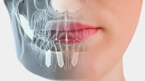
Orthognathic surgery, which represents the highest level of integration between dentistry and oral and maxillofacial surgery (maxillofacial surgery), is a major surgical procedure aimed at correcting severe skeletal jaw discrepancies that cannot be resolved with conventional braces or clear aligners. During the three-dimensional development of the human face, dramatic disproportions can occur between the growth rates and directions of the upper jaw (maxilla) and lower jaw (mandible) due to genetic factors, trauma experienced at an early age, long-term harmful habits such as thumb sucking, or developmental syndromes. These disproportions not only create an aesthetic concern such as the inability of the teeth to contact properly (malocclusion); they also evolve into a complex systemic problem that profoundly affects an individual’s breathing capacity, speech articulation, chewing efficiency, and even the overall health of the jaw joint (TMJ). With an advanced medical infrastructure and a multidisciplinary vision in the field of Ankara orthognathic surgery, Panorama Ankara aims not only to align the teeth but also to redesign the anatomical position of the entire facial skeleton, enabling patients to escape physiological limitations while experiencing a psychological sense of clarity in their facial profiles.
The most fundamental difference that distinguishes orthognathic surgery from other dental interventions is that the problem lies not in the teeth, but in the bony bases that support them. In cases where one jawbone is excessively forward, excessively backward, or asymmetrically shifted to the right/left relative to the other, achieving a healthy bite solely by moving the teeth is biomechanically impossible. In such skeletal cases, forced orthodontic treatments may cause tooth roots to move outside the jawbone, lead to bone resorption, and result in severe gum recession. The working councils at Panorama Ankara—composed of experienced maxillofacial surgeons and orthodontic specialists—combine the power of surgery and orthodontics into a single treatment protocol after the patient’s facial growth has been completed (generally age 18 and over) to solve skeletal asymmetries at their root. This process involves moving the jawbones to new positions planned with millimetric calculations and stabilizing them with titanium mini-plates and screws.
Skeletal Jaw Anomalies: Biomechanics of Class II and Class III Malocclusions
Patients indicated for orthognathic surgery are classified in medical literature into different categories based on their facial profiles and the sagittal (front-back) relationship between the jaws. Among the most common groups encountered in society that require surgical rehabilitation are Class II (Retrognathia) and Class III (Prognathia) anomalies. In Class II cases, the lower jawbone is positioned much farther back than the upper jaw due to genetic or developmental reasons. When viewed from the side, these patients’ chin appears almost absent, and the neck and submental tissues blend together. A retruded mandible pushes the tongue toward the airway, significantly narrowing airway volume; this is one of the most important skeletal causes of life-threatening respiratory disorders such as obstructive sleep apnea (OSA), particularly during nighttime sleep.
On the other hand, Class III anomalies are severe prognathic cases in which the lower jaw is positioned excessively forward relative to the upper jaw (commonly referred to as a “bulldog bite” or “reverse bite”). In these patients, the front teeth cannot meet edge-to-edge, and the lower front teeth are positioned far ahead of the upper front teeth. The “tearing” action—one of the most critical stages of chewing—cannot be physically performed in these individuals. Because foods are sent to the stomach without being adequately ground, chronic digestive system complaints become inevitable. In addition, disproportionate loads placed on the temporomandibular joint (TMJ) may, over time, cause deformation of the joint disc, clicking sounds during mouth opening and closing, and severe headaches. Panorama Ankara aims to eliminate all these functional and mechanical barriers that shadow patients’ quality of life by repositioning the craniofacial bones correctly within three-dimensional space.
Analysis of Clinical Approaches: Orthodontic Camouflage vs. Orthognathic Surgery
In mild skeletal discrepancies considered borderline, clinicians may attempt to conceal the skeletal problem using only the teeth—tilting them at different angles (tipping the upper teeth backward and the lower teeth forward) without intervening on the bones. This approach is called “Orthodontic Camouflage.” However, when the skeletal problem is severe, camouflage treatment tends to cause more harm than benefit. At Panorama Ankara, all biomechanical processes are transparently discussed so that the patient can fully understand the long-term consequences of the treatment route they choose. The table below summarizes the dramatic medical differences between applying only orthodontic treatment (camouflage) versus surgically assisted treatment (orthognathic) in the presence of a severe skeletal asymmetry.
| Clinical Evaluation Parameters | Orthodontics Only (Camouflage Treatment) | Orthognathic Surgery–Supported Treatment |
|---|---|---|
| Primary Target Tissues | Only the teeth within the alveolar bone are moved. | The upper (maxilla) and lower jaw (mandible) bones are moved directly along with the teeth. |
| Effect on Facial and Profile Aesthetics | Creates almost no change in facial profile, lip support, or chin (menton) support. | Provides a dramatic and fundamental aesthetic change in chin position, cheek fullness, lip support, and facial proportions. |
| Breathing and Airway (Sleep Apnea) | Cannot expand a narrowed airway and does not affect breathing problems. | Physically enlarges airway volume by advancing the upper and lower jaws (forward repositioning). |
| Chewing and TMJ Health | Even if the teeth touch edge-to-edge, skeletal loading on the joint remains imbalanced. | Eliminates destructive stress on the joint by seating the condyle into the fossa at the correct angle. |
Multidisciplinary Treatment Protocol: Decompensation and the Surgical Process
Orthognathic surgery is not an instant operation in which a patient comes to the clinic and undergoes surgery the next day; it is typically a long-term medical journey lasting 1.5 to 2.5 years, in which different specialties work in harmony like a symphony. The treatment process consists of three main phases. The first phase is “Pre-Surgical Orthodontic Treatment” (Decompensation). During this phase, the orthodontist moves the teeth not as if the jawbones were already compatible, but so that each tooth is positioned and angled ideally within the jawbone it belongs to. Because the jaws have not yet been corrected, as the teeth straighten, the skeletal asymmetry in the patient’s face temporarily becomes more pronounced (the bite looks its worst right before surgery). This is an intentional worsening, because once the teeth are placed into their correct anatomical angles within the bone, the upper and lower teeth can lock together perfectly—like puzzle pieces—after the surgeon cuts and repositions the jaws.
The second phase—the surgical operation—is performed in a hospital setting under general anesthesia by fully trained maxillofacial surgeons. The third and final phase, which begins immediately after surgery, is “Post-Surgical Orthodontics.” A few weeks after the operation, the orthodontist takes over again. While the bones heal in their new positions, fine bite adjustments (detailing) are carried out with the help of elastics (rubber bands). At Panorama Ankara, this three-legged framework (orthodontics – surgery – orthodontics) is designed and managed under the same roof without any communication gaps, offering patients a closed ecosystem that inspires confidence from day one until the braces are removed.
Maxillofacial Osteotomy Techniques and 3D Digital Planning
Bone-cutting (osteotomy) techniques used in orthognathic surgery are among the most refined procedures achieved by modern medical engineering. Because all operations are performed intraorally, there are no externally visible surgical incisions or scars on the face, cheeks, or neck. The most commonly used technique for the upper jaw is the “Le Fort I Osteotomy.” In this technique, the upper jaw is completely mobilized horizontally just below the nasal floor and moved into a new position within three-dimensional space (forward, backward, upward, downward, or rotationally). For the lower jaw, the technique called “Bilateral Sagittal Split Osteotomy” (BSSO) is applied. The posterior segments of the mandible (ramus region) are split longitudinally into two thin bony plates. This specialized cut allows the lower jaw to be advanced or set back without compromising bone continuity, while ensuring broad surface contact between the bony plates and thereby accelerating cellular healing.
To manage these highly complex bone movements with millimetric precision, Panorama Ankara uses “3D Virtual Surgical Planning” (VSP) technology. Weeks before the operation, 3D optical scans of the patient’s face and volumetric tomography (CBCT) data are combined within specialized engineering software. The surgeon performs the operation virtually on the computer screen, simulating where the bones will be cut and how far—and at which angles—they will be moved. Based on this digital simulation, “Surgical Guide Splints” that help maintain the bones in the correct position in the operating room are produced as patient-specific devices using 3D printers. This leaves no room for surprises in the operating room, dramatically shortens operating time, and reduces the margin of error to microscopic levels beyond human visual perception.
Post-Surgical Healing Dynamics and the Critical Post-Operative Period
Because orthognathic surgery is a major operation in which the jaw skeleton is structurally rebuilt, the recovery period after surgery requires discipline and patience from the patient. With modern fixation techniques (titanium mini-plates), the old practice of wiring the jaws shut for months has largely been abandoned. However, to support bone union (osteogenesis) and allow the muscles to adapt to the new skeletal structure, mild fixation (restricted movement) with orthodontic elastics may still be applied during the first few weeks. Significant edema (swelling) in the facial region—especially around the cheeks and lips—during the first 48 hours after surgery is part of the body’s natural healing response. To avoid jeopardizing bone healing during this critical post-operative period, Panorama Ankara specialists recommend that patients adhere meticulously to the vital protocols below:
- For the first 4 to 6 weeks after the operation, chewing must be strictly avoided; nutrition should be maintained only with liquids (soup, fruit juice, protein-rich medical supplements) and blended (puree-consistency) foods.
- To prevent increased facial blood pressure from triggering swelling, the head should be kept elevated above the torso (at a 45-degree angle) during sleep in the first week using at least two pillows.
- To prevent infection of intraoral surgical incision (suture) lines, chlorhexidine-containing mouth rinse solutions and oral irrigators prescribed by the clinician should be used regularly during the first days when brushing cannot be performed effectively.
- Smoking and tobacco use, which prevent oxygenation of osteoblast cells responsible for bone union at a cellular level and maximize the risk of bone infection (osteomyelitis), must be completely eliminated throughout the healing period.
Functional and Psychological Harmony in Your Face with Panorama Ankara
Skeletal jaw discrepancies are a heavy burden that individuals carry from childhood, gradually leading to tooth wear, digestive issues, and—most importantly—esthetic dissatisfaction with the facial profile. In Ankara orthognathic surgery services, Panorama Ankara approaches craniofacial biomechanics and facial aesthetics like a high-level medical work of art, freeing patients from these physiological and psychological burdens and offering a new life in which they can breathe comfortably, chew freely, and feel at peace with their facial features when they look in the mirror. The elimination of excessive loads on the TMJ, effortless lip closure, and improved sleep quality due to an expanded airway are among the strongest proofs of how treatment can radically change not only appearance but overall quality of life.
If your bite problems cannot be resolved despite years of orthodontic treatment, if you are dissatisfied with your profile aesthetics due to the position of your upper or lower jaw, and if you struggle to perform chewing functions, you may be facing a skeletal anomaly. You do not have to live with these issues. To examine the three-dimensional structure of your jawbones through detailed tomographic analyses, to see the potential postoperative form of your facial profile via digital simulations, and to design this multidisciplinary treatment route toward renewed health together with our experienced surgeons, you can schedule a detailed consultation appointment at Panorama Ankara clinic.