Ankara Impacted Wisdom Tooth Surgery: Advanced Surgery and Anatomical Precision with Panorama Ankara
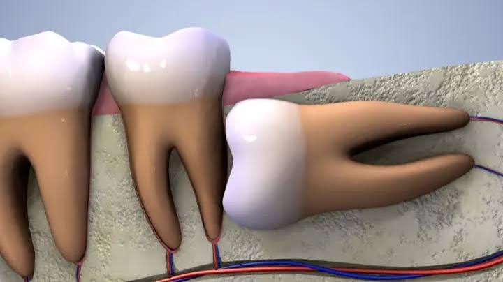
One of the most visible evolutionary traces in human anatomy is the volumetric reduction of the jawbones, which in the modern human mouth often fails to provide sufficient eruption space for the third molars (wisdom teeth). As a result of this anatomical narrowing, these teeth—located at the very back of the dental arch—frequently remain “impacted” (retained) because they cannot break through the dense cortical bone of the jaw or the thick fibrous gum tissue covering them. In dental literature, an impacted wisdom tooth is evaluated not as a simple delay in eruption, but as a complex pathological risk factor that can directly threaten surrounding bone, the roots of adjacent teeth, and the mandibular nerve networks. In the field of Ankara impacted wisdom tooth surgery, Panorama Ankara combines oral and maxillofacial surgical principles with micro-engineering technologies to remove the most challenging impacted tooth cases—fully trapped in bone, inverted, or intertwined with nerve canals—with minimal biological cost to surrounding tissues and maximum surgical safety. Our aim is to protect our patients through a completely painless and predictable clinical process from large cystic lesions that may erode the jawbone in the future and from chronic facial pain of unknown origin.
The silent presence of impacted wisdom teeth does not mean they are harmless. The developmental sac (follicle) surrounding a tooth that is fully embedded in the jawbone (complete bony impaction) and causes no outward symptoms can, over time, undergo cellular mutation and transform into pathological cavities known as “dentigerous cysts.” Over the years, these cysts can erode and thin the jawbone from the inside out through the hydrostatic pressure of accumulated fluid—sometimes to the point where even minor physical trauma (for example, biting something hard or receiving a small blow to the jaw) may cause a spontaneous fracture (pathological fracture). Additionally, as impacted teeth attempt to create space within the bone, they apply continuous and intense pressure to the roots of the healthy second molar directly in front of them. This insidious pressure can lead to root resorption over time, setting the stage for the adjacent tooth—despite being intact and free of decay—to loosen and be lost. Panorama Ankara’s specialist surgical team uses 3D tomographic imaging to map these silent risks with millimetric precision and carefully plans preventive (prophylactic) surgical interventions before bone destruction begins.
Anatomical Classification of Impaction Levels and Surgical Strategies
The only factor that determines the surgical difficulty (degree of invasiveness) of a wisdom tooth operation is the depth and position of the tooth within the jawbone and the thickness of the tissue barriers covering it. For clinicians, every impacted tooth is a unique anatomical puzzle. Teeth trapped in horizontal, inverted, or mesioangular (tilted toward the neighboring tooth) positions cannot be removed in a single piece; to extract them, the surrounding bone must be gently relieved using specialized osteotomy (bone reshaping) techniques, and the tooth must often be sectioned (odontectomy) and removed in parts. To help our patients evaluate their own operation process more clearly according to radiographic findings, we have transparently detailed the retention (impaction) classifications we use in clinical practice and the surgical requirements associated with each category in the table below.
| Retention (Impaction) Classification | Anatomical Condition and Tissue Coverage | Surgical Approach and Operational Dynamics |
|---|---|---|
| Soft Tissue Impaction (Mucosal Coverage) | The tooth has fully erupted from the bone, but is completely covered by a thick gum (mucosal) layer. | The gum tissue is gently reflected with a small incision (flap). Since there is no bony barrier, the tooth is luxated and removed without the need for bone removal. |
| Partial Bony Impaction (Partially Impacted) | Part of the crown remains within the bone, while another part has slightly penetrated the gum and opened into the oral environment. | After the gum is reflected, the bony undercuts holding the tooth are minimally reduced using micromotors or piezosurgery to create an exit path for the tooth. |
| Complete Bony Impaction (Fully Impacted) | The tooth is entirely trapped deep within the jawbone, covered by a thick cortical bone layer and mucosal tissue. | Requires comprehensive microsurgery. A careful bony window is created, and the tooth is typically sectioned into multiple pieces (odontectomy) and removed piece by piece without damaging surrounding tissues. |
Piezosurgery Technology: Shaping Bone with Ultrasonic Vibrations
In impacted tooth surgery, the main determinant of postoperative comfort (the extent of swelling, pain, and bruising) is how the bone surrounding the tooth is removed. In conventional methods, high-speed rotating burs driven by air pressure are used to reduce bone. However, these rotary instruments can create excessive heat (thermal trauma) from friction and may tear soft tissues (gum tissue or nerve sheaths) if they come into contact with them. At Panorama Ankara, in fully bony impacted and anatomically high-risk wisdom tooth surgeries, we use a revolutionary technology known as “piezosurgery” (piezoelectric surgery). Rather than employing a mechanical rotational movement, piezosurgery uses micro-vibrations generated by ultrasonic sound waves to cut bone.
The most remarkable feature of these ultrasonic vibrations is their “tissue selectivity.” Piezo tips cut only hard tissues (teeth and bone); even if they inadvertently touch soft tissues (blood vessels, nerves, or sinus membranes), they do not cut or tear them—only gently displace them. In the lower jaw, when operating in millimetric proximity to the vital inferior alveolar nerve (Nervus Alveolaris Inferior) near the roots of an impacted tooth, piezosurgery dramatically reduces the risk of nerve injury (paresthesia/numbness). In addition, because this technology does not cause thermal burns or cellular trauma in bone, wound healing (osteogenesis) in the surgical area begins much faster compared to conventional methods, and postoperative cheek swelling and edema within the first 48 hours remain minimal for our patients.
Coronectomy: A Safe Alternative for Roots Intertwined with the Nerve
One of the greatest surgical traps in lower-jaw anatomy is that, in some rare but high-risk cases, the roots of an impacted wisdom tooth may hook around the main nerve canal (mandibular canal) supplying sensation to the lower lip and chin, or the nerve may pass directly through the root complex. In these cases of extreme proximity detected via 3D volumetric tomography (CBCT), forcing the tooth out intact can tear or severely crush the nerve, leading to irreversible sensory loss (paresthesia) in the lower lip that may persist for months or even a lifetime. Instead of taking such a neurological risk, Panorama Ankara applies an exceptionally safe surgical alternative known in the literature as “coronectomy” (crown sectioning).
In a coronectomy, the tooth’s upper portion (the crown)—which may be decayed, infected, or exerting pressure on adjacent teeth—is smoothly sectioned at the root level and removed from the bone. The vital root fragments that are intertwined with the nerve are intentionally left in their original socket (within the bone) to avoid applying any traction force to the nerve. The roots are covered with the jawbone’s natural blood clot and, if necessary, bone graft materials, and the gum tissue is fully closed with sutures. Because these healthy root fragments retain pulpal vitality and are isolated from the oral environment, they usually do not cause infection; over time, they can fuse with surrounding bone cells and become part of the jawbone as a calcified tissue. This microsurgical strategy relieves the patient from the pain and pressure caused by the impacted tooth while providing 100% security for lip sensation.
Postoperative Period: Edema and Complication Management
Removing a fully bony impacted wisdom tooth from the jawbone creates a large operative field at the cellular level, and the body’s physiological healing response—accelerating blood flow to the area—typically manifests in the first few days as edema (swelling) and mild hematoma (superficial bruising). Although atraumatic microsurgical techniques at Panorama Ankara minimize these side effects, the postoperative care routines patients follow at home directly determine the comfort of the recovery period. Key instructions our patients should follow to support rapid tissue recovery and healthy closure of the suture line include:
- Intermittent cold compress (ice) application starting immediately after surgery and continued for the first 24–36 hours constricts blood vessels (vasoconstriction) and prevents excessive blood influx to the surgical region. This simple but vital step can prevent much of the swelling and bruising before it develops.
- To prevent the blood clot (alveolar dressing) from being dislodged—leaving bone exposed and triggering severe pain (dry socket / alveolitis)—patients should strictly avoid hot foods for the first 48 hours, avoid drinking through a straw (which creates negative pressure in the mouth), and take an uncompromising break from smoking, which impairs tissue oxygenation in the wound area.
Safely Overcome Impacted Anatomical Limits with Panorama Ankara
Impacted teeth trapped deep within the jawbone are like timed biological “bombs,” as it is impossible to predict when or how severely they may cause problems. In searches for Ankara impacted wisdom tooth surgery, Panorama Ankara transforms even the most difficult surgical cases into controllable, safe, and comfortable procedures by combining advanced radiological diagnostic methods with ultrasonic bone-cutting technologies (piezosurgery). While safeguarding vital structures such as the mandibular nerve, our clinical environment—supported by painless local anesthesia or conscious sedation—helps you leave surgical fears behind entirely.
Do not wait for your impacted teeth to initiate cyst formation in your jawbone or to push your front teeth from behind and reintroduce crowding that you worked hard to correct. With our 3D volumetric tomography scans, we can create a virtual map of your nerve canals and impacted tooth roots, and together with our expert team, plan the safest and most minimally invasive (tissue-friendly) surgical approach for you. You can schedule a detailed oral and maxillofacial surgery consultation at Panorama Ankara and take safe steps without putting your health at risk.