Ankara Jaw Cyst Treatment
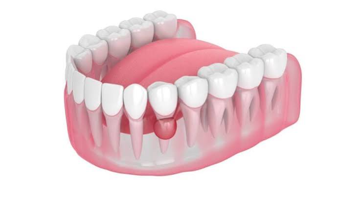
The jawbones (maxilla and mandible), among the most dynamic and complex bony structures in human anatomy, can harbor various pathological lesions as a result of embryological cellular remnants, chronic trauma, or genetic mutations whose causes are not fully understood. Major jaw cysts—forming a clinical picture far beyond simple root-tip (radicular) cysts caused by tooth decay—are aggressive cavitary (hollow) formations filled with fluid, semi-fluid material, or gas, surrounded by a thick epithelial lining, and capable of silently enlarging deep within the jawbone. Their most dangerous and insidious feature is that they can expand slowly within the medullary (spongy) bone marrow for months or even years without producing any pain, aching, or numbness. They are often diagnosed incidentally during a routine panoramic X-ray, or when the cyst inflates the jawbone like a balloon and creates a visible facial asymmetry. In the field of Ankara jaw cyst treatment, Panorama Ankara applies advanced maxillofacial (jaw and facial) surgery protocols to completely remove these aggressive lesions that erode bone from within—without harming adjacent vital structures (nerves and sinus cavities)—and to reconstruct the resulting massive bony defects using advanced tissue-engineering methods.
The growth mechanism of a cyst within the jawbone occurs through hydrostatic pressure generated by fluids secreted by the epithelial cells inside it. As the cyst expands volumetrically, it exerts enormous mechanical force from within against the outer walls of the jawbone (cortical bone). This constant pressure causes the outer bony walls to thin to the level of paper (cortical expansion) and, over time, to erode and disappear entirely. A jawbone that has become thinned and has lost its spongy structure becomes biomechanically so weak that biting a hard food, yawning, or even a minor impact to the jaw may lead to sudden spontaneous fracture (pathological fracture). In addition, massive jaw cysts can severely displace the roots of adjacent healthy teeth while creating space within the bone, leading to tooth movement, root resorption, or compression of the main sensory nerve passing through the lower jaw (Nervus Alveolaris Inferior), which can cause permanent numbness in the lower lip. Panorama Ankara’s experienced maxillofacial surgery board develops centimeter-precise surgical strategies tailored to each patient’s unique radiological map to stop this destructive process and preserve the structural integrity of the jaw skeleton.
Biological Behavior and Aggressive Growth: The Anatomy of Multilocular Lesions
Jaw cysts encountered in maxillofacial surgery are classified by radiographic appearance as unilocular (single-chamber) lesions or multilocular (multi-chamber) lesions, which present a “soap-bubble” or “honeycomb” pattern. In particular, multilocular pathologies such as the Odontogenic Keratocyst (OKC) or Ameloblastoma (a locally aggressive benign tumor) exhibit cellular behavior very different from standard cysts. These lesions do not merely expand like a balloon; they also infiltrate into the depths of the spongy medullary bone, branching through marrow spaces like tree roots. This microscopic infiltration tendency makes it extremely difficult to separate the cyst surgically from bone as a single intact unit (together with its lining).
If aggressive jaw cysts of this nature are managed with a routine approach and simply scraped (curettage) and left behind, even a single microscopic epithelial cell remaining in the deeper bone can lead to recurrence (aggressive relapse/recurrence) within months—often growing faster than before. Recurrence rates of odontogenic keratocysts are considered high in global medical literature. To reduce this relapse risk to near zero, Panorama Ankara does not end the procedure after removing the cyst from its bony cavity. Our clinicians additionally reduce the walls of the cyst cavity by 1–2 millimeters using specialized motorized burs, ultrasonic piezosurgery tips, or chemical cauterization solutions. This critical step, called “peripheral ostectomy,” destroys any potential residual pathological cell remnants that may have infiltrated bone porosities and is the most vital surgical measure to prevent the cyst from ever returning in the same area.
Clinical Classification: Major Cyst Types Seen in the Jawbones
A definitive diagnosis of jaw cysts cannot be made solely by examining X-ray images; it is confirmed when the tissue removed during surgery is sent to pathology laboratories and analyzed under the microscope (histopathological examination). However, for surgical planning, a preliminary diagnosis based on radiological and clinical data is essential. Panorama Ankara provides transparent clinical information so that patients can understand this complex medical terminology and the nature of the pathology within their own jawbones. The table below details the biological characteristics of major cyst types that develop within the jawbone itself, independent of tooth-related cysts.
| Pathological Classification (Cyst Type) | Clinical and Radiological Characteristics | Growth Rate and Recurrence Risk |
|---|---|---|
| Odontogenic Keratocyst (OKC) | Most commonly seen in the posterior mandible with a multilocular “soap-bubble” appearance. It tends to excavate bone in an anteroposterior (front-to-back) direction. | Grows very aggressively. Due to its special cellular structure, recurrence risk is very high if not thoroughly eradicated. |
| Traumatic Bone Cyst (TBC) | A pseudocyst that is empty or filled with blood (no true epithelial lining). It may develop years after significant jaw trauma. | Slow-growing. Surgical curettage to induce bleeding is sufficient for healing, and recurrence is not expected. |
| Globulomaxillary Cyst (Developmental) | A lesion in the upper jaw that grows in an inverted pear shape between the roots of the lateral incisor and canine, arising from embryological fusion errors. | Grows slowly but can forcefully separate tooth roots and compromise aesthetics. It does not recur when removed as a single piece. |
| Residual Cyst | A previously extracted tooth site where an inflammatory lining was left behind and later evolves into a large cyst. | Enlarges silently within the extraction area and weakens the jawbone. Complete removal of the lining yields full success. |
Advanced Diagnostic Protocols and Jaw Reconstruction
Surgical success in jaw cyst treatment consists of two equally important phases: safe removal of the cyst (enucleation) and restoration of jaw integrity by refilling the large void (defect) created in the jawbone (reconstruction). After a cyst is removed, the remaining cavity can be the size of a walnut—or sometimes even as large as a tangerine. The human body cannot regenerate such a large bony void on its own simply by filling it with a blood clot. If the cavity is left to heal without reconstruction, the gum tissue collapses into the space and a permanent, large depression forms in the jawbone. This weakness can predispose the jaw to fracture in later years.
To prevent this anatomical collapse and restore the jaw skeleton to its original strength, Panorama Ankara employs advanced tissue-engineering and bone grafting protocols. After the cyst cavity is thoroughly cleaned and decontaminated, it is densely filled with sterile, biocompatible bone graft particles (allografts) mixed with healing factors derived from the patient’s own blood (PRF – Platelet-Rich Fibrin). To prevent graft dispersion and to ensure that only bone cells proliferate in the area, the graft is covered with resorbable or titanium barrier membranes. If the cyst has thinned the lower border of the jawbone to a paper-like level—creating a fracture risk even on the operating table—our surgeons reinforce the jaw externally by fixing medical “titanium reconstruction plates” with screws, forming a biomechanical splint (support). After a months-long healing period, the grafted material transforms into the patient’s own living jawbone, providing a solid biological foundation for future implant placement if needed.
Postoperative Recovery and Fracture Prevention
Surgery for a large-volume jaw cyst is a major intervention that temporarily alters the structural resistance of the jaw skeleton. For transplanted bone graft particles to become vascularized (angiogenesis), mature into solid bone, and regain full load-bearing strength, a prolonged cellular maturation process of approximately 6 to 8 months is typically required. During this critical recovery period, protecting the jaw from external trauma and excessive chewing forces is essential to preserve the integrity of the treatment. Depending on the size of the cyst cavity, Panorama Ankara specialists provide patients with the following key postoperative biological and biomechanical warnings:
- In the first weeks and months following surgery, patients must strictly avoid hard foods that require forceful biting and could overload the affected jaw region (hard bread crust, apple, quince, nuts). Chewing should be performed on the opposite (healthy) side, and the diet should remain soft (purees, boiled foods).
- After operations for lesions with a high genetic recurrence tendency—such as odontogenic keratocysts—even if there are no symptoms, the area should be closely monitored by the clinician with panoramic X-rays or tomography every 6 months for the first 3 years.
Secure Your Facial Symmetry and Jaw Health with Panorama Ankara
A cyst growing deep within the jawbone may provide no warning until you notice swelling or facial asymmetry, or until it is discovered during a routine dental check-up. This silent progression is the most dangerous aspect of maxillofacial pathologies. By combining advanced surgical expertise with tissue engineering in Ankara jaw cyst treatment services, Panorama Ankara removes these aggressive lesions threatening your facial skeleton with a high level of medical safety—without putting your jaw integrity or the vital nerves responsible for lip sensation at risk.
If you notice unexplained asymmetry (swelling) in your jaw, feel aching in the site of a tooth extracted in the past, or if a dark cavity has been detected within the bone on a routine X-ray, do not allow these cavitations to continue eroding your bone from within. To create a 3D tomographic map of your jawbone, determine the lesion boundaries with millimetric precision, and design a safe surgical route to reconstruct your jaw with our expert physicians, you can schedule a detailed maxillofacial consultation appointment at Panorama Ankara and secure both your facial aesthetics and biological strength.