Ankara Jaw Surgery: Orthognathic Surgery, Skeletal Harmony, and Holistic Facial Aesthetics with Panorama Ankara
The aesthetic harmony of the human face, respiratory capacity, and chewing function all depend on the flawless skeletal balance that the lower jaw (mandible) and upper jaw (maxilla) establish with each other and with the cranial base in three-dimensional space. However, due to our genetic heritage, childhood traumas experienced during growth and development, chronic habits such as prolonged thumb-sucking, or endocrinological factors, one of the jawbones may overgrow relative to the other, or its development may stop entirely and remain behind. These asymmetric presentations—referred to in the medical literature as “Skeletal Malocclusion” or “Jaw Deformity”—are major anatomical abnormalities that go far beyond simple dental crowding that can be corrected merely by placing braces on teeth; they directly involve the bony structure. In cases where the lower jaw is markedly forward (prognathism), the upper jaw is retruded and the midface appears collapsed (retrognathism), or the jaws shift asymmetrically to the right/left, patients do not only carry an aesthetic psychological burden; they also struggle with inability to grind food, chronic temporomandibular joint (TMJ) pain, and even life-impacting syndromes such as severe Sleep Apnea (OSA) due to a narrowed airway. In the field of Ankara jaw surgery (Orthognathic Surgery), Panorama Ankara applies advanced maxillofacial engineering principles by uniting orthodontics and jaw surgery under a single scientific roof; we reconstruct these challenging cases—whose limits extend beyond skeletal boundaries—at the cellular and anatomical level, offering our patients a lifelong functional and aesthetic rebirth.
The term “orthognathic” originates from the Greek words orthos (straight/correct) and gnathos (jaw), and it literally refers to the correction of the jaws. Although jaw surgeries are often confused with plastic surgery operations performed to beautify facial features, the primary medical purpose of orthognathic surgery is to restore impaired or collapsed chewing and breathing physiology to its proper state; the remarkable aesthetic improvement seen in the facial profile is, in fact, a natural biological consequence of jawbones that have been functionally aligned and repositioned correctly. Panorama Ankara’s experienced maxillofacial surgical board does not merely cut and reshape the patient’s bone structure by millimeters; it also digitally designs in advance—long before the patient lies on the operating table—how the facial soft tissues (lips, nasal tip, cheek muscles) will drape over this new skeletal framework (soft-tissue drape). In this way, the patient can experience step by step, in light of scientific data, how chewing forces will distribute evenly and healthily to the jaw joint (condylar region) once the new bite relationship is achieved, how lips that have not been able to close for years will meet effortlessly, and most importantly how sleep quality can improve dramatically as the airway expands.
Orthodontics–Surgery Synergy: The Preoperative Decompensation Phase
Jaw surgery is not an isolated procedure where the surgeon enters the operating room in a single day and performs miracles alone; on the contrary, it is a multidisciplinary medical journey that lasts for months and requires the patient’s patience, carried out with perfect synchronization by Orthodontics (braces therapy) and Maxillofacial Surgery specialists. In a patient whose lower jaw is skeletally too far forward, the body naturally tries to compensate for the abnormality in order to chew. In other words, the lower front teeth tip backward and the upper front teeth tip forward, attempting to close the large skeletal discrepancy and force contact. For Panorama Ankara physicians to proceed to the surgical stage, this “incorrect but necessary” dental adaptation must be completely reversed. In this first phase—called “Decompensation Orthodontics” in the medical literature—orthodontic braces or clear aligners are applied.
During this months-long preparation stage, the orthodontist moves the teeth back to their original and upright (correct) angulations on the jawbone. As the teeth return to their correct angles, the skeletal discrepancy between the jaws becomes even more obvious—meaning that just before surgery the lower jaw may appear even more forward or more “open” than before. This is a psychologically challenging but absolutely necessary “worsening” phase for surgery. Because when the teeth reach the mathematically correct angles on the bone, and the maxillofacial surgeon cuts and repositions the jawbones in the operating room, the upper and lower teeth lock together with millimetric precision—like the two sides of a zipper or pieces of a puzzle. No skeletal surgical intervention performed without this decompensation phase can remain stable in the long term and may lead to destructive functional problems in the jaw joint.
Clinical Comparison: Skeletal Camouflage (Masking) vs. Orthognathic Surgery
Patients presenting with skeletal jaw abnormalities generally face two main medical pathways. In mild skeletal cases, “Orthodontic Camouflage” (Masking) treatment may be applied by changing only tooth angulations to hide the underlying bony discrepancy; in moderate and severe deformities, physical repositioning of the bones is essential. To help patients fully understand the medical realities and limitations when making this life-changing decision, Panorama Ankara details the anatomical differences between camouflage treatment and true orthognathic surgery in the table below.
| Anatomical and Medical Criteria | Orthodontic Camouflage (Braces Only) | Orthognathic Surgery (Jaw Surgery) |
|---|---|---|
| Target Tissue Addressed | Only the angles and inclinations of the teeth within the jawbone are changed. The bone remains unchanged. | The lower and upper jaw skeleton (bony bases) are directly cut and repositioned in three-dimensional space. |
| Effect on Facial Profile and Aesthetics | Does not create a marked aesthetic change in the outer facial outline, chin point, or lip support. | Facial proportions, the nose–lip angle, and chin profile change dramatically and positively. |
| Anatomical Limits of Treatment | Can be applied only in very mild (1–3 mm) skeletal differences without moving tooth roots outside the bone. | The only solution in severe (10–15 mm and above) discrepancies, asymmetries, and skeletal sleep apnea. |
| Airway and TMJ (Joint) Health | Provides no mechanical benefit for a narrowed airway or chronic joint pain. | Advancing the jaws enlarges the airway (apnea resolves), and the jaw joint seats into the correct position. |
3D Cephalometric Simulation: Digital Reconstruction Before the Operating Room
In the past, jaw surgery planning was prepared manually by drawing on two-dimensional X-rays (cephalometric radiographs) and by cutting and reshaping plaster models. These conventional methods limited the surgeon’s foresight and increased the risk of encountering unexpected anatomical obstacles during surgery. Integrating the highest level of technology into its practice in the field of Ankara jaw surgery, Panorama Ankara completely eliminates these manual uncertainties with a “3D Virtual Surgical Planning” (VSP) protocol. A 3D Volumetric Tomography (CBCT) scan of the patient is taken, and the soft-tissue map of the face is transferred into a digital environment using optical facial scanners. This digital dataset—containing billions of points—is merged in advanced engineering software to create an exact virtual replica (a “digital twin”) of the patient’s skull.
Panorama Ankara’s maxillofacial surgeons perform the surgery virtually on this digital twin. The upper jaw is cut horizontally (Le Fort I Osteotomy), and the exact millimeters it will be moved forward and/or upward are calculated. Simultaneously, the lower jaw is split on both sides while protecting the nerve pathway (Bilateral Sagittal Split Osteotomy – BSSO) and rotated into its new position in harmony with the upper jaw. The software simulates within seconds—and shows the patient—how much the nasal tip may lift, how much the lower lip may become fuller, and how the chin point may gain projection as the jaws are moved. Once the plan is approved, the special surgical guides (splints) that the surgeon will use to fix the jaws in place during the operation are produced with 3D printers. As a result, the actual procedure under general anesthesia becomes a safe engineering application with virtually zero margin of error—rehearsed repeatedly in the digital environment—and the operating time is dramatically shortened.
Rigid Fixation and the Dynamics of the Recovery Process
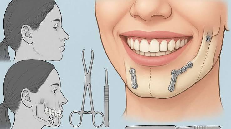
Jaw surgeries are performed in a hospital setting under general anesthesia in fully equipped operating rooms. The greatest aesthetic advantage is that the entire procedure is carried out from inside the mouth (beneath the mucosa), leaving no externally visible surgical incision or scar on the face, cheeks, or neck. Once the bones are positioned correctly, they are firmly stabilized using a method called “Rigid Internal Fixation” in the medical literature: mini titanium plates and micro-screws that are 100% biocompatible and do not oxidize. Thanks to this modern fixation technology, the frightening “jaw wiring for months” (maxillomandibular fixation) nightmare of the past has become history. When patients wake up after surgery, they can open and close their jaws, speak, and yawn.
The first few weeks after surgery constitute an intense biological healing phase in which facial muscles and soft tissues adapt to the new skeletal structure (neuromuscular adaptation). To ensure maximum comfort during recovery, Panorama Ankara’s surgical team follows the implementation of these critical clinical guidelines with great precision:
- A critical period of approximately 6 weeks is required for the repositioned jawbones to fuse at the cellular level (bone consolidation). During this phase, to avoid mechanical load on the muscles and bones, nutrition should be maintained entirely with blenderized foods that do not require chewing (liquid and puree consistency), and a gradual transition to solid foods should be made.
- To reduce physiological cheek edema (swelling) and intratissue hematoma as quickly as possible, cold compress should be applied at intervals recommended by the clinician in the first days; while sleeping, the head should be kept above heart level using two pillows; and oral hygiene must be maintained rigorously with special chemical mouth rinses to prevent infection of intraoral surgical sutures.
Rebuild the Natural Harmony of Your Face with Panorama Ankara
Jaw asymmetries or skeletal bite discrepancies create a deep aesthetic and functional burden that can undermine a person’s self-confidence from childhood, leading them to avoid mirrors or cameras. In the field of Ankara jaw surgery, Panorama Ankara addresses not only the teeth but the entire facial structure and breathing dynamics with a holistic approach, removing this burden from your shoulders with the most advanced technology modern medicine and digital engineering can provide. Managed through the flawless integration of orthodontics and surgery, this process brings facial proportions closer to universal golden-ratio harmony while restoring your breathing and chewing functions—which you may not have used efficiently for years—back to their original health.
No matter how well-aligned your teeth are, if your skeletal bases are not in the correct position, it is not possible to speak of an ideal smile or a comfortable life. To have your facial structure mapped millimetrically in a 3D digital environment, personally experience the remarkable postoperative change through orthognathic surgery simulations, and discover your customized safe surgical pathway, you can schedule a detailed maxillofacial consultation appointment at Panorama Ankara—becoming the aesthetic and strong architect of your own story.