Ankara Wisdom Tooth Treatment
The thousands-of-years-long biological journey of human evolution—along with changes in our dietary habits—has gradually caused our jaw skeleton to become smaller (narrower). However, the number of teeth encoded in our genetic blueprint (32 teeth) has not adapted to this narrowing at the same pace. The cost of this anatomical mismatch is most often paid by the “Third Molars,” known popularly as wisdom teeth, which attempt to erupt last and cannot find adequate space at the very back of the jawbone (the retromolar region). Typically entering an active eruption period between the ages of 17 and 25, these teeth may become trapped within the bone in a horizontal, inverted, or angled position when they cannot find sufficient jaw width (ridge volume), remaining as “Impacted” teeth (embedded in bone) or as “Partially Impacted” (Semi-Impacted) forms that can only partially break through the gum tissue. Integrating the most up-to-date, tissue-friendly (minimally invasive) protocols of modern medicine into its clinical practice in the field of Ankara wisdom tooth treatment and maxillofacial surgery, Panorama Ankara relieves patients from days-long jaw swelling, severe ear pain, and difficulty swallowing. Our expert oral and maxillofacial surgery team removes these anatomical abnormalities—often sitting in bone like a time bomb and decaying the roots of neighboring healthy teeth—with centimeter-level precision guided by 3D tomographic mapping, thereby safeguarding both the structural integrity of the jaw skeleton and the patient’s functional comfort.
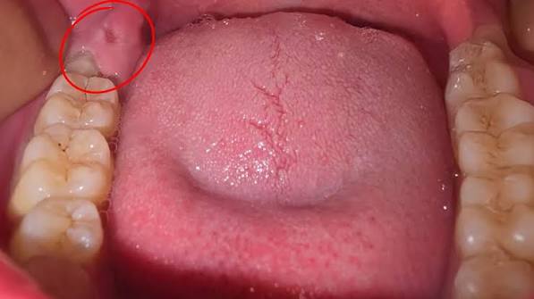
The clinical problems caused by wisdom teeth are not limited to simple “eruption pain.” In particular, the loose gum tissue flap (“operculum” or “hood”) around partially impacted wisdom teeth becomes an ideal, nearly impossible-to-clean incubation site for oral bacteria and food debris. Food trapped under this tissue rapidly ferments and can lead to an acute infection called “Pericoronitis,” which may cause massive swelling visible from the outside of the cheek, limited mouth opening (trismus), a stabbing sensation in the throat during swallowing, and enlarged lymph nodes beneath the jaw. At Panorama Ankara, when patients present with an acute pericoronitis episode, any purulent (infected) fluid in the area is drained using specialized irrigation (rinsing) solutions and the infection is brought under control before surgical intervention. On the other hand, wisdom teeth that remain completely inside the bone (fully impacted) and are never visible can exert significant horizontal pressure on the roots of the second molar in front of them as they try to create space. This pressure may cause both root resorption of that healthy tooth and relapse of orthodontic alignment—undoing years of orthodontic (braces) treatment and creating crowding in the front teeth again. For this reason, Panorama Ankara clinicians evaluate wisdom teeth not only when they cause pain, but proactively—whenever they pose a radiographic risk.
Impacted Third Molars: Positional Anatomies
How long a wisdom tooth extraction will take, the biological burden of the procedure, and the surgical technique required depend directly on the tooth’s three-dimensional angulation within the jawbone (mandible or maxilla). In dental literature, these angulations are among the most critical navigation data that determine the surgeon’s operative roadmap. Positions in which the crown of the wisdom tooth tips forward toward the adjacent tooth and presses against its root are called “Mesioangular” (forward-tilted) impactions; these are the most commonly encountered cases in clinical practice and the ones most likely to contribute to orthodontic crowding.
By contrast, “Horizontal” impactions—where the tooth lies completely parallel to the ground within the jawbone—or “Distoangular” impactions—where the tooth tilts backward toward the mandibular ramus (jaw angle area)—are complex cases in which surgical access is most difficult and advanced maxillofacial expertise is required. Sometimes the tooth is in a fully vertical position, yet cannot reach the oral cavity because the overlying bone layer is very thick. After determining this anatomical angulation, Panorama Ankara’s advanced surgical board avoids forcing the tooth out in one piece (which could damage the alveolar ridge). Instead, with a tissue-friendly approach known in the literature as “Odontectomy,” the tooth is sectioned into two or three parts within the bone and removed like a pearl from its socket—without applying traumatic pressure to the bone.
Clinical Classification: Wisdom Tooth Anatomy by Degree of Eruption
Answers to common pre-operative questions such as “Will my tooth be cut to remove it?” or “Will stitches be placed?” depend on the thickness of the bone and mucosal (gum) barrier over the tooth. To help patients manage both the surgical process and the recovery (healing) period transparently, Panorama Ankara details the eruption positions of wisdom teeth in the table below.
| Status of Eruption | Clinical & Anatomical Characteristics | Surgical Approach & Healing Dynamics |
|---|---|---|
| Fully Erupted (Visible in the Mouth) Wisdom Teeth | The tooth has fully passed through bone and gum and occupies its place in the oral cavity like a normal tooth; the roots are often curved. | The gum is not incised (no flap is raised). The tooth is usually removed like a routine extraction (with forceps and elevators), typically without stitches. Healing is very fast. |
| Partially Impacted (Semi-Impacted) Wisdom Teeth | Only part of the tooth or its cusp tips are visible. A thick, hood-like gum tissue covers it. | A small incision is made in the gum; a small amount of surrounding bone is typically removed to gain access, and stitches are placed. This group carries the highest risk of pericoronitis. |
| Fully Impacted (Intraosseous / Impacted) Wisdom Teeth | The tooth is completely inside the jawbone, under a thick layer of gum and bone. It is never visible from the outside. | The gum is fully opened surgically (mucoperiosteal flap). A bone window is created (osteotomy), and the tooth is removed in sections (odontectomy). This is the most challenging surgery. |
Advanced Diagnosis and Surgical Planning: Protecting the Inferior Alveolar Nerve
What transforms fully impacted lower (mandibular) wisdom tooth surgery into a major procedure—rather than an ordinary extraction—is the most critical anatomical barrier: the main sensory nerve that passes just beneath the roots. Known in the medical literature as the “Inferior Alveolar Nerve (Nervus Alveolaris Inferior),” this thick nerve bundle is a major communication line that carries all touch, temperature (hot-cold), and pain sensations from the lower lip, chin, and lower teeth to the brain. In many patients, the hook-like curved roots of a wisdom tooth can wrap around this nerve canal like a vise, or even pass through the canal itself. On traditional two-dimensional X-rays (panoramic films), it may only be seen that the nerve and root are “close,” but it is not possible to determine in three-dimensional space whether the tooth lies in front of, behind, or within the nerve.
To protect patients 100% from irreversible neurological complications such as persistent lower-lip numbness (paresthesia) after surgery, Panorama Ankara maps the tooth pre-operatively using 3D Volumetric Tomography (CBCT). With this high-resolution digital dataset, our surgeons identify—three-dimensionally—the critical millimeters where the nerve contacts the roots before ever entering the operation. During surgery, while bone is removed or the tooth is sectioned, the nerve pathway is kept under full protection (isolation). The angulation of the surgical burs and the extraction vector (direction of removal) are planned millimetrically so that not even a micron of pressure is applied to the nerve. This advanced radiological vision and microsurgical approach allows our patients to leave the chair with maximum safety—without having to experience the fear of “Will my lip go numb if my tooth is extracted?”
Post-Operative Healing Dynamics and Swelling Control
Impacted wisdom tooth surgery (especially in cases requiring bone removal) is a biological trauma involving intervention in the structural tissues of the jaw skeleton. The body’s first physiological response is to accelerate blood flow to initiate repair—creating edema (swelling) at the surgical site. The higher the surgeon’s respect for tissue during the procedure, the less swelling occurs. However, the wound-care discipline the patient follows during the first 48 hours after leaving the clinic is essential to crown that surgical success. Panorama Ankara oral and maxillofacial surgery specialists recommend the following two strict and vital rules to minimize swelling and experience a comfortable, low-pain recovery period:
- To stop internal bleeding (hematoma) and physiologically suppress cheek swelling, apply continuous “Cold Compress” (Ice Therapy) from the outside of the cheek for the first 24 hours starting immediately after the procedure—using 15-minute cycles (apply for 15 minutes, rest for 15 minutes).
- To prevent the vital “blood clot” that forms in the extraction socket (alveolus) and covers the bone from being dislodged (which can cause the extremely painful condition known as Alveolitis/Dry Socket), do not vigorously rinse the mouth, do not spit forcefully, and avoid any actions that create negative pressure (vacuum) in the mouth—such as drinking through a straw—for the first 72 hours.
Pain-Free Farewells and a Preserved Jaw Structure with Panorama Ankara
Wisdom teeth are often unpredictable pathologies that can remain silent within the bone for years and then suddenly flare up at the worst possible times (during exam periods, vacations, or business trips), dropping your quality of life to zero. By bringing the most up-to-date microsurgical protocols of maxillofacial engineering into its clinical practice for Ankara wisdom tooth treatment, Panorama Ankara removes this hidden threat without harming neighboring teeth, without risking your lip nerves, and without exposing you to severe post-operative swelling. Even in the most challenging anatomies, our surgeons maximize comfort by minimizing procedure time through bone-respecting (atraumatic) techniques.
If you want to protect the effort you invested in orthodontic treatment (braces), eliminate chronic pericoronitis swelling that can make the mouth impossible to open, and remove that impacted “time bomb” while preserving the sensation in your lower lip, you can schedule a detailed surgical consultation at Panorama Ankara to obtain a 3D CBCT map of your wisdom teeth and plan the safest surgical route with our specialist surgeons—so you can say goodbye to your wisdom teeth safely and painlessly.