Ankara Impacted Tooth Extraction
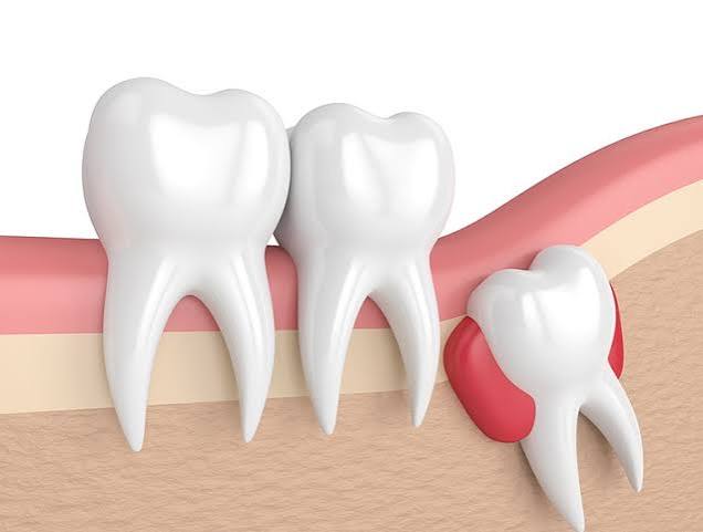
In dental practice, when the public hears the term “impacted tooth,” they almost exclusively think of wisdom teeth. However, maxillofacial anatomy can host far more complex and diverse developmental anomalies. Any tooth in the human jaw may become trapped deep within the jawbone due to genetic factors, premature loss of primary (baby) teeth leading to space deficiency, excessive densification of bone tissue, or the presence of a physical tumor/cyst along the eruption pathway. After wisdom teeth, the most commonly impacted teeth in the mouth are the maxillary canines (upper “eye teeth”)—the keystones of smile aesthetics and tearing function—and supernumerary (extra) teeth that do not belong to the normal dental arch and develop as additional teeth within the jawbone. In the field of Ankara impacted tooth extraction and oral and maxillofacial surgery, Panorama Ankara embraces not only separating teeth from bone, but also performing a tissue-engineering approach integrated with orthodontics. Our specialist clinicians analyze impacted teeth with 3D tomography, deciding whether the tooth should be fully removed (odontecomy/odontotomy and extraction) or whether the overlying bone should be removed to guide the tooth into the oral cavity with orthodontic traction (surgical exposure). In doing so, we design microsurgical routes that eliminate cystic formations and destructive resorption of adjacent tooth roots while preserving aesthetics.
Every impacted tooth that has not completed eruption within the jawbone contains an embryological sac called the “follicle.” When a tooth cannot erupt into the oral cavity on schedule, this follicular tissue is highly prone to degeneration over time, fluid accumulation, and transformation into a destructive bony lesion known as a “Follicular Cyst” (Dentigerous Cyst). These cysts can grow silently and painlessly into massive sizes over months, eroding the jawbone from within like a balloon, displacing neighboring healthy teeth, or dissolving their roots like acid (root resorption), potentially causing the patient to lose otherwise intact teeth. For this reason, the oral and maxillofacial surgery board at Panorama Ankara evaluates impacted teeth not merely as an aesthetic issue, but as potential pathologies that can threaten the structural integrity of the jaw skeleton. Especially palatally or labially deep-impacted canines and extra teeth hidden between the anterior incisors (mesiodens) are often positioned within millimetric proximity to adjacent roots; therefore, to avoid injuring healthy roots during removal, an extraordinary level of operative precision is achieved using advanced “Piezosurgery” (ultrasonic bone cutting) devices.
Impacted Canines (Maxillary Canines): Extraction or Orthodontic Guidance?
Canines are the longest, strongest, and most vital teeth in the human mouth; they support the corners of the lips, define the facial “smile arc,” and guide lateral movements in the chewing system (canine-protected occlusion). When a canine remains impacted toward the palatal vault or the floor of the nose, extracting it directly—like a routine wisdom tooth—can lead to irreversible aesthetic collapse in the facial profile and lip support. Therefore, at Panorama Ankara, once an impacted canine is detected, the oral and maxillofacial surgeon and the orthodontist convene to create a multidisciplinary rescue plan. The goal is not to remove that precious tooth, but to restore it to its rightful place in the dental arch.
This rescue procedure is referred to in the literature as “Surgical Exposure and Orthodontic Traction.” Under local anesthesia, the surgeon creates a small window in the palatal or labial mucosa to expose the enamel surface of the impacted canine. After bleeding is fully controlled, the orthodontist bonds a special gold chain or a miniature bracket onto the exposed enamel. The mucosa is then re-closed, and the end of the gold chain is connected to the orthodontic wires. Through a meticulous biomechanical process lasting months, the deeply impacted tooth is pulled millimeter by millimeter via the gold chain and successfully positioned into the designated space within the normal tooth alignment. If the tooth’s angulation is too unfavorable to be rescued, if the root is severely curved (dilaceration), or if the tooth is fused to bone (ankylosis), then as a last resort, the decision is made to remove the tooth using advanced surgical techniques.
Clinical Classification: Surgical Options for Impacted Teeth
The medical pathway for hidden teeth within the jawbone is shaped by the anatomical value of the tooth and the risk level of its location. To clarify the surgical experience and the functional outcome, Panorama Ankara transparently compares the fundamental maxillofacial approaches used in impacted tooth cases in the table below.
| Clinical & Operational Dynamics | Surgical Removal (Odontecomy / Extraction) | Surgical Exposure (Orthodontic Guidance) |
|---|---|---|
| Specific Case Type (Indication) | Extra (supernumerary) teeth; ankylosed/severely curved-root teeth; or wisdom/canine teeth associated with cyst formation. | Canines with a favorable angulation and healthy roots that are trapped in the palate/lip due to space deficiency. |
| Maxillofacial Surgical Goal (Purpose) | Completely remove the pathology, cyst, and nonfunctional tooth from the jawbone while protecting adjacent roots. | Not to remove the tooth from bone; to expose its surface and bond a chain to incorporate it into the dental arch. |
| Procedure Duration & Healing (Recovery) | A single short surgical operation: the tooth is removed and sutured; the wound typically heals fully within 1 week. | The surgical step is brief, but orthodontic traction to bring the tooth into place may take months/years. |
| Aesthetic & Skeletal Outcomes | An implant may later be needed for the resulting space, or the gap may be closed orthodontically. | Because the patient’s natural tooth is retained, optimal lip support and natural gingival papilla aesthetics are achieved. |
Supernumerary (Extra) Teeth and Mesiodens Syndrome
After wisdom teeth and canines, the impacted-tooth scenario that most frequently requires clinical intervention involves supernumerary (extra) teeth that occur due to a genetic coding error. In some cases, in addition to the normal 32 teeth, one or more additional tooth germs develop in the jawbone. The most common—and most hazardous—location for these extra teeth is the exact midline between the upper two central incisors; an impacted extra tooth in this region is called “Mesiodens” in the medical literature. Mesiodens typically develops in a pear- or cone-shaped form and can physically block the eruption of the two permanent incisors, creating a large gap (diastema), or forcefully displacing the permanent teeth laterally.
When detected on panoramic radiographs in childhood or young adulthood, these mesiodens and other extra teeth must be removed from their location using a highly precise surgical flap (mucosal incision) before they disrupt root development of the normal teeth in the jawbone. To remove these extra teeth without scraping adjacent healthy roots and without harming the nasopalatine nerves near the nasal floor, Panorama Ankara clinicians map the tooth’s exact position millimetrically using 3D tomography (CBCT). After successful surgical removal, the permanent front teeth often complete eruption in a healthy manner, as the physical obstruction is eliminated and they can return to their natural eruption pathway.
Post-Operative Tissue Maintenance and Complication Prevention
Regardless of the anatomical region involved (palate, cheek, or chin) and the objective (extraction or orthodontic exposure), impacted tooth operations are major interventions on the jawbone and the mucosal layer covering it (periosteum). Following a procedure performed with atraumatic techniques (without overheating bone), the patient’s wound-care discipline at home is vital for ensuring that the raised soft-tissue flaps (opened gum tissue) reattach and heal over the bone in a healthy way. To prevent suture opening and to keep physiological cheek edema (swelling) under control, Panorama Ankara’s oral and maxillofacial surgeons recommend that patients adopt the following two essential rules as a consistent routine:
- To prevent wound edges from stretching and tearing (dehiscence) where the opened gum tissue has been sutured back to bone—especially after lip or palatal surgeries—avoid opening the mouth excessively wide, avoid eating large bites (for example, hamburgers or whole apples), and pause physical activities that heavily strain facial muscles during the first week.
- To protect the vital blood clot formed within the socket (cavity) and to avoid “vacuum” effects that can dislodge it, expose bone to the oral environment, and cause severe pain (alveolitis/dry socket), do not use straws, do not rinse the mouth forcefully, and strictly refrain from smoking—which poisons healing cells—until sutures are removed, especially during the first 72 hours.
Overcome the Hidden Obstacles in Your Jaw Skeleton with Panorama Ankara
You may be carrying an impacted tooth deep in the jawbone—possibly unnoticed for years—that is silently transforming into a cyst or pushing against adjacent roots and disrupting orthodontic alignment. By combining orthodontic refinement with advanced oral and maxillofacial surgical discipline in Ankara impacted tooth extraction and orthodontic exposure procedures, Panorama Ankara removes these bone-embedded “pulled-pin” threats with exceptional respect for tissue biology. Beyond wisdom teeth, our expert team identifies and manages your most valuable canines and anterior extra teeth (mesiodens) with a safe, aesthetics-preserving roadmap that protects the biological architecture of your smile.
If you feel a swelling in your palate, if an impacted canine has been detected during orthodontic treatment, or if a routine radiograph shows an oddly angled tooth inside the bone, you can schedule a detailed surgical consultation at Panorama Ankara. Together with our specialist oral and maxillofacial surgeons, we will analyze the tooth with 3D tomography and make the nuanced medical decision between extraction and orthodontic rescue—so you can safely eliminate hidden threats to your jaw skeleton.