Ankara Jaw Cyst Treatment Center
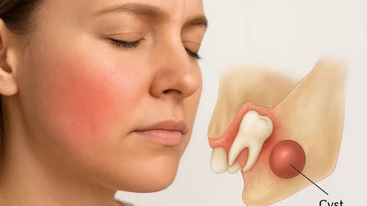
Cysts that develop within the jawbones are pathological cavities filled with fluid or semi-fluid material, classified in medical literature as “Odontogenic” (tooth-related) or “Non-odontogenic.” These lesions often progress silently; without routine panoramic X-ray screenings, they can destroy large areas of jawbone, resorb tooth roots, or weaken the mandible to the point of causing “pathologic fractures.” Redefining Ankara jaw cyst treatment center standards with a vision of “Biomimetic Reconstruction” and “Tissue-Friendly Cystectomy,” Panorama Ankara offers a comprehensive protocol that not only removes the cyst, but also reconstructs the large bone voids (cavities) created by the lesion using advanced grafting systems. The surgical techniques applied in our clinic protect vital anatomical structures—such as nerve canals and sinus cavities—with millimetric precision, minimizing the risk of recurrence (relapse) at a cellular level.
The core engineering behind jaw cyst surgery is based on two fundamental biomechanical approaches known in medical literature as “Enucleation” and “Marsupialization.” In Panorama Ankara’s advanced surgical and pathology unit, the cyst wall (luminal structure) and its boundaries with surrounding tissues are mapped using 3D Volumetric Tomography (CBCT). In small and medium-sized cysts, the “Enucleation” technique is used to separate and remove the cyst in one piece together with its lining, completing the “Cystectomy” procedure. However, in very large cysts adjacent to vital nerves, the “Marsupialization” method is preferred to reduce intracystic pressure and shrink the lesion. Performed with piezosurgery (ultrasonic) technology to avoid damaging bone, these interventions ensure complete removal of the cystic epithelium from within the bone, allowing healthy bone cells (osteoblasts) to migrate back into the area. This vision is the most advanced approach, combining surgical success with pathological safety.
Clinical Classification: Types of Jaw Cysts and Surgical Intervention Dynamics
The treatment protocol for jaw cysts is determined by the cyst’s origin, growth rate, and destructive potential within the bone. While some cysts arise from a simple dental infection, others can behave aggressively like a tumor. To help patients clearly understand this complex pathological process, Panorama Ankara presents the most common jaw cyst classifications within the scientific framework below.
| Cyst Type and Origin | Clinical Behavior and Risk Analysis | Panorama Ankara Surgical and Reconstructive Solution |
|---|---|---|
| Radicular (Apical) Cyst | Originates from a neglected dental infection; resorbs bone slowly but steadily. | Enucleation is performed together with apical resection (root-end surgery); the tooth is preserved. |
| Dentigerous (Follicular) Cyst | Forms around impacted wisdom teeth or canines; can displace the tooth within the bone. | The impacted tooth is removed, the cyst lumen is completely cleared, and the resulting void is grafted. |
| Odontogenic Keratocyst (OKC) | An aggressive, fast-growing type; has a very high tendency to recur due to its epithelial structure. | A protocol of cauterizing the cyst lining with chemical agents (e.g., Carnoy’s solution) or laser is applied. |
| Residual (Remnant) Cysts | Develop years after extraction when apical infection remnants are left within the bone. | The cyst is meticulously cleared together with pre-implant bone reconstruction (augmentation). |
Bone Reconstruction: Bioactive Rebuilding of Post-Cyst Cavities
The most critical stage of cyst surgery is how the large cavity left in the bone after cyst removal is managed. Large voids left untreated may heal with weak scar tissue, leaving the jawbone fragile in that region. In the “Bone Regeneration” protocols applied at Panorama Ankara, the cavity is filled with PRF (Platelet-Rich Fibrin) derived from the patient’s own blood and bioactive bone grafts (bone particulate). Barrier membranes placed over the site prevent soft tissue from entering the cavity while allowing bone cells to multiply freely inside. Thanks to this “Tissue Engineering” approach, the cyst site does not merely heal—it becomes a hard, vital, mineral-rich bone structure within months, suitable for future implant placement. This transforms cyst treatment from simple “cleaning” into full “functional rehabilitation.”
Follow-Up After Cyst Surgery and Biological Safety Discipline
After jaw cyst operations, histological evaluation of the tissue and radiological follow-up are the most important processes determining the final success of treatment. The patient must adhere strictly to clinical discipline so that cellular healing can be completed and bone maturation (remodeling) can occur. Panorama Ankara’s advanced surgery board presents the following two essential rules as a life guideline to ensure permanence and reduce recurrence risk to zero:
- All removed tissue fragments must be sent for “Pathological Examination,” and until benign results are confirmed, periodic radiological controls (CT/Panoramic) at the 3- and 6-month intervals determined by the clinician must be followed without interruption.
- Smoking and tobacco use—which impair vascularization of bone cells at the cleaned site, halt healing, and increase infection risk—must be completely discontinued, especially during the first 3 months of the bone-forming period; oral hygiene must be maintained at the highest level.
Solve Your Jaw Cyst Concerns with Technological Confidence at Panorama Ankara
Cystic formations in your jawbone are not obstacles you must live with in fear or accept tooth loss as inevitable; rather, they are professional surgical conditions that should be treated by expert hands using piezosurgery and advanced grafting technologies. In your search for an Ankara jaw cyst treatment center, Panorama Ankara combines “Microsurgical Enucleation” and “Bioactive Reconstruction” protocols, freeing you from uncertain pain, bone destruction, and recurrence anxiety with scientific elegance. We protect your jaw skeleton down to its deepest cells and rebuild your bone with technology.
To map your cyst’s borders and its proximity to vital nerves with our 3D CBCT technology, determine the most suitable “Laser-Assisted” or “Piezosurgery” route for you, and achieve a pain-free future today, you can schedule a detailed jaw surgery and pathology consultation appointment at Panorama Ankara Clinic—and entrust your health to the safe hands of experience and modern science.