Ankara Intraoral Scanning: 3D Optical Impressions and a Digital Workflow with Panorama Ankara
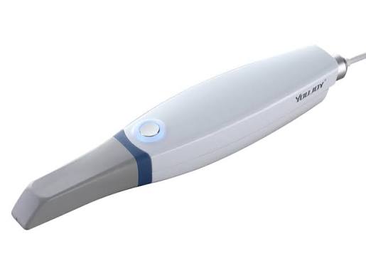
In modern aesthetic and prosthetic dentistry, traditional silicone/alginate impression materials—packed into metal trays, left to set for minutes, and often triggering severe nausea—have been completely replaced by “Optical Intraoral Scanners” (IOS). Referred to in medical literature as “Digital Optical Impressions,” this system creates a true-to-life, full-color, three-dimensional replica on a computer screen by scanning the teeth, gums, and bite (occlusion) with laser or structured-light sensors that capture thousands of frames per second. Redefining Ankara intraoral scanning standards with the vision of “Stereophotogrammetry” and “CAD/CAM Integration,” Panorama Ankara provides patients with a professional digital diagnostic and design rehabilitation experience featuring zero contact, zero gagging, and micron-level precision. While transforming the impression procedure into a comfortable video capture that takes seconds, these systems physically eliminate human-related casting and expansion errors at the laboratory stage—an unshakable achievement of clinical engineering.
The true scientific success behind intraoral scanning lies in the principle known in medical literature as “Polygonal Mesh Modeling,” which converts physical surfaces into virtual coordinates. In the advanced digital unit of Panorama Ankara, as the clinician guides the thin, ergonomic scanner tip over the teeth, confocal microscope sensors inside the device transform the tooth’s topography (its peaks and valleys) into a micron-level data cloud. These data simultaneously become a three-dimensional model on screen. The system records not only tooth shape, but also the true shade of enamel and the tissue structure of the gums (tissue characterization) in HD quality. This digital model is transmitted within seconds via the internet to the clinic’s production laboratory (CAM) milling units. This vision frees impression-taking in Ankara from sticky, suffocating materials and turns zirconia or porcelain production into a process that can be completed the very same day—an advanced discipline of “Digital Replication.”
Clinical Classification: Impression Systems and Biomechanical Precision Analysis
The laboratory success of a prosthetic treatment (crowns, implants, clear aligners) depends on the dimensional stability of the impression taken from the patient’s mouth (its ability to resist distortion). While conventional impressions can shrink during transport to the lab due to heat or time, digital data preserve the same coordinates indefinitely. To help patients clearly understand this advanced digital replication process, Panorama Ankara presents the technological evolution used in our clinic within the scientific framework below.
| Impression Protocol | Biomechanical Method and Patient Experience | Panorama Ankara Clinical Transition Advantage |
|---|---|---|
| Traditional Alginate / Silicone Impression | Pressing a paste-like chemical material into the mouth with metal trays and waiting for it to set. | Discontinued in our clinic; risks of tearing, saliva-related distortion, and gag reflex have been reduced to zero. |
| Optical Intraoral Scanning (Digital) | Capturing a video record by moving a pen-sized optical reader over the teeth. | Does not restrict breathing; the possibility of tearing or deformation is physically eliminated. |
| Virtual Articulation (Bite Registration) | Digitizing the “bite” (occlusion) moment when upper and lower jaws contact each other using the scanner. | Replicates jaw joint (TMJ) dynamics with millimetric precision, ending the “high bite” problem after delivery. |
| Orthodontic Clear Aligner Scan | Scanning the current crowding and instantly simulating the corrected post-treatment outcome. | Shows on screen within seconds how the patient’s smile will look after orthodontic (Invisalign) treatment. |
Eliminating the Gag Reflex and Achieving Marginal Fit
In traditional dentistry, the greatest phobia that makes patients avoid treatment is the intense “gag reflex” triggered the moment impression material touches the palate. This physiological reaction not only traumatizes the patient but can also cause muscular tension that distorts the impression. With intraoral scanning systems used at Panorama Ankara, there is no chemical material touching the palate or flowing toward the throat. The slim scanner tip completes the procedure within seconds without compressing tissues. The true medical power of this digital data lies in creating “marginal fit” (border adaptation). The microscopic boundary where porcelain crowns meet the gumline is copied so clearly by the scanner that the zirconia produced in the lab seats onto the tooth like a puzzle piece—with a “click” and with zero gap. This is the highest level of digital architecture, completely blocking bacterial leakage and decay beneath the gums.
Prosthetic Adaptation After Digital Impressions and the Maintenance Discipline
Digital impressions captured within seconds by intraoral scanners are processed with millimetric precision in the clinical studio, and production of your restoration (porcelain or an implant-supported structure) begins. Throughout this fast and comfortable technological process, while your new teeth are digitally milled in the laboratory, the Panorama Ankara digital dentistry board presents the following two core rules as a “treatment constitution” for patient adaptation:
- Immediately after scanning prepared teeth, “temporary crowns” produced in our clinic with 3D printers and placed on the teeth are used to prevent sensitivity. To avoid fracture or dislodgement until your definitive porcelain restorations are produced, you should not chew sticky (gum, caramel) or extremely hard foods on that side.
- To ensure that definitive restorations—integrated onto the tooth with perfect marginal adaptation via digital scanning—do not accumulate plaque at the gumline and maintain their biological lifespan, “water flosser” use must be added to the daily routine in addition to conventional brushing.
Discover the Future of Your Smile on a Digital Screen with Panorama Ankara
The feeling of being unable to breathe when impression trays enter your mouth, the sticky materials that must be endured for minutes, and the repeated impression try-ins afterward are not torments you must accept for an aesthetic smile. On the contrary, in expert hands, they become a professional process that can be transformed into a comfortable video lasting only a few minutes with light-speed optical scanners. In your search for Ankara intraoral scanning, Panorama Ankara combines “Polygonal Modeling” and “CAD/CAM Workflow” protocols to free you with scientific elegance from traditional molds, nausea, and casting procedures vulnerable to human error. We respect the biological topography of your teeth 100% and replicate nature’s flawless contours with the millimetric power of digital pixels to build your new smile.
To generate the three-dimensional map of your oral anatomy within seconds using our confocal sensors, simulate the most suitable “Digital Zirconia” or “Clear Aligner” pathway on screen before treatment even begins, and feel that perfect harmony each time you look in the mirror, you can schedule a detailed digital diagnostic consultation at Panorama Ankara and entrust your health to the safe hands of experience and modern technology.