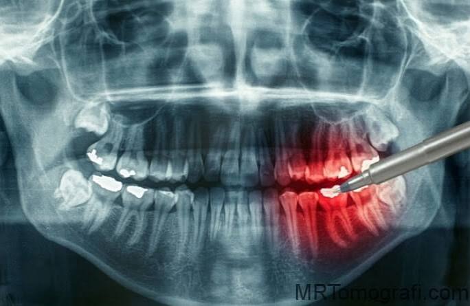
Ankara Dental X-Ray
In modern stomatology and oral surgery, the first step of a flawless treatment protocol is the ability to analyze—at micron-level precision—the
depths of the teeth and surrounding tissues that cannot be seen with the naked eye. A dental X-ray, referred to in medical literature as
“dental radiography”, is an advanced imaging science that maps pathological conditions within the oral cavity onto a digital
sensor by using the differing transmission rates of X-rays through tissues of varying density (enamel, dentin, bone, cysts). Redefining Ankara
dental X-ray standards through the vision of “Digital Radiovisiography (RVG)” and “Cephalometric Analysis”, Panorama Ankara
provides patients with a professional diagnostic infrastructure that eliminates the time loss and higher radiation risk of old-style processed films,
delivering HD-quality images within seconds. The systems used in our clinic represent an unshakable clinical navigation discipline, detecting
early-stage interproximal caries, silent infections hidden within bone, and root asymmetries before any pain appears.
The fundamental scientific success behind digital X-ray technology lies in the principle known in medical literature as
“phosphor plate and RVG sensor dynamics”: minimizing X-ray dose while maximizing image clarity. The digital sensors used in Panorama
Ankara’s advanced radiology unit transfer images instantly to a computer screen while using 80% to 90% less radiation compared to
conventional films. This technology enables the clinician to perform digital manipulation (colorization, magnification, density
measurement) on the image, illuminating microscopic cracks or anatomical variations inside root canals—details that may be missed on classic films—
within seconds. This vision elevates radiological diagnosis in Ankara from the simplicity of “taking a film” into an advanced
diagnostic engineering center that processes the patient’s anatomical dataset (Big Data).
Clinical Classification: Types of Dental Radiography and Diagnostic Focus
Depending on the nature of the issue inside the mouth, the type of X-ray used differs in beam angulation, focal distance, and sensor positioning.
Each radiographic technique highlights a different anatomical layer of the tooth. To help patients clearly understand this advanced imaging
hierarchy, Panorama Ankara presents the radiological methodology used in our clinic within the scientific framework below.
| Radiography Type (Technique) | Biomechanical Imaging Field | Panorama Ankara Clinical Application Goal |
|---|---|---|
| Periapical Radiography (RVG) | Small-format, high-resolution images that show all details of a single tooth (or two adjacent teeth) from the root tip to the crown. | To determine root-end lesions and the anatomical working length of root canals with millimetric precision during endodontic treatment. |
| Bitewing X-Ray | A dedicated technique that shows the opposing occlusion of posterior teeth and especially the contact (interproximal) surfaces between adjacent teeth. | To detect “hidden interproximal caries” concealed between teeth at the earliest stage—lesions that may not be noticed by the naked eye or standard radiographs. |
| Cephalometric Radiography | An orthodontic mapping method capturing the craniofacial skeleton and soft tissues from the lateral or frontal profile in a wide-angle view. | To compare jaw relationships and dental alignment with the facial profile and design orthodontic treatment (braces/aligners) with maximal accuracy. |
| Hand–Wrist X-Ray | A radiograph taken to determine bone age and skeletal maturity in children whose growth and development are ongoing. | To synchronize the timing of orthodontic intervention with the child’s growth spurt (peak period) using scientific data. |
Radiation Safety: Ionizing Dose Management and the ALARA Principle
“Radiation exposure,” the concern patients most often have in radiological imaging processes, is managed at Panorama Ankara with scientific discipline
and ethical obligation. In accordance with the ALARA principle (As Low As Reasonably Achievable), imaging is limited strictly
to the region required for diagnosis. Thanks to the speed of digital sensors, the dose received from a dental X-ray can be lower than the cosmic
radiation exposure during an intercontinental flight or a day spent walking under the sun. Lead aprons and thyroid-protective collars used in our
clinic physically block ionizing radiation from reaching non-target tissues. This level of precision represents the highest biosafety standard,
protecting patient safety at the cellular level without compromising diagnostic quality.
Data Integration and Maintenance Follow-Up After Radiographic Diagnosis
Having your X-ray images—obtained in HD resolution in our clinical radiology unit—processed within seconds in our digital archive and delivered
immediately to your clinician ensures your treatment becomes predictable. To maximize the impact of this digital data on your health,
the Panorama Ankara diagnosis and radiology board presents two essential rules as a diagnostic constitution:
- Because radiographic data reflects the static status of your teeth at that moment (caries, cysts, bone loss), a general screening should be performed
with a control panoramic X-ray at least once a year—even if you have no complaints—to monitor insidiously progressing pathologies
(such as peri-implant bone loss or recurrent caries beneath restorations). - In children being monitored for growth and development, or in individuals undergoing orthodontic treatment, follow-up
cephalometric or panoramic X-rays should be taken at intervals determined by the clinician (every 6 months or 1 year)
without delay, and digital comparison (superimposition) should be performed to observe changes in eruption paths and jaw structure.
Illuminate the Hidden World of Your Teeth with Panorama Ankara
Vague pains you feel in your teeth, the condition of the bone beneath your gums, or the pressure impacted wisdom teeth place on neighboring teeth are
not situations to be risked through guesswork. On the contrary, in expert hands, they are a professional diagnostic process that can be clarified
within seconds using low-dose digital sensors and HD imaging technologies. In your search for dental X-rays in Ankara, Panorama Ankara combines
“Digital Radiovisiography” and “Ionizing Safety” protocols to free you from outdated, ambiguous films, high-radiation
anxiety, and diagnostic errors with scientific elegance. We respect 100% the microscopic architecture of your jaw and teeth, initiating your treatment
journey with digital clarity by searching for health in every pixel.
To create the radiographic map of your oral anatomy within seconds using our digital sensors, determine the most suitable diagnostic route—whether
bitewing or periapical—without missing a single detail, and see everything in its clearest form on the screen before
treatment begins, you can schedule a detailed radiological consultation appointment at Panorama Ankara and entrust your health to the safe hands of
experience and modern radiology science.