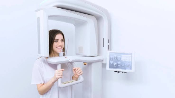
Ankara 3D Dental Tomography
Accurate diagnosis in modern dentistry is the unshakable foundation of successful treatment; however, traditional two-dimensional X-rays cannot always provide sufficient depth because they present the complex jaw anatomy as overlapping shadows. Dental 3D tomography, referred to in medical literature as ‘CBCT’ (Cone Beam Computed Tomography), is an advanced ‘Volumetric Imaging’ technology that sends X-rays in a conical beam to replicate the patient’s jaw skeleton, tooth roots, nerve canals, and sinus cavities in three-dimensional slices at millimetric precision. Redefining the standards of Ankara 3D dental tomography with a vision of ‘Voxel-Based Precision’ and ‘Advanced Surgical Planning’, Panorama Ankara provides patients with a professional diagnostic infrastructure that presents every detail—from the internal structure of a tooth to bone density—through a 360-degree perspective. The systems used in our clinic form a robust clinical navigation discipline that eliminates surprises in surgical procedures, verifies implant positioning at the micron level, and elevates the patient’s biological safety to the highest standard.
The true scientific success behind three-dimensional imaging technology lies in the ability known in medical literature as ‘Multiplanar Reconstruction’ (MPR), which allows the jawbone to be sliced into horizontal, vertical, and front-to-back sections (axial, sagittal, coronal). In the advanced radiology unit of Panorama Ankara, once the patient’s scan—lasting only a few seconds in the tomography device—is completed, the resulting digital data cloud is divided into microscopic three-dimensional pixels called ‘Voxels’. Thanks to the high resolution of these voxels, the true contact between an impacted wisdom tooth and the mandibular nerve can be precisely evaluated, and the bone where an implant will be placed can be analyzed not only for its height but also for its width and quality (bone density). This perspective transforms radiological practices in Ankara from a two-dimensional estimation process into an advanced discipline of ‘Virtual Surgery’, where every surgical step can be simulated digitally in advance.
Clinical Classification: 3D Volumetric Analysis and Diagnostic Superiority Parameters
The clinical value of a 3D dental tomography examination (CBCT) emerges particularly at the points of ‘Depth Perception’ and ‘Measurement Accuracy’, where traditional methods remain limited. In conventional X-rays, there may be up to a 30% risk of magnification or distortion, whereas the measurements obtained through 3D tomography correspond to real anatomical dimensions with a 1:1 ratio. To help patients clearly understand the diagnostic advantages of this advanced technology, Panorama Ankara presents the radiological evolution used within the clinic in a scientific framework through the following table.
| Diagnostic Feature and Parameter | Conventional Panoramic (2D) X-Ray | Panorama Ankara 3D Tomography (CBCT) |
|---|---|---|
| Image Dimension and Depth | Two-dimensional; tissues overlap and there is no depth information. | Three-dimensional; bone volume, width, and anatomical relationships are clearly visible. |
| Measurement Accuracy and Safety | High risk of magnification; may contain error margins in implant planning. | Provides true 1:1 measurements; minimizes surgical risks (such as nerve injury). |
| Root Canal Treatment and Cyst Analysis | Small cysts or fourth canals (MB2) may not always be visible. | Clearly maps microscopic lesions at the root apex and anatomical variations. |
| Implant and Sinus Planning | Shows sinus floor height but cannot analyze width. | Calculates bone volume with millimetric precision in sinus lifting (bone augmentation) procedures. |
Digital Navigation: Mandibular Canal Mapping and Surgical Safety
The most critical role of 3D dental tomography in implant surgery is the mapping of the sensitive area known in medical literature as the ‘Mandibular Canal’ (the main nerve canal of the lower jaw). Since contact between an implant and this nerve may lead to difficult complications, ‘Virtual Navigation’ is performed via tomography in all advanced surgical procedures at Panorama Ankara. In the digital environment, our physician positions the implant within the bone while measuring the safe distance to the nerve canal at millimetric accuracy and adjusts the implant’s angle according to the densest region of the bone. This ‘Guided Surgery’ approach shortens the duration of the procedure while opening the door to a stitch-free and painless recovery process (flapless technique). It represents the highest level of diagnostic and treatment protocol where technology and biological safety are harmonized.
Radiation Safety and Cellular Protection with the ALARA Principle
Although 3D dental tomography systems operate with significantly lower radiation doses (approximately 90% less) compared to medical body CT scans, radiation safety at Panorama Ankara is a strict clinical ethical standard. The process of balancing the immense diagnostic power of three-dimensional data with the patient’s overall health is regulated in our radiology unit according to the following two fundamental rules:
- According to the core principle of medical ethics, ‘ALARA’ (As Low As Reasonably Achievable), the scanning area (FOV – Field of View) is digitally restricted only to the region of suspicion, preventing surrounding tissues from receiving unnecessary radiation.
- To provide cellular-level protection, next-generation digital sensors and lead protective equipment are used during scanning, ensuring that the received dose remains close to the natural background radiation levels encountered in daily life. After the scan, all data is transferred to high-resolution digital archives as soon as the diagnosis is confirmed.
With Panorama Ankara, Entrust Your Treatment Not to Chance but to Millimetric Data
Silent infections in your teeth, the risky positions of impacted teeth, or the uncertainty about whether your bone structure is sufficient for implants should not be left to guesswork in the dark; instead, they are professional processes that can be illuminated within seconds through CBCT technology and three-dimensional analyses in expert hands. In your search for Ankara 3D dental tomography, Panorama Ankara combines ‘Volumetric Analysis’ and ‘Digital Navigation’ protocols to scientifically free you from incorrect diagnoses, unexpected surgical outcomes, and insufficient radiological data. We respect the hidden architecture of your jawbone and teeth with absolute precision, seeking perfection in every slice and from every angle to secure your health.
To map the volume of your jawbone and the pathways of your nerves through our three-dimensional tomography analyses, to determine the most suitable ‘Digital Surgery’ or ‘Advanced Endodontic’ treatment route without taking any risks, and to feel the comfort of confidence every time you look in the mirror, you can schedule a detailed radiological consultation with the Panorama Ankara clinic and entrust your health to the safe hands of experience and modern surgery.