Ankara CBCT Imaging
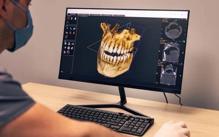
In modern dental implantology and maxillofacial surgery, the key to success is the ability to analyze bone tissue and anatomical relationships as realistically as possible before surgery, without any image distortion. The imaging technique referred to in medical literature as “CBCT” (Cone Beam Computed Tomography) is an advanced “Volumetric Imaging” discipline that scans the entire jaw volume in a single rotation by sending X-rays in a conical beam and combines this data into three-dimensional pixels called “voxels.” Redefining Ankara CBCT imaging standards with the vision of “Isotropic Resolution” and “Low-Dose Radiation Protocol,” Panorama Ankara provides patients with a professional diagnostic infrastructure that goes far beyond the limited two-dimensional information offered by panoramic X-rays, presenting the depth of bone, the inclination of the sinus floor, and the true path of nerve canals with a full 360-degree perspective. The systems implemented in our clinic represent a robust clinical navigation workflow that eliminates surgical risks in the digital environment before the patient even reaches the operating chair.
The true scientific success behind CBCT technology lies in the principle known in medical literature as “Isotropic Voxel Geometry,” which constructs images with equal resolution in every direction (X, Y, and Z axes). In the advanced radiology unit of Panorama Ankara, the “Raw Data” obtained during scanning is processed using advanced algorithms and converted into bone density maps. Metal reflections (artifacts) commonly seen in traditional tomography are eliminated with our next-generation CBCT software, maximizing clarity—especially in tissues surrounding implants. This allows our clinicians to analyze not only the height of the bone when virtually placing implants, but also the internal trabecular density of the bone structure. This vision elevates radiological diagnostic practices in Ankara far beyond the simplicity of “taking a film,” transforming them into an advanced “Bio-Engineering” center where the patient’s anatomy is examined within a digital laboratory environment.
Clinical Classification: CBCT Indications and Surgical Planning Parameters
The clinical value of a CBCT scan depends on the selected “Field of View (FOV)” size and the capability of the data to provide “Multiplanar Reconstruction.” From implant surgery to impacted tooth operations, each case requires a different volumetric analysis. To help patients clearly understand this advanced three-dimensional scanning process, Panorama Ankara presents the technological hierarchy used in the clinic within the following scientific framework.
| Clinical Field and Application | Biomechanical and Diagnostic Analysis Objective | Panorama Ankara Clinical Application Advantage |
|---|---|---|
| Advanced Implantology (Dental Implants) | Millimetric measurement of bone volume (height/width) and mapping of the mandibular nerve canal. | Preventing unexpected bone defects during surgery by positioning implants at the ideal angle through “Virtual Surgery.” |
| Oral and Maxillofacial Surgery (Impacted Teeth) | Determining the true three-dimensional relationship of wisdom teeth with neighboring tooth roots and sinus cavities. | Minimizing the risk of postoperative numbness (paresthesia) by detecting whether roots are in contact with nerves at the micron level. |
| Endodontics (Complex Root Canals) | High-resolution cross-sections to detect canal calcifications and additional canals (such as MB2). | Analyzing root fractures and the true size of periapical lesions at a cellular level—details invisible on traditional X-rays. |
| TMJ (Temporomandibular Joint) Analysis | Determining the position of the condyle within the joint socket and identifying erosions on joint surfaces. | Providing volumetric documentation of joint space and disc positioning in patients experiencing jaw joint pain and clicking. |
Virtual Surgical Navigation: Integration of CBCT Data with Guided Surgery
The most revolutionary outcome of CBCT imaging is the process known in medical literature as “Guided Surgery,” where the entire procedure is completed digitally before any surgical intervention begins. In the protocols implemented at Panorama Ankara, bone data from CBCT scans is combined with soft tissue data obtained from intraoral scanners (3D scanning). This “Digital Fusion” allows patient-specific “Surgical Guides” to be produced. With these guides, implants can be placed directly into the safest point within the bone without the need for incisions or sutures (flapless technique). This process transforms treatment from a surgical intervention into the highest level of “Precision Navigation,” where every step is digitally verified.
Radiation Safety and Data Archiving After CBCT Imaging
Your CBCT scans—completed within seconds in our clinical studios and providing HD-quality volumetric data—enhance treatment safety with the vast amount of information they provide, while radiation dose management is maintained at the lowest possible level according to the “ALARA” principle. The radiology board of Panorama Ankara presents the following two fundamental rules as a diagnostic constitution to ensure that your three-dimensional data becomes a protective shield for your health:
- Since the “Cone Beam” technology of our CBCT devices emits up to ten times less radiation than medical computed tomography, the Field of View (FOV) should be limited only to the target area, and the patient’s overall radiation exposure should be minimized through digital filtering methods.
- The three-dimensional data cloud obtained in DICOM format should be preserved in the patient’s digital archive for a lifetime and should be retrievable within seconds as “Reference Data” for future implant revisions or monitoring changes in jaw structure.
Experience the Clarity of Science and Technology with Panorama Ankara
Uncertainties about whether your bone structure is sufficient for implants, fears of nerve injury during impacted tooth surgery, or mysterious pain during repeated root canal treatments are not matters that should be guessed in the darkness of two-dimensional imaging. On the contrary, in expert hands, they are professional processes that can be clarified within seconds using CBCT technology and volumetric analysis protocols—transforming surgery into a work of precision. In your search for Ankara CBCT imaging, Panorama Ankara combines “Voxel Precision” and “Isotropic Resolution” protocols to free you from unpleasant surgical surprises, insufficient radiological data, and diagnostic errors with scientific elegance. We respect the unique three-dimensional architecture of your jaw skeleton with absolute care, reconstructing your health and safety in every cross-sectional image.
To map the true volume of your jawbone and the millimetric pathway of your nerve canals through our CBCT analyses, determine the most suitable “Navigation-Guided Surgery” or “Advanced Endodontic” treatment path without any risk, and experience the peace of mind that comes with treatment planned with zero margin for error, you can schedule a detailed CBCT and 3D radiology consultation appointment at Panorama Ankara clinic and entrust your health to the safe hands of experience and futuristic medicine.