Ankara Jaw X-Ray
In modern dental and maxillofacial surgery, the unshakable foundation of a successful treatment protocol lies in being able to clearly interpret the comprehensive anatomy of the lower and upper jawbones—going beyond the teeth themselves. A jaw X-ray, referred to in medical literature as “Maxillofacial Radiography,” is an advanced “Skeletal Imaging” discipline that presents changes in jawbone density, the symmetry of joint sockets, the fullness of the sinus cavities, and the skeletal position of impacted teeth on a single panoramic plane. Redefining Ankara jaw X-ray standards with the vision of “Extraoral Scanning Precision” and “Temporomandibular Joint (TMJ) Analysis,” Panorama Ankara provides patients with a wide-angle diagnostic infrastructure that covers the entire facial skeleton, not just tooth-based findings. The systems used in our clinic deliver a robust clinical navigation workflow that maps the jaw’s anatomy in 360 degrees via digital sensors and illuminates hidden pathologies within seconds.
The core scientific success behind jaw X-ray technology lies in the principle known in medical literature as the “Tomographic Focal Trough,” which enables the curved jaw structure to be transferred onto a flat digital screen without loss of clarity. In Panorama Ankara’s advanced radiology unit, while the device rotates around the patient’s head along a synchronized trajectory, the visual noise created by “superimposed” overlapping images is eliminated through digital filtering methods. This allows the pathway of the “Mandibular Canal,” which passes through the lower jawbone and carries sensation to the teeth, the mental foramina (the nerve exit points near the chin), and the functional mobility of the jaw joint to be monitored with millimetric precision. This vision elevates radiological diagnostic practices in Ankara far beyond a simple “film-taking” procedure, transforming them into an advanced “Maxillofacial Engineering” center where the patient’s facial skeleton is examined within a digital laboratory environment.
Clinical Classification: Jaw X-Ray Techniques and Advanced Diagnostic Fields
The clinical value of a jaw X-ray lies not only in showing the condition of existing teeth, but also in offering a broad capability for “Differential Diagnosis”—from jaw joint pain to toothaches originating from sinus conditions. Each anatomical region requires a different focusing approach and beam angulation. To help patients clearly understand this advanced imaging process, Panorama Ankara presents the technological hierarchy used in the clinic within the following scientific framework.
| Radiographic Focus and Technique | Biomechanical and Diagnostic Analysis Objective | Panorama Ankara Clinical Application Advantage |
|---|---|---|
| Temporomandibular Joint (TMJ) Radiograph | Analyzes the movement capacity of the condyle (the head of the jaw joint) in open- and closed-mouth positions, and evaluates joint wear. | Determining the condition of the joint disc and bony surfaces in patients with jaw locking or clicking (“crackling”) complaints. |
| Maxillary Sinus Imaging | Visualizes the volume of the air spaces (sinuses) within the upper jawbone and mucosal thickening. | Identifying the need for a “Sinus Lift” before implants and diagnosing tooth-origin sinus infections (sinusitis). |
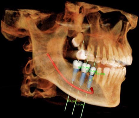
| Mandibular Canal Mapping | Determines the vertical and horizontal position of the main nerve canal running through the lower jawbone. | Creating a “Safety Navigation” pathway that reduces the risk of nerve injury to near zero during wisdom tooth surgery and implant placement. |
| Cephalometric Lateral Analysis | A wide-angle side-profile radiograph of the craniofacial skeleton showing soft tissue and bone relationships. | Determining the position of the jaws relative to each other and to the cranial base in orthognathic surgery and advanced orthodontic planning. |
Extraoral Scanning: Patient Comfort and Wide-Angle Data Acquisition
The greatest advantage of a jaw X-ray is that it is an “Extraoral” (outside-the-mouth) system; this feature provides a comfortable diagnostic process especially for patients with a strong gag reflex or limited jaw opening. In the protocols implemented at Panorama Ankara, digital sensors scan the entire jaw arch within seconds without touching the patient’s face. This system reveals not only teeth-related issues, but also silently enlarging cysts within the jawbone, pathological fracture risks associated with bone loss, and developmental asymmetries. This process expands diagnosis from a narrow scope into the highest level of “Holistic Radiology” practice that encompasses the entire facial anatomy.
Post-X-Ray Diagnostic Monitoring and Radiation Safety
To ensure that your jaw X-ray images—completed within seconds in our clinical studios and processed in high resolution—support the long-term success of your treatment and protect your health, systematic follow-up is essential. Panorama Ankara’s radiology board presents the following two fundamental rules as a diagnostic constitution to preserve the unshakable clarity offered by advanced imaging technologies:
- To detect early mineral loss in the jawbone and micro-changes in joint structure (such as osteophyte formation), a “General Jaw Screening” should be performed at least once a year even without pain or complaints, and a “Comparative Analysis” (Superimposition) should be conducted using prior data stored in the digital archive.
- In areas that have undergone implants, prosthetics, or surgical interventions, focused follow-up radiographs should not be neglected at the intervals determined by the clinician to verify the continuity of “Biological Compatibility” between bone tissue and materials, and radiation dose should be kept to a minimum according to the ALARA principle.
Clarify the Future of Your Jaw Health with Panorama Ankara
Sounds coming from your jaw joint, unexplained facial pain, or pressure from impacted teeth on the jawbone are not matters that should be left uncertain or risked. On the contrary, in expert hands, they are professional processes that can be clarified within seconds using digital jaw X-ray technologies and holistic diagnostic protocols—transforming surgery into a work of precision. In your search for an Ankara jaw X-ray, Panorama Ankara combines “Extraoral Scanning” and “TMJ Diagnostics” protocols to free you from undiagnosable cramps, insufficient radiological data, and surgical surprises with scientific elegance. We respect the unique architecture of your jaw skeleton with absolute care, rebuilding your health from every angle and every image.
To map the true volume of your jawbone and the pathway of your joint mechanics within seconds using our digital sensors, define the most suitable “Maxillofacial Diagnostic” route without missing a single detail, and experience the peace of mind that comes from treatment that “sees the full picture,” you can schedule a detailed jaw and joint examination appointment at Panorama Ankara clinic and entrust your health to the safe hands of experience and modern radiology science.