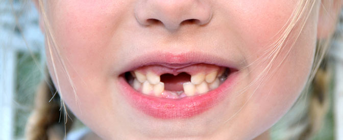
What is Amputation Treatment?
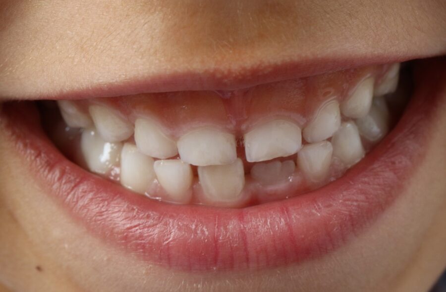
Amputation treatment (pulpotomy) is a micro-invasive endodontic treatment discipline aimed at preserving the vitality of the tooth, which is called “regional or partial pulpa removal” in clinical dental literature. In the anatomical internal structure of the tooth, there is a vascular and nerve network called pulpa that nourishes the tooth. While this network forms a wide chamber in the crown portion (coronal), it extends in the form of narrow canals in the root portions (radicular). Amputation engineering is based on the principle of cutting only the inflamed upper chamber tissue in the early stages when the infection has not surrounded all the tooth nerves, and keeping the underlying root nerves alive so that the tooth continues to be nourished. In this way, the tooth remains in the mouth without losing its bio-integrity.
- Partial Pulpectomy: Removing only the infected upper chamber without touching the living root bridges of the tooth.
- Bio-Active Barrier Construction: Deploying smart medical cements that will weave fresh bone bridges (reparative dentin) on the cutting line.
- Vital Preservation Philosophy: Mechanically preventing the tooth from evolving into a brittle, lifeless mass by maintaining its blood circulation uninterrupted.
In Which Situations is Amputation Treatment Applied to Teeth?
Intraoral partial nerve cutting surgeries are applied at moments of clinical certification when bacterial caries or mechanical trauma pierce the roof boundary of the pulpa tissue, but the inflammation has not yet leaked into the root ends and jawbone. The fact that root canals are anatomically highly tortuous, thin, and insidiously porous in primary teeth makes traditional complete root canal treatments technically very risky. The physician rationally decides on this preventive approach in order to prevent the infection from damaging the permanent tooth bud beneath the tooth roots and to keep the tooth in the mouth in the safest corridor until the exfoliation age.
The primary clinical situations where amputation treatment is strictly preferred in teeth are as follows:
- In cases where the pulpa chamber is mechanically or bacterially exposed by exceeding the enamel and dentin boundaries while cleaning deep caries,
- When fracture lines that expose the nerve on the anterior tooth crown occur as a result of sudden trauma such as falls or impacts experienced by the child,
- In cases where no bone resorption, abscess shadow, or leaking inflammatory sac is radiologically detected at the root ends,
- When it is mandatory for the tooth pulp to remain alive so that the root end can close in young permanent teeth that have not yet completed their root development.
Who is Amputation Treatment Suitable For?
This advanced technology restorative pedodontics process is medically suitable for pediatric patients with widespread caries, youngsters who cannot tolerate long-session treatments due to needle and chair anxiety, and adult individuals with open root-end young permanent teeth. Since the system relies on the self-repair potential of the living intraoral tissues (cellular homeostasis capacity), it is bound by the rule that the vitality reflexes given by the tooth to stimuli must be intact. If there are persistent throbbing pains that start spontaneously at night in the tooth or if an abscess discharge (fistula) is certified at the root end, it means the nerve is completely dead, and these patients are not suitable candidates for the amputation process.
- Children with Compliance Issues: Little ones who cannot stay in the chair for long hours and need quick and practical solutions.
- Youngsters Carrying Young Permanent Teeth: Individuals in the 7-12 age range whose newly erupted permanent molars decay rapidly and whose roots are still in the developmental stage.
- Individuals Who Have Suffered Anterior Tooth Trauma: Sensitive patients whose tooth nerve was exposed naked to the outside world as a result of an accident but did not catch an infection.
Why is Amputation Treatment Preferred in Children?
The primary choice of the pulpotomy method with full sealing quality in pediatric dentistry practices stems both from radically optimizing session durations and from preventing dental phobia breakthroughs that will be permanent in the child for a lifetime. In the lower layers of primary teeth, there are living buds of permanent teeth that will erupt into the mouth in the future. If an inflamed primary tooth is not treated and is extracted, the jawbone narrows; if a complete root canal treatment is attempted, the underlying bud may suffer chemical damage due to root resorptions. Amputation acts as a biological bridge by absorbing all these risks in a single session.
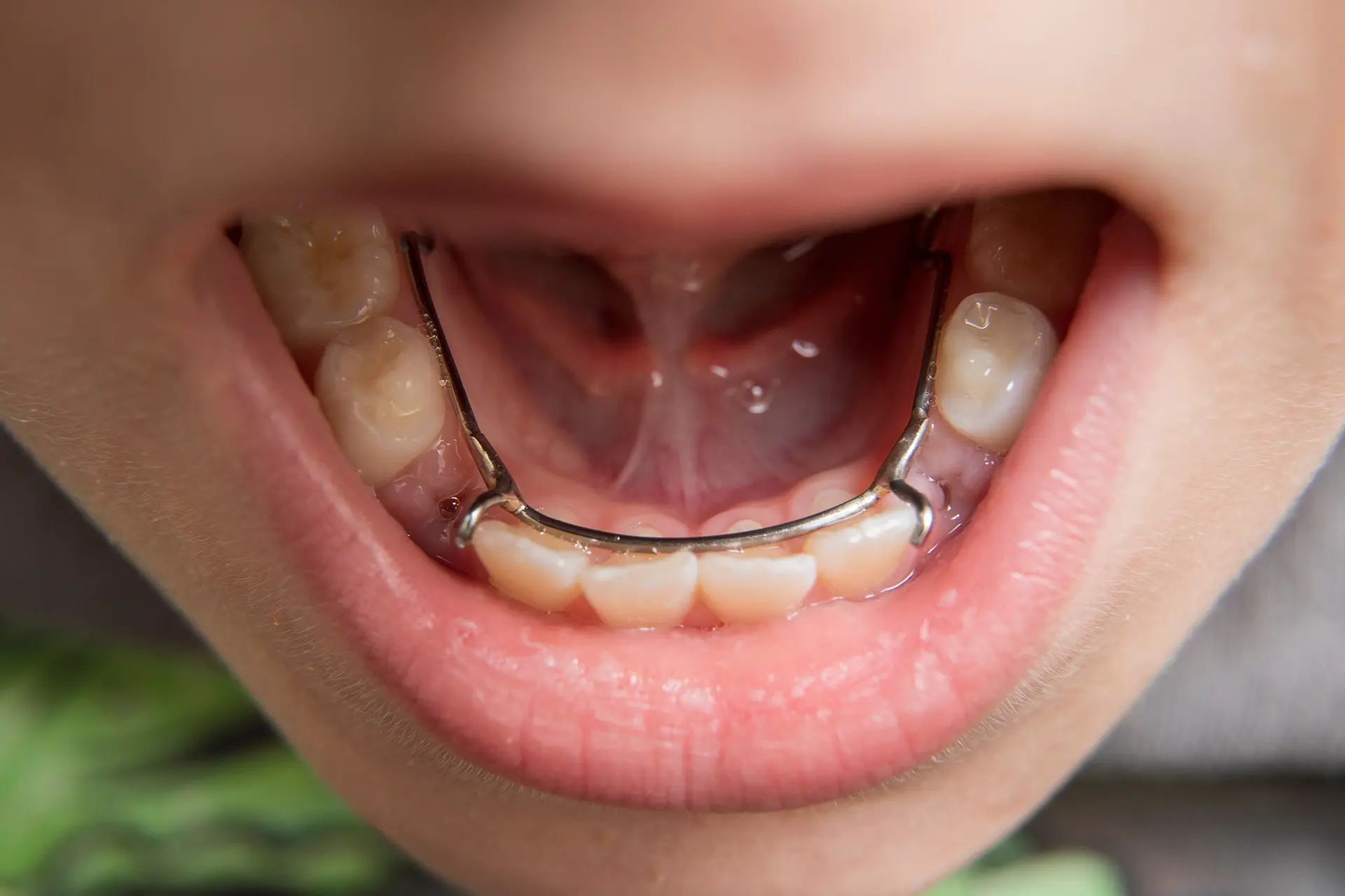
- Preservation of Tooth Lifespan: Protecting the space maintainer mission of the primary tooth in the jaw, thereby preserving the orthodontic alignment of the permanent tooth alignment lines.
- Success in a Single Session: The luxury of breaking the entire infection chain in a single chair sitting of approximately 20 minutes, without cargo or laboratory cycles.
- Jaw Bone Development Support: Contributing to the volumetric growth rhythm of the jaw skeleton thanks to the preservation of the living periodontal fibers of the tooth.
How is Amputation Treatment Applied to Primary Teeth?
The primary tooth amputation operation performed in pedodontics clinical rooms is a multi-step adhesive and endodontic procedure executed with micron precision under a full sterilization chain, accompanied by the comfort of digital anesthesia. First, the carious tissue is completely cleaned, and the roof of the pulpa chamber is entirely removed with diamond burs. The exposed coronal nerves are cut and removed using sterile excavators. After the bleeding that occurs at the entry points of the root canals is controlled with the help of sterile cotton pellets and medications, the base of the cavity is coated with bio-compatible sealing cements. The process is concluded with an aesthetic filling or stainless steel crown assembly that will protect the upper crown integrity of the tooth.
- Coronal Amputation: Cutting the inflamed nerve mass in the upper chamber with high-speed water-cooled handpieces that will not damage the root entries.
- Hemostasis (Bleeding Control): The phase of monitoring the leakage bleeding to stop spontaneously within 2-3 minutes to certify the vitality of the root nerves.
- Sealing of Root Entries: Closing the root canals seamlessly with medical materials containing MTA or Formocresol.
What are the Differences Between Amputation Treatment and Root Canal Treatment?
The most fundamental difference between amputation treatment and complete root canal treatment (pulpectomy) lies in the removal depth of the nerve tissue, whether mechanical intervention is made inside the root canals or not, and the biological completion timeline architecture of the procedures. In amputation, the nerves inside the roots are strictly not touched, files or reamers are not inserted into the canals; only the damage in the upper chamber is cleaned. In root canal treatment, however, the entire nerve network inside the tooth is scraped and removed down to the root end and filled with artificial fillings. In the comparison table below, the clinical parameters of the two superior endodontic treatment classes are listed:
| Clinical, Histological, and Technical Comparison Criterion | Amputation Treatment (Partial Pulpotomy) | Root Canal Treatment (Total Pulpectomy) |
|---|---|---|
| Removal Depth Area of Nerve Tissue | Only the nerves in the crown (coronal) chamber are cut; the root nerve remains alive. | The entire nerve and vascular network inside the tooth is completely removed down to the root end. |
| Mechanical Intervention Inside Root Canals | Strictly not applied; mechanical files or reamers do not enter into the canals. | Mandatory; canals are shaped and widened by scraping with diamond or nickel-titanium rotary files. |
| Vitality Status of the Tooth After Treatment | The tooth preserves its vitality; blood flow and nutrition continue linearly from the roots. | The tooth passes into the devital (lifeless) phase; its brittleness increases over time as its nutrition stops. |
| Cavitation and Session Completion Time | It is lightning fast; it finishes in a single chair sitting of an average of 15-20 minutes. | It is longer; it may require consecutive sessions of 45-60 minutes depending on the status of the infection. |
| Most Frequently Preferred Tooth Group / Class | Primary molars and young permanent molars whose root ends have not yet closed. | Highly destroyed permanent teeth whose roots are fully matured and where night pain has started. |
How is the Tooth Evaluation Performed Before Amputation Treatment?
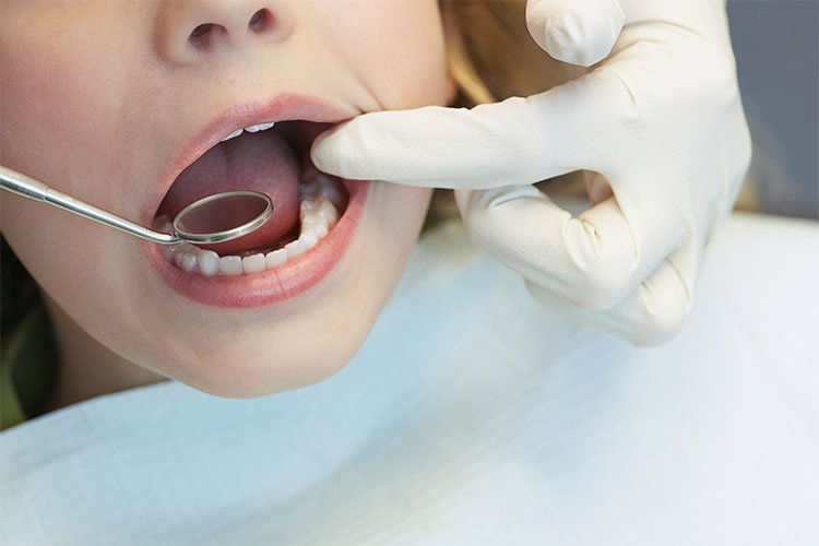
The clinical evaluation performed before proceeding to the restoration and surgical cutting stages is the most vital pre-operative analysis phase that maps out the boundary of infection inside the tooth on a millimeter basis and certifies the innocence of the root canals. During the examination, the physician inspects the integrity of the gingival margins and whether there is an abscess pimple (fistula opening) on the mucosa adjacent to the tooth with the naked eye. The greatest assurance at this stage is the layer-by-layer scanning of high-resolution digital periapical or bite-wing radiology data, which clearly scans insidious bone destructions in invisible root bifurcation areas.
- Physical Mobility Scan: Determining the scale of periodontal ligament destruction by measuring the mobility degree of the tooth within the jawbone.
- Percussion and Palpation Reflexes: Analysis of the pain perception awakened when the tooth is tapped with a micro mirror, in terms of whether the inflammation has leaked into the bone.
- Radiological Root Resorption Limit: Certification of the physiological resorption levels of primary tooth roots; if more than half of the roots are resorbed, treatment is contraindicated.
How is the Amputation Treatment Process Planned?
The planning of the regional nerve cutting workflow is carried out through a gradual clinical algorithm based on the depth index scores of the caries, the chair anxiety threshold of the patient, and the vertical distance of the permanent tooth bud to the mucosal boundary. While planning the process, hasty decisions are not made; all steps are shaped with full loyalty to the vitality stability of the root nerves. In planning, thanks to the ability to log a large number of fillings and amputations into the common schedule within the same session, the patient’s total number of days in the clinic is radically optimized.
- Stage 1 (Clinical and Digital Diagnosis): Certification of the relationship of the caries with the pulpa chamber through intraoral visual scanning and periapical film analyses.
- Stage 2 (Infection Clearing): The period of completely removing the damaged tissues in the upper chamber with caries cleaning moves.
- Stage 3 (Bio-Sealing): The phase of spreading and hardening smart mineral fillings at the entry points of the root canals.
- Stage 4 (Permanent Crown Construction): Placing the upper structure of the tooth under permanent protection by weaving it with seamless composites or prefabricated crowns.
Which Methods are Used in the Amputation Procedure?
In combined pedodontic restoration practices, different pharmaceutical methods and bio-material catalysts certified in medical literature are deployed to protect the cut root nerve surfaces from infection and keep them alive. The primary function of these methods is to trigger the underlying cells to produce living mineral tissue while forming a controlled sterilization layer on the wound surface. Which method to choose is calibrated by the surgeon according to the bleeding color of the tissue and the enamel biotype of the patient.
- MTA (Mineral Trioxide Aggregate) Protocol: The premium bio-ceramic method that has the highest sealing quality, shows molecular harmony with the jawbone, and weaves fresh bone bridges.
- Calcium Hydroxide Application: The conventional system that kills bacteria within seconds thanks to its high alkaline pH degree (approximately 12.5) and stimulates fresh dentin construction.
- Formocresol Technique: The mummification technique phase used in pedodontics for many years, which deactivates cyst cells at the cyst and root canal margin boundaries by fixing them.
- Laser-Assisted Desensitization: Scanning the root ends with low-dose laser beams to seal them bloodlessly and accelerating the cell stimulation speed.
How Does Amputation Application Contribute to Dental Aesthetics?
The greatest support of amputation and adhesive filling processes carried out in primary and young permanent tooth groups on facial and smile aesthetics is that it preserves facial bone and lip frame symmetry by preventing early tooth losses (edentulous spaces) that develop in the anterior region. If an inflamed anterior primary tooth is not saved and is extracted early, the child will display asymmetric dark spaces while talking or smiling. The treatment prevents the child from experiencing smile anxiety among peers by keeping the tooth’s own original crown morphology in the mouth; it instantly brings a pure illumination, smooth joy, and high self-confidence relief to the looks.
“Amputating treatment, instead of extracting your teeth from the root, is to offer a smooth and aesthetic right to life to the tooth in its own biological nest by clearing the internal cloud of inflammation.”
The Relationship Between Repairing Broken or Worn Teeth with Bonding
Especially in childhood, bonding (composite lamina) and amputation technologies are deployed with a simultaneous synergy in cases where deep fracture lines occur on the anterior incisor margins and the pulpa chamber is exposed as a result of accidents, falls, or school sports traumas. The surgical team first seals the impacted open nerve endings under a microscope with bio-active materials; immediately after, they rebuild the broken crown body layer by layer on the tooth with the help of nanohybrid ceramic pastes. Since that broken transition line where the filling merges with the tooth is erased with special polishing, it can definitely not be understood from the outside that the tooth suffered a trauma.
- Pulpal Protection Shield: Resetting post-operative sensitivity shocks by closing the exposed micro nerve channels.
- Knife-Edge Boundary Integration: Full adaptation to masticatory pressures thanks to the flexibility coefficient of the resin being equivalent to natural dentin tissue.
12. How is Bonding Application Used in Smile Design?
In smile design architecture, bonding and micro-restorations are the most visionary and instantaneous cosmetic instruments that smoothen the micro form defects and local color flaws of the teeth by applying zero trauma to the tooth. While making the design on the computer screen, the physician calibrates the bonding finish boundaries according to the patient’s lip movement arc, nose tip distance, and lower facial oval. This method ensures that a personalized authentic and pure smile expression is certified intraorally on the same day, without the need for expensive and long-term prosthetic crown sessions.
13. How Long Does the Bonding (Composite Lamina) Procedure Take?
Bonding (composite lamina) treatment is at the forefront of the fastest and most practical processes that adapt perfectly to the intense time limitations of modern metropolitan life, taking almost none of the patient’s time in the medical aesthetic schedule. The stages of cleaning the caries of a tooth, shaping it by curing it layer by layer, and polishing it with polishing discs are usually completed with full comfort in just 20 to 30 minutes per tooth, depending on the scale of the case; the process is highly dynamic as laboratory cargo waiting times are eliminated.
14. What Should Be Considered After Bonding?
There are some practical rules for the patient to follow in the home environment after the procedure in order to make the aesthetic success of the restoration session permanent, to support the rapid repair of the stimulated periodontal fiber networks, and to protect the final hardening balance of the injected polymer bonds. Even if composite resins are polished non-porously, they may pass into a temporarily more sensitive phase against external stimuli in the first hours; it is essential for the patient to act meticulously in terms of preserving the balance of homeostasis.
- No food should be eaten for the first 2 hours until the effect of local anesthesia (numbness) completely disappears, in order to prevent tongue and lip biting injuries.
- In order to preserve the glass smoothness of the filling surface and prevent micro stain absorption in the first days, staining substances such as tea, coffee, cigarettes, and wine should be avoided for the first 24 hours.
- Hard-shelled nuts such as pistachios or hazelnuts must strictly not be cracked with the anterior teeth, and packaging lids should not be forced to open with teeth (shearing stresses must be eliminated).
15. What are the Factors Affecting the Service Life of Bonding Application?
The ability of composite lamina restorations to remain in the mouth for long years without breaking, falling off, or causing marginal leakage is directly dependent on the technical workmanship quality at the moment of restoration working in full coordination with the functional home hygiene diligence of the patient. New generation nanohybrid resins structurally show high resistance to wear; however, the unique factor determining the long-term success of the prosthesis is that not even a microscopic level of saliva moisture was leaked into the cavity during the bonding moment.
16. What are the Differences Between Bonding and Porcelain Lamina?
The most radical difference between composite bonding applications and E-Max porcelain laminas (porcelain veneers) is the distribution of the production techniques, tissue reduction volumes, material lifespans, and budget parameters of the restorations. Porcelain lamina is a high-temperature glass ceramic produced by milling in the laboratory and its structural resistance is maximum; therefore, it does not stain and its lifespan is longer. Bonding, on the other hand, is a resin-based micro design woven manually directly by the physician in the clinic; while offering the flexibility of finishing in a single session, it is more economical compared to porcelain.
17. What are the Advantages of Bonding Application?
The certified advantages of nanotechnological composite veneer systems compared to traditional porcelain crowns not only create an instantaneous visual whiteness but also place the biological integrity of the tooth tissues under protection at the highest level. The tooth circumferential cutting harrassments in traditional restorations are completely eliminated in these smart microscopic materials. The chemical locking power offers the patient a lifetime permanent masticatory comfort and tissue hygiene success.
18. How Should Bonding Maintenance Be Done?
Since composite restorations are artificial nanohybrid ceramic structures, they cannot biologically show a decay reaction; however, that micron-level lower boundary line where the filling ends and merges with your own natural tooth enamel is the most sensitive corridor inside the mouth for bacterial plaque accumulation. If home hygiene is neglected at these marginal boundaries, plaque deposits may leak under the filling over time and initiate insidious secondary caries; a disciplined cleaning schedule should be implemented in the home environment to reset these risks.
19. What are the Frequently Asked Questions About the Initial Consultation and Examination?
The most common practical anxieties arising in the minds of clients who want to certify their oral health status or apply for a clinical examination for the first time and their polyclinic medical counterparts are presented below:
At the initial consultation and examination appointment, will my teeth be cut immediately at the exact second I sit in the chair?
No, absolutely not. The initial consultation and examination session is entirely a “diagnosis, mapping, and informing” sitting. Without obtaining your written and verbal consent, and without reporting the pathologies in your mouth and treatment alternatives to you in a clear language via digital screens, no scalpel, drill, or cutting move is strictly applied to your teeth; you leave the chair comfortably only having learned about your oral health.
Will the radiation dose of the digital panoramic X-ray to be taken during the examination harm my general body health?
No, this is an anxiety that is strictly not experienced in these new generation green technology digital radiology units. The radiation coefficient emitted by low-dose digital panoramic X-ray devices used in modern clinics is at a micron level, much less than the natural background radiation dose a human is exposed to from the sun in normal daily life or during a single intercontinental flight; furthermore, safety is kept at a maximum by putting on lead apron shields.
I cannot even sit in the examination chair due to my heavy fear of needles and drills (dentophobia) that I experienced in the past, what can I do?
This situation is a process managed with the highest sensitivity and empathy in our modern oral surgery department. If your chair anxiety is at a very advanced note, our initial consultation meeting is conducted not in the clinical room, but entirely in a sterile hospital office environment with the comfort of a coffee chat; at this stage, it is rationally decided whether the examination and all subsequent filling and extraction steps will be managed with the comfort of sedation (semi-sleep) or general anesthesia (narcosis) under the supervision of our anesthesiologist while you are asleep.
Can porcelain lamina or zirconium crowns be applied in the following years to a tooth that has previously undergone bonding?
Yes, this situation is one of the greatest biological flexibility advantages that bonding application offers to the patient. Since permanent cutting and harrassment are not applied to the tooth enamel during the bonding process, porcelain lamina or zirconium crown processes can be safely constructed from scratch in the following years.
Can I return to my normal daily and business life immediately at the exact second my initial examination and consultation appointment is completed?
Yes, initial consultation and examination sessions are completely non-invasive visual and digital scanning steps that do not involve injection syringes, surgical scalpel wounds, sutures, or numbness that lasts for hours on the face; therefore, you can immediately return to your normal daily, professional, social, or academic life rhythm from where it left off, uninterrupted and with high consciousness alertness, by getting up from the chair the exact second your examination is over; there is no obstacle to driving a car.
20. Who Are We? & Panorama Ankara Oral and Dental Health
Panorama Ankara is a licensed oral and dental health center that offers corporate health assurance to its patients by combining an academic expert staff and the latest global bio-technological digital infrastructure under a single roof in all specialty branches of dentistry, primarily oral, dental and maxillofacial surgery, advanced implantology, microscopic endodontics, periodontology, pediatric dentistry (pedodontics), and aesthetic dentistry. Our center, which eliminates traditional rule-of-thumb estimations and human error risks with technology in treatment practices, operates 3D Digital Diagnosis Modeling and Initial Consultation – Examination ecosystems without compromise with schedule discipline from the very beginning to the end of the clinical operation workflow.
In all diagnosis, radiological screening, design, and microscopic restoration interventions performed under the roof of Panorama Ankara; absolute sterilization, high patient safety, transparency, and honesty principles are operated in full compliance with medical regulations by virtue of the charter. In our clinical practices, while analyzing the vertical boundaries of the jawbone and lesion depths with millimetric sections via 3D tomographies, proprietary intraoral scanner cameras that transfer the intraoral twin to the computer in seconds without causing gag stress to the patient and spectrum-beam digital color analyzers are applied with millimetric precision under the supervision of our expert physician staff. Our aim is not to impose old-fashioned, slow, tooth-over-cutting analog examination molds that harrass teeth unnecessarily for crowns because it cannot relieve the ache; but to analyze the jaw skeleton quality, occlusion mechanics, and facial aesthetic golden ratio boundaries of each individual on the virtual screen in the preliminary examination in 3D, and to obtain smooth oral functions that you will use with maximum session comfort, same-day dentistry standards, and lifelong unshakable confidence without damaging the surrounding soft tissues and sensitive vascular lines. If you postpone your check-ups due to dental chair fear, injector phobia, or time constraints despite being tired of your missing teeth, old worn fillings, or crowding when you look in the mirror, and you want to start your smart digital treatment process from start to finish with scientific assurance in an expert corporate center environment; you can safely protect your health and smile integrity by contacting the expert staff of Panorama Ankara for your detailed 3D intraoral scan and digital planning analyses.