What Is Primary or Permanent Tooth Discoloration?
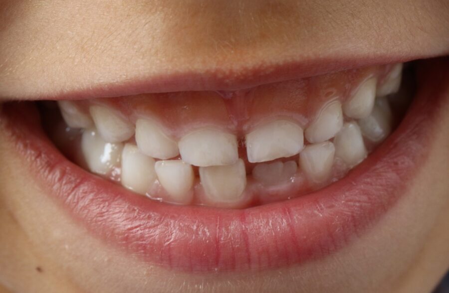
Tooth discoloration (dental discoloration) is the pathological color change of the crystallized enamel, which is the outermost protective surface of primary and permanent teeth, and the underlying dentin structure, causing them to lose their natural pale white or ivory tones. In a healthy tooth structure, light filters through the semi-permeable enamel prisms, refracts off the dentin, and reflects back, giving the tooth a natural brilliance. Dental discoloration is not merely an aesthetic defect; it is a morphological manifestation on hard tissues of a disruption in the mineralization matrix, microbial plaque accumulation, a past trauma, or a systemic metabolic stimulus.
- Textural Deformation: The chemical locking of foreign pigments (chromogens) into the porous structure of enamel prisms.
- Developmental Signal Character: The inscription of systemic disorders during pregnancy or early childhood onto the tooth matrix as color lines.
- Biological Tissue Reaction: Hemoglobin degradation products in the pulp (nerve) chamber that has lost its vitality staining the tooth from the inside.
Why Does Tooth Discoloration Occur?
Color changes occurring in intraoral hard tissues represent a multifactorial pathology formed as a result of the direct interaction between external environmental factors and the internal biochemical dynamics of the organism. Primary initiating causes include chromogenic substances in food, insufficient oral hygiene, and alkaline fluctuations in salivary pH levels. The uncleaned, transparent bacterial plaque accumulating on the tooth surface absorbs iron and calcium salts from saliva over time, hardening and turning into dark-colored tartar towers. At the cellular level, excessive fluoride exposure or heavy medical drug compounds experienced during tooth development permanently disrupt the weaving geometry of enamel prisms, producing insidious pores and stains.
- Chromogenic Bacteria: The colonization of sulfur-producing microorganisms that breed in the oral flora and secrete black/orange stains,
- Food and Beverage Pigments: The infiltration of heavily consumed black tea, espresso, filter coffee, tomato paste, and nicotine tar into the enamel pores,
- Systemic Drug Exposure: Tetracycline group antibiotics used during pregnancy or early childhood binding to calcium and staining the tooth,
- Metabolic Fluctuations: Childhood high-fever illnesses, neonatal jaundice (bilirubin accumulation), and erythroblastosis fetalis.
In Which Situations Are Tooth Whitening and Discoloration Treatments Preferred?
The inclusion of stain removal protocols in the clinical workflow is indicated based on the depth and chronicity of the staining, the patient’s biological age lines, and the extent of tissue loss in the hard tissue. If there is no deformity in the anatomical shape, width, or length of the tooth, carving and cutting the tooth for a zirconium crown merely to lighten its color is a medical waste of tissue. The physician rationally decides on this protective technological power to brighten the smile within the framework of holistic tissue preservation rules, without disrupting the natural vitality (vitality) of the tooth.
The primary clinical situations where tooth discoloration treatments are definitively preferred are:
- When unaesthetic black rings (black stains) around the tooth necks become chronic in children due to the secretions of chromogenic bacteria,
- In cases where tissue remnants in the internal chambers of root-canal-treated teeth turn the tooth purple and gray from the inside (requiring devital bleaching),
- In cases where the tooth enamel harbors chalky matte white spots due to fluorosis or hypoplasia, sabotaging facial symmetry,
- When the thinning of the enamel layer due to aging causes the underlying yellow dentin layer to completely dull the smile line.
In Which Situations Is Discoloration Observed in Primary Teeth?
Color deviations occurring in the primary dentition represent a clinical picture frequently observed in early childhood, as they harbor a more delicate and porous organic matrix compared to permanent teeth. The most common finding is the dark brown or black sulfur stains formed by “iron-containing syrups” used in the treatment of upper respiratory tract infections during infancy adhering to the enamel surface. In addition, early childhood caries (nursing bottle caries) initiate insidious yellowish-brown color destruction on the interproximal surfaces of the teeth, dragging down the recovery resistance of primary teeth.
- Demergent Drug Precipitations: Iron-supplemented suspension liquids reacting with saliva and staining the enamel wall,
- Nursing Bottle Caries Onset: Acidic matte yellowing forming on the anterior incisors due to the consumption of milk with honey/molasses during night sleep,
- Soft Tissue Trauma: The scenario where capillary vessels inside the primary tooth burst as a result of falling or impact, staining the tooth gray from the inside.
Why Does Discoloration Occur in Permanent Teeth?
Discoloration processes in the permanent tooth series show a direct correlation with thecumulative accumulation of environmental irritants the teeth are exposed to in the mouth for decades and the physiology of aging. When brushing discipline is neglected over the years, chromogenic microscopic coloring agents in food infiltrate down to the deepest layers of enamel prisms through diffusion. Furthermore, as age advances, the outermost white enamel layer of the tooth mechanically wears down and thins; since the underlying dark yellow dentin layer reflects outward more, the teeth naturally take on a darker, matte, and lifeless tone.
- Tobacco and Caffeine Accord: Nicotine tar (tar molecules) embedding permanently into enamel pores and weaving brown lines,
- Pulp Calcification Infiltration: The living chamber of the tooth narrowing by producing new dentin from the inside as it ages, reflecting a dark yellow color outward,
- Leaking Restorations: The phase where silver ions from old-type amalgam fillings leak into dentin tubules, staining the tooth gray-black.
For Whom Should Tooth Discoloration Be Evaluated?
Hard tissue color analysis protocols are medically suitable for all pediatric and adult patients who are aesthetically unhappy with the color of their teeth when looking in the mirror, hide dimness or localized stains in their smile line, and therefore restrict their facial expressions in social environments. Early diagnoses made under pedodontist supervision in children are critical for protecting the health of permanent tooth buds. In adults, adhesive or chemical color lightening courses with the highest safety profile are planned for each individual by registering systemic drug histories and enamel thickness parameters.
- School-Age Children: Little ones who are exposed to peer bullying due to black stains on their teeth and whose psychology is affected,
- Heavy Tea-Coffee Consumers: Professionals whose smile arc is dimmed due to all-day caffeine exposure in their professional and social lives,
- Smile Design Candidates: Those who aim to lighten their natural teeth to the maximum shade before having porcelain veneers or zirconium crowns.
How Is Primary and Permanent Tooth Discoloration Diagnosed?
The diagnosis of tooth discoloration carried out in clinical environments is an analytical periodontal examination phase that determines the histological location depth and source of the staining, rather than making random guesses. With the help of the “Vita Shade Guide” and digital spectrophotometer devices, the physician measures and numerically records the current shade of the tooth in the daylight spectrum. Subsequently, through high-intensity transillumination rays projected onto the tooth, whether an insidious crack line or a sub-filling leak exists in the enamel is registered at micron resolution.
- Spectrophotometric Analysis (Easyshade): Electronic intelligence that digitally registers the exact color code of the tooth, resetting the light illusions of the human eye.
- Aspiration and Vitality Tests: Scanning whether the pulp chamber of discolored individual teeth is alive using thermal and electrical tests.
- Radiological Infrastructure Audit: The process of monitoring with panoramic X-rays whether an insidious internal resorption is occurring inside the tooth root.
What Evaluations Are Made Before Tooth Discoloration Treatment?
The clinical evaluation performed before the operation or whitening schedule is finalized is the most vital pre-operative analysis phase that scans the structural quality of the enamel layer and zeroes out the risks of permanent sensitivity after the session. During the examination, the physician inspects the health of the gingival margins; for the cleaning and whitening steps to be performed in the presence of a bleeding or edematous mucosal border will fail chemically. In this phase, the presence of exposed dentin areas (abfraction notches) opened up due to brushing in the cervical region of the teeth is meticulously scanned.
- Enamel Thickness Control: Determining the tissue volume through which chemical whitening gels can be absorbed without damaging the enamel crystal structure.
- Drug History Charting: Inquiring into the patient’s history of tetracycline use during childhood or systemic fluoridated water consumption.
- Caries and Leakage Isolation: The calibration of identifying leaking filling margins to prevent whitening chemicals from seeping into the nerve chamber.
What Are the Types of Tooth Discoloration?
Dental discoloration cases are medically classified into two main groups according to the pathways of color pigments into the dental tissues and their structural or morphological localization in the internal or external layers of the tooth structure. The type of staining is the primary factor determining the limits of the clinical purification technique to be applied at the chairside and the density of the raw materials to be selected. The physician maps out the pathology based on the shade character observed with the naked eye (yellow, brown, gray, black, chalky white).
The main stain types registered in clinical periodontology and restorative practices are:
- Chromogenic Superficial Stains: Black, brown, or green bacterial stains adhering to the pellicle layer on the outermost surface of the tooth enamel,
- Fluorosis Stains: Opale chalky white discolorations appearing as vertical or horizontal lines in the enamel due to excessive fluoride intake,
- Systemic Metallic Stains: Black line markings formed on the tooth necks as a result of iron syrups or inhalation of industrial metal dusts,
- Pulpal Necrosis Darkenings: The variation where tissue fluids stain the tooth gray from the inside out when the nerve network inside the traumatized tooth dies.
What Is the Difference Between Intrinsic and Extrinsic Tooth Discoloration?
The most radical difference between intrinsic tooth discoloration and extrinsic discoloration processes is the localized position of the color pigments within the deep layers of the tooth tissue and the management mode of the treatment. Extrinsic stains accumulate on the enamel surface entirely due to environmental factors (tea, coffee, smoking, coloring foods) and can be removed in seconds with a comfortable mechanical cleaning (polishing/Airflow). Intrinsic stains, on the other hand, are embedded at the molecular level inside the enamel and dentin skeleton during the development of the tooth; they definitely do not come out by brushing and require deep chemical or adhesive interventions. The clinical parameters of the two major discoloration classes are listed in the comparison table below:
| Clinical, Technical, and Histological Evaluation Criterion | Extrinsic Tooth Discoloration (Extrinsic) | Intrinsic Tooth Discoloration (Intrinsic) |
|---|---|---|
| Histological Localization Layer | Only the organic pellicle membrane on the outermost surface of the tooth enamel. | The deep prisms of the tooth enamel and the underlying dentin matrix. |
| Main Etiological (Trigger) Cause | Tea, coffee, smoking, colored foods, and neglect of insufficient tooth brushing. | Tetracycline antibiotics, excessive fluoride, root canal treatment, and dental trauma. |
| Removability with Standard Dental Cleaning | Possible; instantly removed with scaling and Airflow sweeping. | Impossible; superficial scraping or brushing has absolutely no effect on the stain. |
| Primary Preferred Clinical Treatment Method | Ultrasonical polishing, Airflow prophylaxis, and regular home maintenance. | Laser whitening (bleaching), devital internal whitening, or bonding. |
| Long-Term Color Stability Lifespan | Depends on the patient’s home hygiene quality; can recur if plaque accumulates. | High; provides permanent brightness as deep chemical bonds are broken. |
What Are the Causes of Tooth Discoloration in Children?
In pedodontics (pediatric dentistry) practices, discoloration cycles observed in primary and young permanent dentitions are directly related to children’s daily dietary habits and early childhood medication schedules. The finding encountered most frequently in children that causes panic among parents is the insidious “black stains” appearing close to the gum line. These stains are not cavities; they are mechanical accretions formed as a result of hydrogen sulfur substances secreted by chromogenic bacteria in the mouth combining with iron ions in saliva to form iron sulfur precipitates.
- Pediatric Iron Syrup Courses: Iron drops used in the treatment of anemia (iron deficiency) staining tooth enamels dark brown,
- High Fluoride Water Consumption: Excessive fluoride taken from drinking water during the ages when tooth buds are woven, spotting the enamel in patches,
- Insufficient Brushing and Plak Accumulation: Plaque absorbing yellow food dyes as a result of children avoiding brushing their posterior teeth.
How Can Dental Traumas Cause Color Changes?
Mechanical shock impacts delivered to anterior incisor tooth groups during falls, bumps, accidents, or sports competitions trigger permanent histological damage in the vital nerve and vascular pathways (pulp) within the tooth pulp, even if they create no fracture on the outer enamel; this trauma is the main trigger of discoloration. At the moment of impact, micro-capillary vessels inside the tooth rupture, and widespread blood leaks into the pulp chamber. When the red blood cells inside the blood cells disintegrate, the released iron and hemoglobin breakdown products (hemosiderin crystals) seep into the dentin tubules, staining the tooth from the inside out first pink, then gray-brown, or an insidious leaden black.
- Pulp Necrosis (Nerve Death): The tooth whose vessels are severed losing its vitality and initiating an insidious decay infection inside,
- Temporary Pink Tooth Stage: The acute internal bleeding silhouette formed due to vascular rupture in the first days following the trauma,
- Intracanal Calcification Bridge: The process where the tooth fills the inside of the canal with bone as a defense reflex against trauma, staining the tooth matte yellow.
How Are Fluoride and Mineral-Sourced Discolorations Evaluated?
Excessive fluoride minerals taken into the organism from outside or via systemic water sources during skeletal and dental development stages disrupt the working biology of enamel-forming ameloblast cells, producing “Fluorosis” pathology; this condition is the most established form of mineral-sourced discolorations. The physician bases the evaluation of these cases on “Dean’s Index” criteria. Chalky matte white lines, which begin as light glints on the enamel surface, give way to hard tissue losses and widespread dirty brown pits (pitting) across the jaw in advanced stages.
- Dean’s Classification Registration: Drawing the treatment route by registering the degree of discoloration (mild, moderate, advanced).
- Transillumination Scan: The phase of mapping the depth boundary lines of enamel mineralization quality by measuring light transmittance.
How Is Discoloration in Primary Teeth Treated?
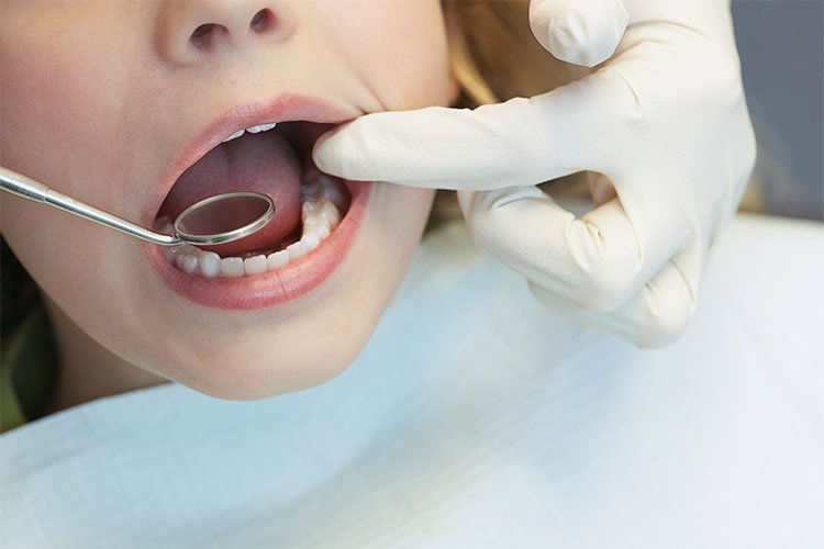
In pediatric dentistry, the treatment of superficial discoloration processes appearing in the primary dentition is managed with completely non-invasive protective prophylaxis methods that will not harm the underlying permanent tooth buds and will not increase the child’s dental phobia coefficient. Aggressive acid-containing chemical whitening (bleaching) gels are definitely not applied to primary teeth in children as they are in adults; because primary tooth enamel is thin and the nerve chamber is wide. The aim of the treatment is to instantly boost the child’s social self-confidence by removing superficial bacterial fortresses.
- Micronized Prophylaxis (Polisaj): Sweeping away iron and bacterial stains within seconds with the help of special rotating felt brushes and fruit-flavored polishing pastes,
- Airflow Bio-Film Cleaning: Blasting away black lines between teeth using hydraulic power without mechanical contact via pressurized air, water, and micronized glycine powders,
- Micro-Abrasion (Microabrasion): Polishing the surface at the micron level with very light hydrochloric acid and pumice mixtures that do not damage the enamel in localized white spots caused by fluorosis.
Which Treatment Methods Are Applied in Permanent Tooth Discoloration?
In persistent intrinsic and extrinsic discoloration pictures registered in the adult permanent tooth series, evidence-based mechanical, chemical, and adhesive combination protocols are deployed according to the depth of the stain in the dentin layer. A single device is not used randomly when planning treatment; the goal is to achieve maximum pink-white symmetry illumination with minimum tissue loss. Which method will be chosen is registered according to pre-operative mapping results.
The main treatment methods actively applied in permanent tooth discoloration are:
- Office-Type Laser Teeth Whitening (Bleaching): Breaking down internal dark carbon rings by activating concentrated peroxide gels with dental laser beams,
- Devital (Internal) Whitening: Whitening the tooth from the inside out by placing whitening gel inside teeth that have turned purple due to trauma or root canal treatment,
- Aesthetic Composite Bonding (Composite Veneer): Camouflaging severe antibiotic stains by crafting aesthetic resin veneers over them without damaging the tooth,
- E-Max Ceramic Veneers: The technique of smoothly covering deep mineralization defects with porcelain veneers, which have the highest light transmittance.
How Long Does Tooth Discoloration Treatment Take?
Thanks to the speed of modern clinicians and the practicality of digital units, dental discoloration interventions are extremely fast and flexible processes that perfectly integrate into the time constraints of modern metropolitan life. Sweeping away superficial black bacterial stains in children with polishing and Airflow is generally completed in just 10 to 15 minutes for a single jaw arch. Laser-supported holistic whitening (bleaching) sessions in adults consist of a single appointment session lasting at most 45 to 60 minutes, including preliminary preparation.
- Children’s Polishing Session: 15 Minutes (A practical cleaning time completed painlessly, without needles, which children complete while having fun).
- Laser Office-Type Whitening: 60 Minutes (A schedule that lightens the tooth shade to a bright white phase by 2 to 4 shades in a single session).
- Internal Whitening (Devital Bleaching): Short dressing appointments involving the renewal of the gel placed inside the root-canal-treated tooth every 2–3 days.
What Should Be Considered After Tooth Discoloration Treatment?
The care discipline that the patient will follow in the home environment is of vital importance to preserve the aesthetic quality of restoration and chemical whitening sessions over the long term without recurrence, and to keep safe those critical first 48 hours when enamel pores are most open to leaks. Since newly cleaned or opened enamel prisms display high absorption permeability almost like a sponge against external coloring irritants in the first days, full compliance with “white diet” rules is a clinical necessity.
The medical rules that must be meticulously followed at home during the sensitive post-procedure period are:
- To prevent stain remnants from forming in the enamel tubules, tea, coffee, wine, cola, and foods with tomato paste must absolutely not be consumed for the first 48 hours following whitening sessions.
- Smoking and tobacco products should be discontinued for at least 1 week to prevent nicotine molecules from seeping into the pores and creating permanent dark yellowing.
- After iron syrup stains are cleaned in children, the medicine should be administered by leaking it behind the tongue with the help of a medical pipette to prevent the syrup from directly touching the enamel.
- A slight adaptation sensitivity around the filling edges and mucosal margins is normal on the day of the procedure; this process is comfortably soothed with simple fluoride courses.
How Does Oral Care Affect Tooth Discoloration in Children?
The quality of oral hygiene in the early childhood phase is the main biological shield that preserves the smooth polished brilliance of primary and young permanent tooth enamel and prevents chromogenic bacteria from adhering to the tooth walls. If brushing routines are neglected in children, the soft bacterial plaque layer covering the tooth surface thickens; this porous matrix structure acts like a magnet, attracting artificial dyes in food and iron salts in saliva, fueling the chronicity of stains, while brushing breaks the stain chain by removing the plaque.
- Bacterial Flora Management: Deactivating the colonization speed of sulfur microbes that produce black stains through uncompromising brushing twice a day.
- Salivary pH Stabilization: Keeping the oral environment in an alkaline phase thanks to care, preventing enamel pore openings due to acid erosions.
What Habits Should Be Developed to Prevent Tooth Discoloration?
Keeping the bright white shade obtained in the mouth for years without chipping, cracking, or yellowing depends directly on the patient calibrating their daily lifestyle and nutritional reflexes according to preventive medicine rules. Since tooth enamels are not like lifeless artificial porcelain and carry dynamic absorption characteristics, limiting intraoral acid attacks and tobacco exposure is the key to a lifetime of smooth smile success.
- Straw Utilizing Habit: Leaking liquids containing heavy chromogens, such as coffee, cola, or iced tea, directly into the pharynx without touching the anterior surface of the teeth,
- Rinsing with Water Reflex: Washing the enamel pores by rinsing the mouth with pure water immediately after consuming coloring or acidic foods,
- Correct Brush Selection: Switching to soft brushes with micro-filaments instead of hard-bristled brushes that scratch and sand the tooth enamel, accelerating stain retention,
- Routine Polishing Control: Applying to regular physician checks every 6 months to remove plaques with clinical technologies before they turn into calculus.
What Are the Frequently Asked Questions About Primary or Permanent Tooth Discoloration?
The most common practical concerns arising in the minds of patients planning to get rid of yellowing and black stains on their primary or permanent teeth and gain a smooth pink-white aesthetic harmony and their clear medical counters are presented below:
Are those black lines and stains on my child’s teeth a form of tooth decay?
No, those persistent black stains appearing as lines around the necks of children’s teeth are definitely not active tooth decay. They are harmless mineral precipitations formed as a result of harmless but color-secreting chromogenic bacteria inside the mouth combining with iron ions in saliva. In fact, in medical literature, since the saliva coefficients of children with these stains are alkaline, their decay index scores are found to be lower; however, they should be removed in seconds with Airflow in the clinic for aesthetic reasons.
Do charcoal powders or activated carbons sold in pharmacies or on the internet completely remove tooth stains at home?
Absolutely not; quite the contrary, charcoal powders sold uncontrollably on the internet or granular hard pastes under the name of whiteners are not licensed under medical standards and harbor very aggressive abrasive crystals in their contents. Rubbing these powders onto the tooth permanently scratches and sands the tooth enamel, exposing the yellow dentin layer beneath the enamel, making your tooth even more yellow and sensitive in the future; tooth shade can only be safely lightened in the clinic under physician supervision.
Does a tooth whitening (bleaching) session permanently thin my teeth and wear down my enamel?
No, this is a piece of misinformation that is definitely not true according to medical and periodontology standards. The office-type laser whitening process is not a sanding, scraping, or acid-melting procedure. The medical gels used do not touch the calcium crystal structure of the tooth enamel; they only seep into the enamel pores and break down foreign organic stain pigments that darken the color of the tooth via chemical oxidation. Therefore, thinning, erosion, or permanent enamel wear definitely does not occur in your tooth; it is completely safe.
My child’s anterior primary tooth, which they fell and bumped, suddenly turned gray months later; does this situation damage permanent tooth buds?
The graying of a traumatized primary tooth is a major clinical sign indicating that the vessels inside the tooth have ruptured and the nerve chamber has lost its vitality, entering necrosis. This root tip inflammation that has lost its vitality can sabotage the enamel development (Turner’s hypoplasia) of the permanent tooth bud growing insidiously in the lower layer; therefore, either a microscopic pediatric root canal treatment should be applied to the graying primary tooth by our pedodontist or the tooth should be safely removed to protect the bud.
Can I return to my normal social and professional life immediately on the day the discoloration cleaning and whitening treatment is finished?
Yes, dental discoloration purification interventions are a completely local and non-invasive prophylaxis craft that does not disrupt consciousness, does not affect motor manual skills, and does not involve surgical scalpel incisions or sutures; therefore, you can get up from the chair the exact second your teeth are polished and continue your normal daily, professional, social, or academic life rhythm uninterruptedly from where it left off; there is no clinical barrier to driving or participating in business meetings immediately after the procedure.
Who Are We? & Panorama Ankara Oral and Dental Health
Panorama Ankara is a licensed oral and dental health center offering institutional health assurance to its patients by combining an academic specialist staff and the latest global bio-technological digital infrastructure under a single roof in all specialty branches of dentistry, primarily oral, dental, and maxillofacial surgery, advanced implantology, microscopic endodontics, periodontology, pedodontics (pediatric dentistry), and aesthetic dentistry. Instead of masking hereditary darkenings occurring in primary and permanent teeth, persistent stain matrices caused by smoking and caffeine, and mucosal asymmetries sabotaging children’s life comfort with scraping analog scrapings that scratch the tooth, our center attaches vital importance to stopping them at the molecular level with evidence-based medical disciplines, and operates seamless Digital Diagnostic Modeling and Laser-Supported Teeth Whitening (Bleaching) / Bonding ecosystems with calendar discipline without compromise from the very beginning to the end of the clinical operation workflow.
Under the roof of Panorama Ankara, absolute sterilization, high patient safety, transparency, and honesty principles are operated with full compliance with medical codes in all diagnostic, radiological scanning, design, and restoration interventions. In our clinical practices, while focusing on enamel integrity with micron-level resolution, proprietary Erbium and Diode laser units that protect tissues with photo-thermal effects, spectrophotometer digital color analyzers, and absolute rubber dam moisture isolation systems that keep resin bond strength at maximum are applied with milimetric precision under the supervision of our expert aesthetic dentist and pediatric dentistry staff. Our aim is not to impose old-fashioned, painful analog acid molds on our patients that wear down their enamel or burn their gums; but to analyze each individual’s enamel density and stain Miller class degree with the naked eye during the preliminary examination, achieving maximum session comfort, application practicality lasting seconds without damaging surrounding soft tissues and vascular pathways, and healthy bright white smile designs that you will use with a high sense of self-confidence and freshness for a lifetime. If you are tired of your teeth yellowing when looking in the mirror, your fading smile integrity, or the black stains on your child’s teeth, yet postpone your check-ups due to fear of sensitivity or time constraints, and wish to start your smart digital treatment process from beginning to end with scientific guarantee in an expert corporate center environment; you can safely protect your health and smile integrity by contacting the expert staff of Panorama Ankara for a detailed examination.