
What Is the Initial Consultation and Examination Service?
The initial consultation and examination service, referred to as a “comprehensive oral evaluation” in dental medicine practices, is the most vital orientation phase where the patient’s intraoral hard and soft tissue integrity is registered. This service is not merely the reshaping of a single tooth causing a complaint; it is a discipline of screening the entire oral ecosystem as a whole, including the tongue, palate, mucosa, temporomandibular joint (TMJ), and salivary glands. In this session, the patient’s general systemic health profile, genetic tendencies, and oral hygiene index score are registered, and a holistic treatment barrier is constructed against caries or bone resorption foci that progress insidiously and cannot be noticed with the naked eye.
- Holistic Oral Screening: Biological examination of each segment, mucosal folds, and hard tissues in the oral cavity.
- Anamnesis and Risk Indexing: Recording the patient’s medical drug history, allergy map, and hereditary dental defect risks.
- Clinical Communication Bridge: The process of overcoming dental anxiety barriers and establishing a trust profile for the treatment between the patient and the physician.
Which Evaluations Are Performed During the Initial Dentist Examination?
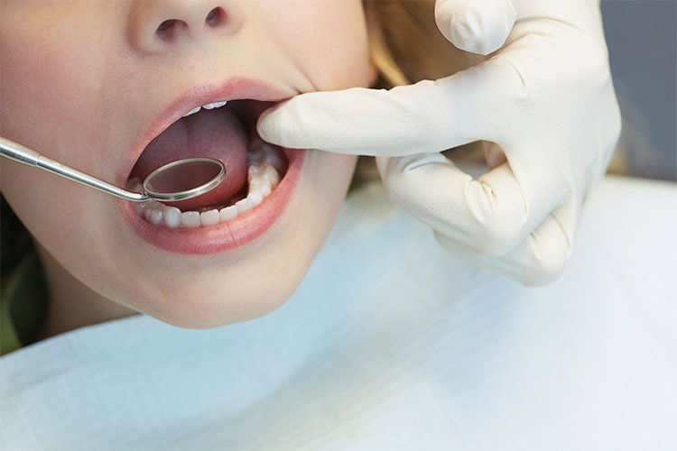
The medical protocols carried out on the initial examination chair involve analytical phases aimed at registering the current map of oral health with micron-level resolution. First, the integrity of the tooth enamel, the marginal sealing quality of old fillings, and the depth of the periodontal pockets are screened with the help of periodontal probes. At this stage, the muscle balance in the patient’s lower facial oval, the hydraulic tension coefficients exerted on the temporomandibular joint during chewing, and the presence of insidious cysts or impacted teeth hidden beneath the mucosa are passed through a versatile diagnostic filter.
- Hard Tissue Enamel Screening: Detection of micro-cracks and demineralization (incipient caries) lines on tooth surfaces.
- Periodontal Tissue Indexing: The phase of indexing the gums in terms of color, consistency, bleeding tendency, and alveolar bone levels.
- Soft Tissue Oncological Screening: Macroscopic control of the tongue, buccal mucosa, and floor of the mouth for benign or malignant lesions.
How Does the Initial Consultation and Examination Process Progress?
The initial examination workflow carried out in the clinic room progresses through a sequential and strict time hierarchy that completely eliminates micro-diagnostic deviations that human error might bring. The process absolutely does not contain analog harshness such as the patient sitting on the chair and immediately encountering a drill; the steps are executed synchronously according to the patient’s psychological relaxation rhythm and tissue analysis needs. Each phase is locked to reinforce the accuracy of the subsequent treatment planning stage.
The fundamental hierarchical steps applied in a corporate initial dental examination workflow are as follows:
- Medical Anamnesis Collection: Registering the patient’s systemic chronic diseases, coagulation times, and allergy card,
- Clinical Intraoral Examination: Screening all teeth and the mucosal bed with the help of mirrors, probes, and digital intraoral cameras,
- Radiological Screening Phase: Transferring intra-bone lesions to the screen by taking a digital panoramic X-ray in the diagnostic room,
- Design and Consultation: The physician reporting custom treatment alternatives and budget parameters to the patient in the light of the obtained data.
For Whom Is the Initial Examination Necessary?
The initial dental examination and consultation service is medically mandatory for acute patients with active toothache, abscess, or fracture complaints in their mouth, as well as for children, young people, and adult individuals of all age groups who have no symptoms or pain but aim to protect their oral health. As per medical standards, since the decay of teeth or resorption of gums has an insidious and painless character, the perception of “if I have no pain, I have a healthy mouth” is a major medical misconception; therefore, it is essential for every individual to pass through this analytical screening to maintain intraoral homeostasis balance and prevent early tissue loss.
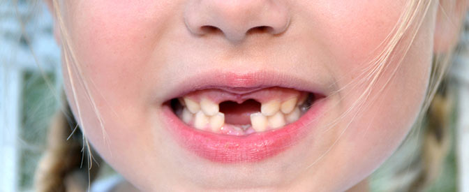
- Pediatric Age Group Children: Little ones whose milk teeth need to be supervised for their quality of guiding permanent tooth seeds,
- Those with Chronic Diseases: Clients with systemic diseases such as diabetes and osteoporosis that directly resorb the jawbone,
- Those Wishing to Renew Their Smile: Individuals with crowding, yellowing, or form defects in their teeth who desire a permanent aesthetic transformation.
What Preparations Should Be Made Before the Initial Examination?
The simple steps to be completed by the patient personally in the calendar leading up to the appointment morning are safety steps that increase diagnostic accuracy and ensure the physician logs the medical history flawlessly. It is important for the patient to bring epicrisis reports of past jaw operations or systemic medical interventions for rational process management. Bringing the inside of the mouth in a clean phase to the chair directly reinforces the point cloud capture accuracy of optical scanners.
- Preparing the names and dosage lists of regularly used medications (especially blood thinners or bone medications) to be recorded on the surgical card.
- Retrieving past X-ray films belonging to surgeries or old dental treatments from the archive to perform dimensional change tracking.
- The obligation of carefully brushing the teeth and clearing food debris with interdental floss before the session for operating room and examination comfort.
How Are Patient Expectations Evaluated in the Initial Consultation Meeting?
Screening the patient’s aesthetic and functional expectations in the initial consultation session is an artistic and medical calibration phase that identifies the patient’s psychological anxiety thresholds, chair anxiety, and facial change desires instead of imposing rote package programs. The physician examines how many millimeters of teeth are visible between the upper and lower lips during speech expressions (the visibility score). The patient’s past harrowing dental experiences (fear of needles, drill phobia) are questioned plainly, and it is rationally decided at this stage whether the processes will be managed with the comfort of sedation or general anesthesia.
- Facial Expression Tracking: Registration of the stretching coefficients of lip corners while speaking and gummy smile (gingival smile) risks.
- Aesthetic Vision Matching: Comparing the bright white shade envisioned by the patient with the golden ratio boundaries of their face on a digital screen.
How Is the Oral and Dental Health Evaluation Performed?
The holistic oral examination process goes beyond merely screening teeth for caries and is a versatile medical supervision lane that measures the histological health of all hard and soft tissues surrounding the oral cavity. The physician measures the depth of the pockets where the gums meet the bone one by one with the help of millimetric measuring tools called “Periodontal Probes.” If deepening over 3 mm or spontaneous bleeding is detected in these pockets, it is registered that insidious periodontitis inflammation has begun to resorb the bone, and treatment priority is shifted to drying this periodontal field.
- Hard Tissue Examination: Screening micro-cracks, wear notches, and cusp filling fractures on the enamel with diamond probes.
- Soft Tissue Mucosa Registration: Screening the tongue, inner cheek, and palatal mucosa for precancerous (pre-malignant) lesions or lichenoid structures.
- Salivary Flow Analysis: Algorithmic calculation of insidious caries breeding rates by measuring the presence of dry mouth (xerostomia).
How Is the Tooth and Gum Control Performed in the Initial Examination?
The tooth and gum margin control performed in the initial consultation session involves sequential mechanical and chemical control steps to zero out the margin of error that human hands and eyes might bring. The physician can make bacterial strongholds missed by the patient while brushing visible in vivid colors on the screen by applying “Plaque Disclosing Tablets” to the tooth surfaces. At this stage, while the millimetric dimensions of gingival recessions are mapped, the sealing quality of filling margin lines is registered with the help of micro mirrors.
| Control Area / Segment | Applied Clinical Protocol and Method | Targeted Tissue and Medical Outcome |
|---|---|---|
| Gum Margin Line (Marginal Mucosa) | Registration of pocket depth and bleeding index with periodontal probing. | Flawless pre-operative detection of insidious bone resorption (periodontitis) foci. |
| Tooth Wall Surfaces (Enamel) | Tactile surface screening with the help of micro diamond-tipped explorers (probes). | The phase of clear detection of early-stage insidious interproximal caries and enamel cracks. |
| Old Filling and Crown Margins | Transillumination light leakage and microscopic margin control. | Drying secondary caries foci breeding under fillings that wear out over time. |
Evaluation of Tooth Shape Deformities
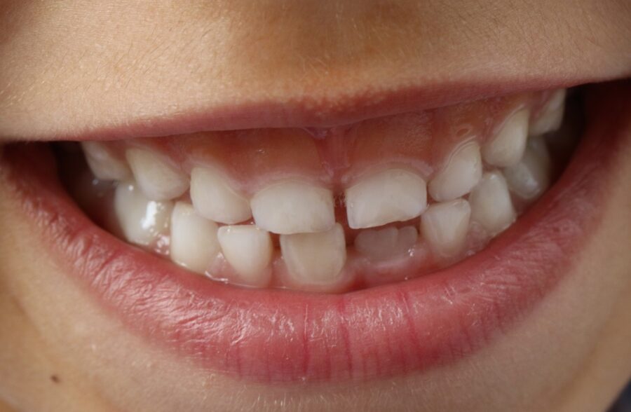
The analysis of geometric and facial shape deformities, crowding, or malocclusion in the teeth is an artistic and medical phase that examines the millimetric alignment variations of the teeth on the dental arch. The physician measures the symmetry relation of the lower and upper jaw anterior incisors with the facial midline axis. The level of form defect in the tooth alignment, and whether it requires orthodontic clear aligner treatment or micro-invasive composite bonding applications, is indexed according to the golden ratio parameters of the face and recorded on the treatment planning card.
- Diastema Volume Measurement: Registering the space distances between separated teeth on a mm basis and drawing the closure vector.
- Peg Lateral Analysis: Screening the capacity to bring developmentally small lateral incisors to aesthetic form dimensions.
Determining the Need to Close Tooth Gaps
While planning to close the gaps (diastemas) existing between front teeth due to genetic reasons, not only aesthetic concerns but also the functional damage these gaps create on speech phonetics and periodontal tissues are laid on the table. When tooth gaps remain open, food morsels strike the gum (papilla tissue) directly during chewing, producing chronic crushing edemas and bone pockets there. In the evaluation, how the gums will be protected from bacterial plaque accumulation risks by closing the diastema is simulated with mathematical clarity.
Structural Analysis of Fractured or Worn Teeth
The analysis of deep cracks, serrated margin lines, and wear notches occurring on incisal edges due to accident, trauma, or bruxism (teeth grinding) is a critical phase aimed at protecting the integrity of the living pulpal chamber (nerve network) inside the tooth. The physician calculates the awakening coefficient of thermal sensitivity shocks in the tooth by measuring the depth of exposed dentin tubules; if the fracture line exceeds the enamel boundary and descends to the dentin, the schedule for sealing open tunnels with bio-active adhesives is accelerated.
- Dentin Tubules Openness Degree: Closing exposed micro nerve endings to prevent post-procedure sensitivity shocks.
- Fracture Resistance Coefficient: Drawing adhesive restoration boundaries by measuring the thickness of remaining sound tooth walls.
Evaluation of Wisdom Teeth and Impacted Teeth
The medical evaluation of third molars (wisdom teeth) squeezed in the rearmost terminals of the dental arch is a vital surgical check-up step that detects lateral shearing tensions applied by faulty eruption angles onto adjacent tooth roots. Impacted or semi-impacted wisdom teeth can lean against the cervical margin of the perfectly sound molars in front of them and initiate insidious root caries; pre-registering these skeletal destructions and pericoronitis abscess risks is the only way to stop permanent tooth loss.
Why Are Digital Imaging Methods Important for Wisdom Teeth?
Digital 3D imaging systems in wisdom tooth surgery register surgical safety at the highest level by completely eliminating the depth blindness and risk of superimposed illusions brought by classical two-dimensional X-rays. Especially the relationship of lower wisdom tooth roots with the lower jaw sensory nerve line (inferior alveolar nerve) canal poses a vital risk. Thanks to CBCT 3D dental tomography scanning, the surgeon examines in three axes on a 1:1 scale whether the nerve canal passes between, below, or through the lateral wall of the roots; this digital accuracy prevents blind shots during surgery, definitively breaking the complication chain of permanent lower lip paralysis and loss of sensation (paresthesia).
- Hooked Root (Dilaceration) Registration: Zeroing the risks of fracture during extraction by scanning the curvature angles of micro root tips.
- Sinus Floor Isolation: Tomographic measurement of the proximity degrees of upper wisdom tooth roots to the maxillary sinus floor membrane.
Wisdom Tooth Extraction and Impacted Tooth Operations
The surgical removal of third molars in an impacted or semi-impacted state is a high technical focus jaw surgery operation performed under local anesthesia block in polyclinic operating rooms subject to full sterilization rules. In the first stage, a smooth incision line is drawn with the help of a scalpel considering the topographical projection boundaries of the tooth within the bone, and the mucosal flap is raised over the bone membrane (periosteum) to expose the bone. In order not to apply leverage pressure to the surrounding bone, the crown and root bodies of the tooth are divided into pieces with micro burs (using the separation technique) and removed from the socket with tweezers; the socket is washed with antiseptic solutions and closed tension-free with primary sutures.
How Long Does the Initial Consultation and Examination Process Take?
The initial consultation and comprehensive oral examination session is a highly dynamic and practical process that integrates perfectly with the busy time constraints of modern metropolitan life, barely blocking the patient’s social calendar. Transferring the entire oral map to the screen, including digital panoramic X-ray capture, intraoral clinical scans, and periodontal probing indexing, is usually completed with complete comfort in only 20 to 30 minutes; the detailed consultation and planning presentation to be made by the physician after the session is shaped flexibly depending on the distribution of the patient’s questions.
How Is the Initial Dental Examination Performed in Children?
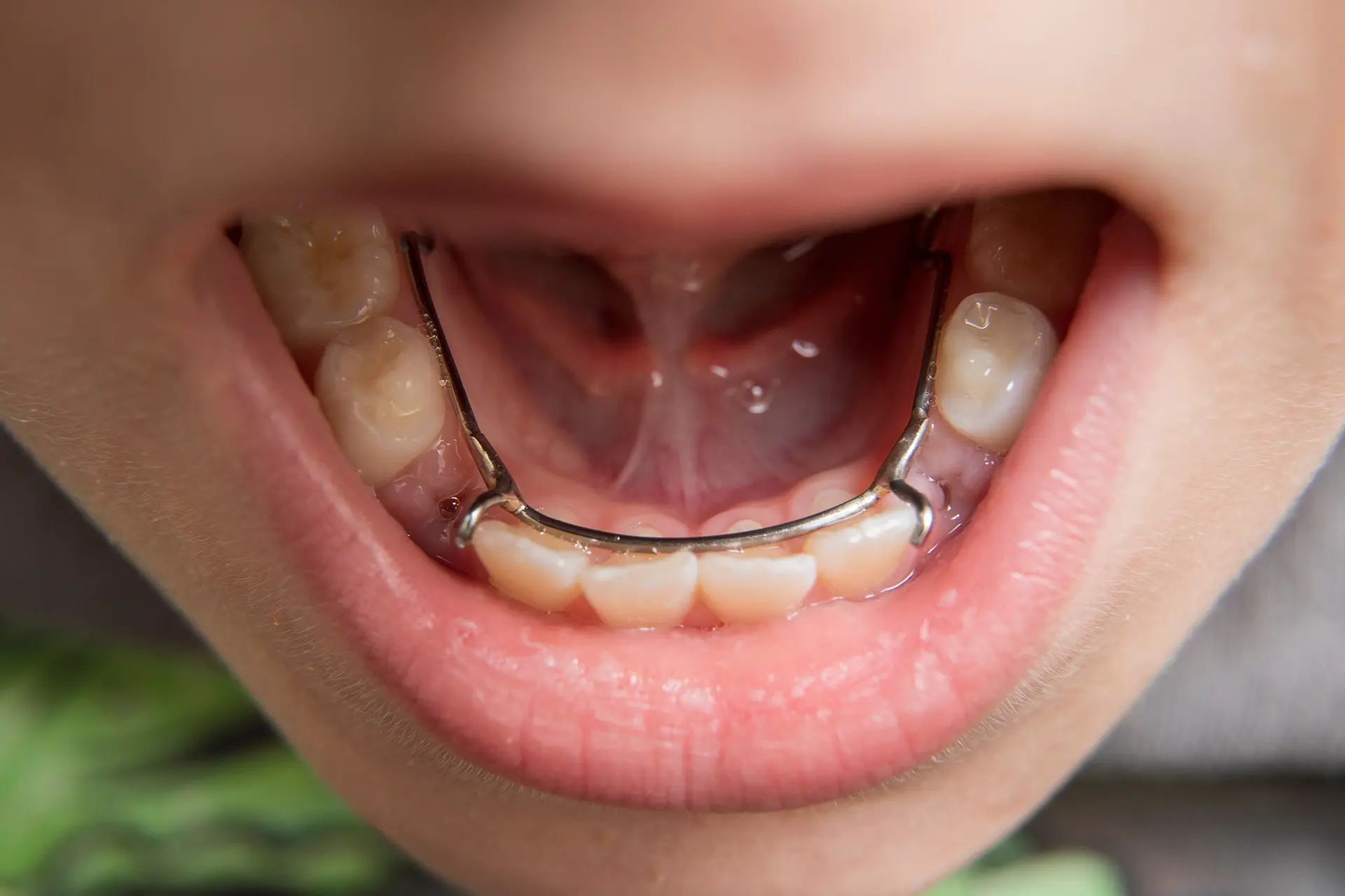
The initial examination steps carried out in the lane of pedodontics (pediatric dentistry) represent the most visionary approach that prevents the child’s dental phobia coefficient from breaking for life and maintains the space maintainer protection mission of primary teeth in the jaw skeleton. The session room is designed with play algorithms suitable for child psychology; the physician completely eliminates drill noise or injector needle taboos while introducing tools to the child. The aim is to ensure the child opens their mouth without fear and to catch bottle caries and primary tooth infection foci at the initial phase.
- Tell-Show-Do: The tactic of teaching the water spraying features of the devices engagingly by spraying them onto the child’s hand.
- Early Orthodontic Screening: Protective detection of jaw narrowness developing in the child due to thumb sucking or pacifier use.
How Are Oral Care Recommendations Determined During the Initial Examination?
The home care program to be prescribed to the patient at the time of examination is a protective medical protocol tailored specifically to the patient’s mucosal structure, salivary pH balance, and current plaque accumulation index score, instead of offering rote package advice. Giving hard brushes to a patient with deep gingival recessions will fuel root abrasions; therefore, care parameters are calibrated completely according to custom tissue resistance lines.
- Brush Hardness Calibration: Registering ultra-soft filament brushes for individuals with thin enamel thickness and medium tips for those with calculus tendency.
- Determining the Interdental Instrument: Prescribing micro dental flosses or pressurized hydraulic oral irrigator (waterpik) devices according to the narrowness of the interdental spaces.
What Is the Importance of Regular Dental Controls for Oral Health?
Routine dental examinations repeated periodically every 6 months are the clearest and most cost-effective protective medicine investment that saves the oral health ecosystem from destruction on a linear line. A microscopic caries focus at the initial phase can be stopped with a simple 15-minute filling without even needing a numbing needle when caught in regular controls; however, if neglected, the insidious infection leaks into the pulpal chamber, leading to root canal treatments, advanced skeletal bone resorption, and high-budget implant surgeries. Regular control definitively breaks this damage chain.
“A regular 6-month dental examination is the smartest medical shield that thwarts pathology strongholds growing insidiously in your mouth right at the beginning of the road, protecting you from heavy surgical operation burdens in the future.”
How Does the Follow-Up Process Progress After the Initial Consultation and Examination?
The customized treatment card created in line with the findings registered in the initial examination is permanently archived in the patient’s cloud data servers for life. After the filling, surgical extraction, or implant phases included in the planning are completed in order with calendar discipline, the mouth is brought to a fully purified phase. Following this permanent delivery phase, the patient is subjected to clinical prophylaxis follow-up with automatic periodic reminder messages every 6 months to maintain the stability of intraoral biology.
What Are the Frequently Asked Questions About the Initial Consultation and Examination Service?
The most common practical concerns arising in the minds of clients who want to have their oral health status registered or apply for a clinician examination for the first time, and their polyclinic medical counters, are presented below:
In the initial consultation and examination appointment, will a procedure or cutting be done to my teeth the exact second I sit on the chair?
No, absolutely not. The initial consultation and examination session is entirely a “diagnosis, mapping, and information” session. Without obtaining your written and verbal consent, and without reporting the pathologies and treatment alternatives in your mouth to you in plain language via digital screens, no scalpel, drill, or cutting move is definitely applied to your teeth; you leave the chair comfortably, having only learned about your oral health.
Will the radiation dose of the digital panoramic X-ray to be taken during the examination harm my general body health?
No, this is a concern that is definitely not experienced in new generation green technology digital radiology units. The radiation coefficient emitted by low-dose digital panoramic X-ray devices used in our modern clinics is in micron levels, much less than the natural background radiation dose a human is exposed to from the sun in normal daily life or during a single intercontinental airplane trip; furthermore, safety is kept at maximum by wearing lead apron shields.
I cannot even sit on the examination chair due to my severe fear of needles and drills (dentophobia) experienced in the past, what can I do?
This situation is a process managed with the highest sensitivity and empathy in our modern oral surgery department. If your chair anxiety is at a very advanced pitch, our initial consultation meeting is not held in the clinical room but entirely in a sterile hospital office environment with the comfort of a coffee chat; the examination and all subsequent filling and extraction steps can be finished painlessly and without needles under the comfort of sedation (semi-sleep) supervised by our anesthesiologist while you are asleep.
Am I obliged to have the procedures in the treatment plan presented to me done immediately after the initial examination, and what is its validity period?
No, you are definitely under no obligation to have the planned procedures done at the clinic that very day; the decision and calendar will belong entirely to you. However, since the living bone and mucosal tissues inside the mouth have a continuously dynamic and variable healing/resorption biology, the medical and technical validity stability period of the presented 3D diagnostic map and budget parameters is registered as an average of 3 to 6 months in medical standards.
Can I return to my normal daily and work life the exact second my initial examination and consultation appointment is completed?
Yes, initial consultation and examination sessions are completely non-invasive, visual, and digital screening steps that do not involve injection syringes, surgical scalpel wounds, sutures, or hours of numbness covering the face; therefore, the exact second your examination is over, you can get up from the chair and return to your normal daily, professional, social, or academic life rhythm from where it left off, instantly and with high clarity of consciousness; there is no obstacle to driving.
Who Are We? & Panorama Ankara Oral and Dental Health
Panorama Ankara is a licensed oral and dental health center offering corporate health assurance to its patients by combining an academic specialist staff and the latest global bio-technological digital infrastructure under a single roof, especially in oral, dental, and maxillofacial surgery, advanced implantology, microscopic endodontics, periodontology, pediatric dentistry (pedodontics), and aesthetic dentistry. Attaching vital importance to completely eliminating rote guesses, risks of oversight caused by human hands, and those old grueling needle moves that create chair anxiety stress in the patient within treatment practices, our center operates seamless 3D Digital Diagnostic Modeling and Initial Consultation – Examination ecosystems with calendar discipline without compromise from the very beginning to the end of the clinical operation workflow.
Under the roof of Panorama Ankara, absolute sterilization, high patient safety, transparency, and honesty principles are operated with full compliance with medical regulations by law in all diagnostic, radiological scanning, design, and microscopic restoration interventions. In our clinical practices, while analyzing the vertical boundaries of the jawbone and lesion depths with 3D tomographies in millimetric sections, proprietary intraoral scanner cameras that transfer the intraoral twin to the computer within seconds without causing nausea stress to the patient and spectrum-beam digital color analyzers are applied with millimetric precision under the supervision of our expert physician staff. Our aim is not to impose old-fashioned, slow, heavy analog examination frameworks on our patients that cut the tooth excessively or harass the tooth for unnecessary crowns because it cannot relieve the ache; but to analyze each individual’s jaw skeleton quality, occlusion mechanics, and facial aesthetic golden ratio boundaries in 3D on a virtual screen during the pre-examination, achieving smooth oral functions that you will use with maximum session comfort, same-day dentistry standards, and unshakable confidence for a lifetime without damaging surrounding soft tissues and sensitive blood vessels. If you are tired of your missing teeth, old worn fillings, or crowding when looking in the mirror, yet postpone your controls due to fear of the dentist chair, injector phobia, or time constraints, and wish to start your smart digital treatment process from beginning to end with scientific guarantee in an expert corporate center environment; you can safely protect your health and smile integrity by contacting the expert staff of Panorama Ankara for your detailed 3D intraoral scanning and digital planning analyses.