How Many Stages Does Implant Treatment Consist Of?
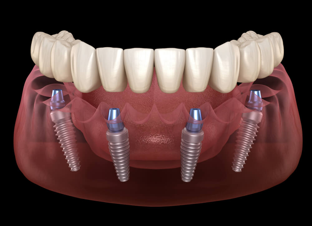
Implant treatment generally consists of four basic stages: a detailed clinical and radiological examination, the surgical operation in which the titanium root is placed into the jawbone, the fusion-with-bone (waiting) process called osseointegration, and finally the prosthesis stage in which the artificial tooth (crown) is fitted. The progression of the process is personalized according to the patient’s anatomical bone structure.
Implant treatment, applied in modern dentistry to compensate for missing teeth, is not simply a matter of placing a screw into the bone; it is a staged treatment as a whole that combines biological compatibility, precise surgical principles, and aesthetic prosthetic engineering. Each stage prepares the foundation for the step that follows it. For the treatment to be long-lasting, for chewing functions to be restored as close to natural as possible, and for the jaw joint to continue functioning healthily, it is of great importance that none of these stages be skipped and that clinical protocols be followed to the letter.
How Is the Clinical and Radiological Examination Performed Before Implant Treatment?
The examination before implant treatment is performed by taking the patient’s systemic general health history, clinically examining the intraoral tissues, and measuring bone volume with the help of three-dimensional jaw tomography (CBCT). In light of this data, the diameter, length, and position of the implant to be used are planned digitally down to the millimeter.
Perhaps the most critical step of the treatment is making an accurate diagnosis and plan. While standard two-dimensional panoramic X-rays provide information about the height of the bone, they cannot offer definitive data about its thickness (width). For this reason, three-dimensional volumetric tomography has become standard in current clinical practice. By examining the tomography cross-sections, the dentist determines the location of the nerves passing through the lower jaw (mandibular canal) or the sinus cavities in the upper jaw, ensuring the implant stays at a safe distance from these vital anatomical structures. In the same session, the medications the patient uses (especially blood thinners or bone-loss medications) and chronic diseases (diabetes, hypertension, etc.) are reviewed to complete the surgical preparation.
How Does the Surgical Stage of Implant Placement Proceed?
The surgical stage proceeds through the steps of numbing the area with local anesthesia to ensure the patient’s comfort during the procedure, appropriately opening the gum tissue and creating a special socket in the jawbone with graduated drills, and placing the titanium implant into this socket. Afterward, the gum tissue is closed with sutures (stitches).
Contrary to popular belief, the implant placement procedure is a highly controlled and quickly progressing surgical procedure. In a clinical setting where the sterilization chain is maintained at the maximum level, the work is carried out under saline solution cooling to prevent the bone tissue from overheating. The surgeon places the titanium implant into the socket, opened according to the density of the bone, with a specific torque (compression force) value. The mechanical compression achieved at this initial placement moment is called “primary stability” and ensures the implant remains stable within the bone without wobbling. Generally, the surgical placement time for a single implant takes between 15 and 30 minutes, excluding the anesthesia and preparation stages.
| Procedure Step | Purpose and Details of Application |
|---|---|
| Local Anesthesia | Temporarily halting nerve conduction in the relevant area. |
| Flap Opening / Incision | Minimally opening the gum (mucosa) to reach the jawbone. |
| Socket (Cavity) Preparation | Gradually creating a bed in the bone matching the implant’s dimensions. |
| Implantation | Fixing the sterile titanium screw structure into the prepared bone socket. |
| Suture Application | Bringing the wound edges together to begin the healing process. |
What Is the Osseointegration (Fusion with Bone) Process and Why Is It Awaited?
The osseointegration process is when jawbone cells attach to the micro-rough surface of the titanium implant, forming a biological and physical whole (fusion). This biological repair and bone-formation period must be awaited so that the implant can withstand the strong chewing forces to come and remain stable.
After the surgical procedure, the implant’s initial retention in the bone is entirely mechanical (like a screw wedged into wood). However, permanent success depends on osseointegration occurring at the cellular level. Titanium is a 100% biocompatible element that the human body does not perceive as a foreign substance. During the healing period, osteoblast cells in the jawbone surround the implant and form new bone tissue. This biological adaptation process generally takes around 2 to 3 months for the lower jaw and around 3 to 4 months for the upper jaw, where the bone density is softer. During this waiting phase, it is essential that no load (chewing force) be placed on the implant.
In Which Cases Is the Gum Shaping (Healing Abutment) Stage Performed?
The gum shaping stage is the procedure of opening the mucosa over implants that have been left to heal in a closed manner (beneath the gum) after the osseointegration process is complete, with a small laser or tissue touch, and fitting healing abutments (gingiva formers). Its purpose is to create a natural and aesthetic gum socket around the porcelain tooth to be made.
In some clinical protocols (single-stage surgery), healing abutments can be fitted on the same day the implant is placed. However, in “two-stage surgery” methods where the implant is kept completely under the gum for protection, this small intervention is needed after bone fusion is complete. After this procedure, performed under local anesthesia and taking only a few minutes, the patient experiences no discomfort at all. The fitted healing abutments remain in the mouth for an average of one week. During this time, the gum surrounds the abutment and takes on a collar form. Thus, when moving to the prosthesis stage, the porcelain tooth to be made displays a flawless aesthetic appearance, as if it were emerging from the patient’s own natural gum tissue.
How Do the Impression and Superstructure Applications of the Prosthesis Stage Proceed?
The prosthesis stage begins with removing the healing abutments and taking an impression, either with digital intraoral scanners (CAD/CAM) or silicone-based impression materials, to transfer the exact position of the implant in the mouth to the laboratory. Then, the zirconium or porcelain crowns prepared in the laboratory setting are fixed onto the implant superstructure components (abutments) by screwing or bonding.
This is the final stage where the patient permanently regains their missing tooth and their chewing functions are fully restored. After the impression procedure is completed, coordinated work begins between the prosthodontist and the dental technician. In applications in the aesthetic zone (front teeth), superstructures are designed to high aesthetic standards, taking into account facial anatomy, lip support, the smile line, and the color shades (translucency) of the adjacent teeth. In the back regions, in addition to aesthetics, durable porcelain or monolithic zirconium designs are preferred that will distribute incoming chewing forces evenly. The prosthesis is delivered to the patient after fit trials, followed by a final polishing procedure.
| Prosthesis Step | Explanation and Process |
|---|---|
| Taking the Impression | Recording the implant’s position with digital scanning or impression trays. (Session 1) |
| Framework Trial | Checking the intraoral fit of the metal or zirconium supporting structure. (Session 2, if needed) |
| Aesthetic and Bite Trial | Testing the color and form of the porcelain and the upper-lower jaw bite relationship. |
| Delivery (Cementation / Screwing) | Permanently screwing or bonding the prepared prosthesis onto the abutment. |
How Are the Stages of Implant Treatment Organized Around Panorama Ankara?
In the select clinical approaches around Panorama Ankara, the stages of implant treatment are coordinated with digital appointment tracking systems suited to the patient’s schedule, and by offering both 3D tomography and digital impression (scanner) capabilities under the same roof, the processes are completed quickly, transparently, and with high infection-control standards, without being split across different centers.
Considering the intensity of regional business life and the pace of patients’ lives, health expectations in the Panorama Ankara location are quite high. Patients prefer treatment plans with predictable start and end dates, rather than uncertainty about the treatment stages. To meet this demand, clinics maintain communication with patients not only during active treatment moments but also during waiting stages such as osseointegration. The use of optical scanners, which eliminate the gag reflex caused by traditional dough-like impression materials during the impression stage, is among the current protocols that maximize patient comfort in this region. All of this organizational capability reduces the time spent without teeth and ensures the patient reaches their permanent tooth in the most comfortable way.
How Do the Stages Change in Cases Requiring Bone Graft?
If there is not enough jawbone volume at the implant site, a “bone grafting” (bone graft addition) stage is included in the process. If the bone deficiency is mild, grafting and implant placement are done in the same session; however, if the deficiency is very severe, bone graft material is placed first, 4-6 months are awaited for the area to turn into bone, and implant surgery is performed as a separate stage after this period.
Many years having passed since tooth loss, gum disease (periodontitis), or traumatic tooth extractions cause bone resorption. It is essential that there be a healthy bone wall of at least 1.5 – 2 mm thickness surrounding the implant. In cases requiring bone augmentation (for example, sinus lifting operations), the process naturally lengthens. This is because an additional expectation of healing from the body arises: the added artificial (synthetic or biological) bone graft material must organize with the patient’s own bone cells and turn into a hard, blood-supplied living tissue. Since proceeding to the implant before this process is complete would risk the biological foundation, the number of stages increases.
How Does Same-Day Teeth (Immediate Loading) Affect the Implant Stages?
The same-day teeth (immediate loading) technique modifies the traditional bone-fusion (waiting) stage, fitting fixed temporary prostheses on the day the implant is placed in patients with suitable bone density. This technique immediately eliminates aesthetic concerns, but the patient must avoid chewing hard foods with these temporary teeth until osseointegration is complete.
The immediate loading technique, frequently applied especially in cases of missing front teeth or All-on-4 complete edentulism concepts, speeds up the treatment stages in terms of time. As the dentist places the implant into the bone, they measure the torque (compression force) value from the device; if this value is sufficiently high, it means primary stability is strong. Using the digital impression taken, fixed teeth prepared from acrylic or temporary resin in the laboratory are screwed onto the implant on the same day or within 24 hours. The patient does not leave the clinic without teeth. However, the nature of osseointegration does not change; after the 2-3 month bone-formation period passes, these temporary teeth are removed and the permanent porcelain/zirconium prosthesis stage begins.
Frequently Asked Questions About the Stages of Implant Treatment
How many months in total do the implant stages take?
In patients with a normal and healthy jawbone, the total time from diagnosis to the fitting of the permanent porcelain tooth is an average of 2.5 to 3 months for the lower jaw and 3 to 4 months for the upper jaw. In complex cases requiring advanced surgical bone graft additions, this period can total 6-8 months.
The timeline depends entirely on the individual’s biological healing potential and their initial anatomical conditions. This process, which requires patience, is essential for laying a healthy foundation that will be used for a lifetime.
Is separate anesthesia applied for each surgical stage?
Yes, during the surgery in which the implant is placed into the jawbone and later during the fitting of the healing abutments (gum shaping) stage, local anesthesia specific to the area being treated is applied so that the patient feels no ache or pain at all. No anesthesia is needed during the prosthesis impression and trial stages.
Since local anesthesia is applied today with quite refined and patient-friendly needles, all intervention processes are managed in a highly comfortable manner.
How are digital scanners used during the impression stage?
Digital scanners used during the prosthesis stage (CAD/CAM systems) transfer a three-dimensional, high-resolution virtual model of the implant and surrounding tissues to the computer screen within seconds, with the help of a small optical camera inserted into the mouth. This eliminates the need to use impression material (silicone).
This method provides great comfort, especially for patients with a gag reflex, while also reducing to zero the millimeter-level margin of error (distortion) that can occur during the mold-casting processes at the laboratory stage.
What should be considered after the treatment stages are completed?
After the prosthesis stage is finished and the permanent tooth is fitted, flawless oral hygiene should be maintained through brushing twice a day and the use of bridge floss (superfloss) or interdental brushes. Regular dentist check-ups every 6 months must be carried out without fail to prevent tartar buildup around the implants and to protect bone health.
Although the titanium roots themselves do not decay, the gum around the implant can become inflamed due to poor oral care, and bone loss (peri-implantitis) can begin. The only way to maintain the success of the treatment for a lifetime is through correct daily oral hygiene habits.