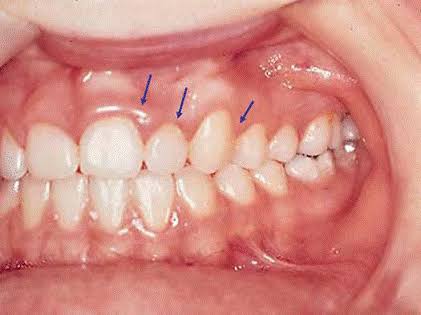
How Does Gingivitis (Gum Inflammation) Develop?
The oral cavity is not only one of the body’s primary gateways to the outside world, but also a dynamic and complex ecosystem hosting millions of microorganisms. Within this ecosystem, a delicate biological balance exists between teeth, gums, bone tissue, and saliva. When this balance is disrupted, it most commonly results in one of the most prevalent oral pathologies: gingivitis, or gum inflammation. Gingivitis should not be perceived merely as a set of simple symptoms such as gum redness or bleeding; rather, it represents a complex immunological battle waged by the body against bacterial invasion. Understanding how this battle begins, how it progresses, and what cellular-level changes occur in the tissues is crucial for maintaining optimal oral health.
The pathogenesis of gingivitis is not a sudden event triggered by a single cause, but a cumulative process driven by a chain of biological reactions. The primary instigator of this process is bacteria adhering to the tooth surface. However, the mere presence of bacteria is not sufficient to explain disease development; the critical factor is their organization into a structured community (biofilm) and the host immune response to this structure. In healthy gums, the gingival tissue tightly embraces the cervical region of the tooth, acting as a protective barrier that prevents bacteria from reaching deeper structures such as the periodontal ligament and alveolar bone. Gingivitis begins when this barrier is weakened and damaged by bacterial toxins and the body’s own immune response. Examining this pathological process at the molecular and cellular level helps explain why gingivitis often progresses silently and unnoticed.
1. The Starting Point: Pellicle Formation and Bacterial Colonization
The journey of gingivitis begins just minutes after tooth brushing, even before bacteria attach to the tooth surface. Glycoproteins present in saliva rapidly adhere to the enamel surface, forming a thin, bacteria-free organic film known as the “Acquired Pellicle.” While this layer plays a protective role by shielding enamel from acidic attacks, it simultaneously provides an ideal adhesion surface for bacteria. Shortly after pellicle formation, pioneer bacteria (primarily streptococcal species) attach to this layer. At this stage, the attachment is weak and can still be disrupted by saliva flow.
However, if mechanical cleaning (brushing) is neglected, these bacteria begin to multiply and adhere more firmly to the surface. At this point, a process known as “co-adhesion” occurs, whereby different bacterial species attach to the already-adhered pioneer bacteria. Much like bricks forming a wall, bacteria stack upon one another. To protect themselves and maintain cohesion, these bacteria secrete a sticky substance called the “extracellular matrix.” This organized microbial community embedded within a self-produced slime layer is known as “Microbial Dental Plaque” or “Biofilm.”
2. Biofilm Maturation and Toxin Production
As plaque accumulation continues, the biofilm layer thickens. The deepest layers of this thickened biofilm become oxygen-deprived, creating an ideal environment for anaerobic bacteria—those most harmful to periodontal health—to proliferate. These gram-negative bacteria produce highly destructive enzymes (such as collagenase and hyaluronidase) and toxins (notably lipopolysaccharides – LPS) as metabolic byproducts. These substances compromise the cellular integrity of gingival tissues and initiate tissue breakdown.
At this stage, gingivitis may still be clinically asymptomatic, yet a microscopic battle has already begun. The plaque starts migrating beneath the gingival margin into the subgingival area. Bacteria that colonize the gingival sulcus find a protected niche beyond the reach of toothbrush bristles, allowing them to multiply rapidly and continuously release toxic byproducts that assault the gingival tissues.
3. The Body’s Defense Response: Inflammation
Gingival tissues do not remain passive in the face of bacterial aggression. Once bacterial products and toxins penetrate the gingival epithelium and reach the connective tissue, the body’s alarm system is activated. The immune system dispatches defensive cells (neutrophils, macrophages, and lymphocytes) to eliminate the invaders. For these cells to exit the bloodstream and reach the target tissue, blood vessels must dilate and become more permeable. The classic clinical signs of gingivitis are direct consequences of this immune response:
- Redness (Erythema): Vasodilation and increased blood flow cause the gums to appear red.
- Swelling (Edema): Increased vascular permeability allows fluid to leak into surrounding tissues, resulting in gingival swelling.
- Bleeding: Newly formed capillaries within inflamed tissue are fragile, and the sulcular epithelium becomes ulcerated. Even minimal trauma (brushing or probing) can cause capillary rupture and bleeding.
[Image of healthy gums vs gingivitis diagram]
4. The Role of Tartar (Calculus) and the Vicious Cycle
In the development and progression of gingivitis, tartar plays a facilitating and exacerbating role. If plaque is not removed, it absorbs calcium and phosphate ions from saliva and undergoes mineralization, hardening into a substance known as tartar (calculus). Tartar itself does not produce toxins and is not directly pathogenic; however, its rough and porous surface dramatically promotes further plaque accumulation.
This irregular surface makes plaque retention extremely easy, while also exerting mechanical pressure on the gums that hinders effective cleaning. In the presence of tartar, no amount of routine brushing can eliminate bacteria hidden within its porous structure. As a result, inflammation becomes chronic and a self-perpetuating vicious cycle ensues—one that can only be broken through professional dental scaling.
Disease Stages and Progression
In periodontology, the histopathological progression of gingivitis is classified into four distinct stages. These stages determine reversibility and the extent of tissue damage:
Stages of Gingivitis Development
The table below summarizes gingival changes over time following plaque accumulation:
| Stage | Timeline (After Plaque Accumulation) | Microscopic and Clinical Features |
|---|---|---|
| Initial Lesion | 2–4 Days | No clinical signs. Microscopically, increased vascular permeability and migration of polymorphonuclear leukocytes (PMNs). |
| Early Lesion | 4–7 Days | Mild gingival redness. Bleeding on probing may occur. Collagen breakdown begins (up to 60–70%). |
| Established Lesion | 14–21 Days | Classic gingivitis presentation: red, swollen, bleeding gums. Ulceration of pocket epithelium. No bone loss yet. |
| Advanced Lesion | Indefinite (If Untreated) | Transition to periodontitis. Inflammation reaches bone, leading to bone resorption and periodontal pocket formation. Damage becomes irreversible. |
Conclusion
In conclusion, gingivitis is a multifactorial condition initiated by microbial plaque accumulation but shaped by host immune response, genetic predisposition, and environmental factors such as smoking and stress. It represents the biological consequence of neglected oral hygiene. Red, swollen, and bleeding gums are not merely cosmetic concerns; they are indicators of an active infection and ongoing tissue destruction. The key to halting this process lies in understanding biofilm formation and disrupting it consistently. Toothbrushes, dental floss, and routine professional check-ups are the most powerful tools available in winning this ongoing microbiological battle.