Ankara 3D Dental Scanning
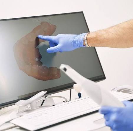
One of the most advanced achievements of modern dental technology is the ability to transfer intraoral tissues and dental alignment into a digital environment with millimetric precision. This process, referred to in medical literature as “Intraoral Optical Scanning,” is an advanced “Data Acquisition” discipline that completely eliminates the volumetric distortion risks caused by traditional impression materials while constructing the three-dimensional topography of teeth on a computer screen within seconds. Redefining Ankara 3D dental scanning standards with the vision of “Laser Triangulation” and “High-Resolution Optical Sensors,” Panorama Ankara offers patients a flawless treatment infrastructure through a comfortable scan completed in seconds—without the discomfort of traditional impression trays. The systems implemented in our clinic represent an advanced engineering workflow that copies intraoral structures at the micron level with digital sensors, eliminating error margins in restorations and orthodontic planning with uncompromising clinical precision.
The primary scientific success behind 3D dental scanning technology lies in the principle known in medical literature as “Active Wavelength Measurement,” which processes the reflection speed of light from the tooth surface at thousands of frames per second. In the advanced technology unit of Panorama Ankara, millions of data points (Point Cloud) are collected while the scanner tip moves inside the mouth, and these points are combined to create the “Digital Twin” of the tooth. While traditional impressions may lead to diagnostic inaccuracies due to micro-shrinkage during plaster hardening, 3D scan data is transferred instantly to our laboratory via cloud systems in “STL” or “PLY” formats without any physical distortion. This vision transforms diagnostic practices in Ankara from the simplicity of “taking an impression” into an advanced “Virtual Prototyping” center that reconstructs the patient’s anatomy within a digital universe.
Clinical Classification: Digital Data Processing and Functional Advantage Parameters
The clinical value of a 3D dental scan lies not only in producing a visual model but also in providing a mathematical infrastructure capable of analyzing chewing movements (dynamic occlusion). Digital scanning ensures “Predictability” at every stage of treatment. To help patients clearly understand this advanced data acquisition advantage, Panorama Ankara presents the technological superiority applied in the clinic within the following scientific framework.
| Technological Parameter | Traditional Impression Tray | Panorama Ankara 3D Dental Scanning |
|---|---|---|
| Precision and Micron-Level Fit | May contain a 100–200 micron margin of error due to expansion or shrinkage risks of plaster and impression materials. | Optical precision reaching up to 20 microns ensures restorations with perfect marginal fit to the gum line. |
| Patient Comfort and Reflex Response | Especially in upper jaw impressions, materials touching the palate may trigger nausea and gag reflex. | Contact-free scanning collects data using only light, allowing the patient to breathe comfortably without causing nausea. |
| Simulation and Preview | Physical procedures such as tooth preparation and temporary restorations must be completed to visualize the final result. | The “Smile Simulator” allows the final treatment outcome to be examined in 3D from every angle before touching the tooth. |
| Archiving and Data Security | Plaster models can break, occupy physical space, and lose dimensional stability over time. | Digital models are preserved indefinitely without distortion and can be accessed within seconds even years later. |
[Image showing the CAD/CAM integration where a 3D scan is used to design a precision crown or bridge]
Virtual Articulation: Digital Analysis of Chewing Movements
The most revolutionary contribution of 3D dental scanning is the millimetric analysis of contact points between the upper and lower jaws, known in medical literature as “Dynamic Occlusal Mapping.” In the protocols implemented at Panorama Ankara, the moment the patient closes their mouth is recorded in 3D. The software then displays pressure points (contact points) between teeth using color-coded maps. As a result, restorations such as porcelain veneers or zirconium crowns can be calibrated during production to ensure that they do not create any occlusal interference (“high spots”) in the mouth. This process transforms treatment from trial-and-error into the highest level of “Digital Occlusion” discipline that guarantees chewing comfort with mathematical precision.
Digital Data Monitoring and Maintenance Discipline After Scanning
For your 3D dental data—copied using futuristic optical sensors in our clinical studios and transferred to our digital archive—to support the longevity of your treatment, periodic monitoring is essential. The digital dentistry board of Panorama Ankara presents the following two fundamental rules as a “data constitution” to preserve the uncompromising clarity provided by technology:
- To maintain the exceptional “Marginal Integrity” between restorations produced through digital scanning (implant-supported or aesthetic crowns) and the gum tissue, interdental brushes should be used during daily care, and annual “Comparative Scanning” appointments should not be neglected to monitor potential gum recession.
- For patients undergoing orthodontic aligner therapy or clear aligner treatment, intermediate scans should be performed during appointments determined by the clinician in order to verify tooth movements according to the digital plan and maintain up-to-date “Progress Tracking.”
Experience the Digital Revolution in Dentistry with Panorama Ankara
The nausea you experience during traditional dental impressions, repeated trial sessions, or restorations that do not properly fit your mouth are not inconveniences you must endure in the age of technology. On the contrary, in expert hands, optical scanners and virtual articulation technologies enable a professional process completed within seconds, allowing you to monitor every stage of your treatment live on the screen. In your search for Ankara 3D dental scanning, Panorama Ankara combines “Laser Triangulation” and “Digital Twin” protocols to free you from old-fashioned impression trays, shrinking impression materials, and diagnostic uncertainties with scientific elegance. We respect the unique geometry of your oral anatomy with absolute care, transforming every pixel of data into a flawless and enduring smile.
To generate the three-dimensional map of your teeth within seconds using our optical sensors, determine the most suitable “Digital Restoration” or “Clear Aligner” path without any margin of error, and experience the comfort of restorations that “fit perfectly the moment they are made,” you can schedule a detailed 3D scanning and digital examination appointment at Panorama Ankara clinic and entrust your health to the safe hands of experience and futuristic digital technology.