Ankara Odontogenic Cyst Surgery
In the disciplines of dentistry and oral surgery, intraosseous cavities originating from embryological remnants of tooth development or chronic infections are referred to as “Odontogenic Cysts.” These cysts often exhibit an insidious progression, growing silently within the jawbone for years without producing any visible external symptoms. Like an inflating balloon, odontogenic cysts increase internal pressure within the jawbone, causing bone resorption from the inside, displacing healthy tooth roots, and potentially compromising the structural integrity of the jaw—predisposing it to spontaneous fractures. Embracing advanced radiological diagnostics and microsurgical protocols in Ankara odontogenic cyst surgery, Panorama Ankara approaches these complex lesions not merely as “cysts,” but as pathological entities with a high potential for recurrence. Our clinic maps the cyst’s boundaries with millimetric precision using 3D panoramic and volumetric tomography (CBCT) data; depending on the lesion type, advanced surgical techniques such as enucleation or marsupialization are performed to eliminate existing bone destruction while preserving healthy dental tissues.
The surgical management of odontogenic cysts is a tissue engineering process shaped by the cyst’s type and biological aggressiveness. While Radicular (root-origin) cysts—the most commonly encountered type in medical literature—can often be resolved with root canal treatment or minor surgery, lesions such as the “Odontogenic Keratocyst” (OKC) demonstrate aggressive behavior, spreading within bone like a malignancy and showing a strong tendency for postoperative recurrence. The specialist maxillofacial surgeons at Panorama Ankara do not consider simple removal (enucleation) sufficient for such aggressive cysts; they further eliminate microscopic remnants by applying chemical agents such as “Carnoy’s Solution” to the cyst cavity walls or performing peripheral osteotomy (bone shaving). This comprehensive surgical vision is our strongest medical assurance against patients facing recurrent cyst problems in the same region years later.
Clinical Classification: Types of Odontogenic Cysts and Their Biomechanical Effects
The type of cyst developing within the jawbone directly determines the extent of surgery and the subsequent rate of bone healing. To help patients clearly understand their radiological findings and pathological risks, Panorama Ankara presents odontogenic cyst classification within a scientific framework in the table below.
| Cyst Type (Pathological Origin) | Clinical Behavior and Radiological Appearance | Panorama Ankara Surgical Approach |
|---|---|---|
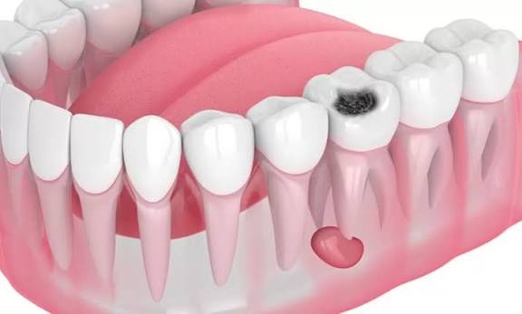
| Radicular (Periapical) Cyst | Develops due to chronic infection at the tooth root. Appears as a dark, round cavity at the root apex. | Root canal treatment or apical resection is attempted first; if unsuccessful, the cyst is surgically removed completely. |
| Dentigerous (Impacted Tooth) Cyst | Encapsulates the crown of an impacted tooth (usually a wisdom tooth) like a sac, causing bone resorption. | The cystic sac is removed in one piece together with the impacted tooth (enucleation). |
| Odontogenic Keratocyst (OKC) | Characterized by aggressive behavior. Spreads into bone with finger-like projections and carries a high recurrence risk. | After cystectomy, the area is irrigated with special solutions and bone margins are carefully shaved (lumen decontamination). |
| Residual Cyst | Occurs when a cyst at the root apex is left behind or incompletely removed after tooth extraction. | The cystic cavity within the bone is accessed using microsurgical techniques and thoroughly debrided. |
Bone Regeneration: Restoration of the Cyst Cavity with Grafting
When a large odontogenic cyst is removed, a substantial defect remains within the jawbone. If left untreated, the body may not always regenerate this area with high-quality bone tissue, leaving the jaw structurally weakened and potentially unsuitable for future implant treatment. At Panorama Ankara Clinic, a “Guided Bone Regeneration” protocol is initiated following cyst surgery. Bioactive bone graft materials (bone substitutes) are placed into the cleaned cavity and covered with specialized barrier membranes. Over the following months, the area previously destroyed by the cyst is regenerated with the patient’s own living bone. This approach not only treats the disease but also restores the structural strength of the jaw skeleton, allowing for safe implant placement in the future.
Postoperative Pathological Monitoring and Healing Discipline
Odontogenic cyst surgery does not end with the removal of the lesion; sending all excised tissue for histopathological examination to confirm the exact cell type is a medical necessity. For healthy bone cells (osteoblasts) to properly regenerate the cavity after surgery, the Panorama Ankara surgical board recommends the following two fundamental healing principles:
- To protect the vital blood clot formed in the surgical area and the placed bone graft materials, avoid hard chewing on that side for the first 2 weeks. Refrain from habits such as pulling the lips forcefully or examining the area with fingers, which may compromise suture stability.
- Smoking, which disrupts oxygenation at the cellular level and impairs bone healing, must be strictly discontinued during the critical “ossification” phase following surgery. Radiological follow-ups (X-ray monitoring) should be conducted at intervals determined by your physician (typically every 6 months) to professionally assess potential recurrence.
Build Your Jaw Health on Scientific Foundations with Panorama Ankara
Odontogenic cysts—often detected incidentally during routine radiographic examinations or presenting as painless swelling in the jaw—are silent threats that can compromise the integrity of your jawbone if not treated in time. Combining microscopic precision with biomimetic bone grafting systems in Ankara odontogenic cyst surgery, Panorama Ankara frees you from these pathological burdens with scientific elegance. We analyze the boundaries of your cyst using 3D CBCT technology, minimize pathological risks, and reconstruct lost bone volume with advanced regenerative systems.
To determine the nature of the suspicious lesion in your jaw, map the relationship between your tooth roots and the cyst with millimetric precision, and explore our advanced surgical techniques that eliminate recurrence risk, you can schedule a detailed maxillofacial surgical consultation at Panorama Ankara Clinic—and entrust your health to the safe hands of experience and science.