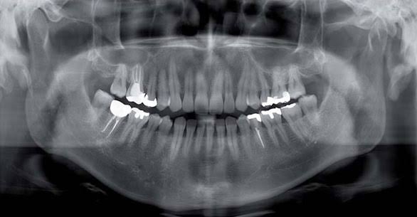
Ankara Intraoral X-Ray
In modern dental diagnostic protocols, intraoral X-rays are a “Micro-Radiological” diagnostic discipline used to analyze the finest details of teeth and surrounding tissues—details that may be lost in macroscopic images—at the micron level. This process, referred to in medical literature as “Intraoral Radiography,” involves placing digital sensors or phosphor plates directly inside the mouth to visualize the anatomical structure of a single tooth or a group of teeth from the root apex to the crown without any distortion. Redefining Ankara intraoral X-ray standards with the vision of “Digital Radiovisiography (RVG)” and “Lamina Dura Analysis,” Panorama Ankara provides patients with a professional diagnostic infrastructure capable of presenting early-stage cavities, canal calcifications, and micro-lesions within bone—often too small to be detected on panoramic films—in HD quality within seconds. The systems used in our clinic represent an advanced data-processing discipline that eliminates diagnostic error through intelligent sensors that minimize X-ray dosage with uncompromising clinical precision.
The true scientific success behind intraoral X-ray technology lies in the principle known in medical literature as the “Parallel Technique and Angular Precision,” where X-rays strike perpendicular to the long axis of the tooth to produce a true-scale image. In the advanced radiology unit of Panorama Ankara, “Digital Sensor (RVG)” technology enables images to be transferred instantly to a computer screen without the need for chemical processing. This technology allows the clinician to perform “Digital Contrast Adjustments” and “Millimetric Measurements” on the image, illuminating the exact length of root canals or the bone density around implants (densitometry) within seconds. This vision transforms intraoral imaging practices in Ankara from the simplicity of “taking a film” into an advanced “Diagnostic Engineering” center that converts the microscopic architecture of the tooth into a digital dataset.
Clinical Classification: Intraoral Radiography Techniques and Diagnostic Focus
The clinical value of an intraoral X-ray depends on which part of the tooth—root, crown, or surrounding bone—the selected technique emphasizes. Each radiographic methodology is optimized to detect a different pathology. To help patients clearly understand this advanced micro-imaging process, Panorama Ankara presents the technological hierarchy used in the clinic within the following scientific framework.
| Radiography Type and Technique | Biomechanical Imaging Area | Panorama Ankara Clinical Application Objective |
|---|---|---|
| Periapical Radiography (RVG) | Displays the tooth root apex and surrounding supporting bone tissue in high resolution. | Mapping root-end infections and periapical lesions with millimetric precision during root canal treatment processes. |
| Bitewing Radiography | Focuses on the occlusion of upper and lower teeth and the interproximal contact areas between them. | Detecting “hidden interproximal cavities” at their earliest stage—cavities invisible to the naked eye and often concealed on panoramic films. |
| Occlusal Radiography | Scans the entire dental arch from above or below with a wide-angle view. | Analyzing the position of impacted teeth in the palate, salivary gland stones, and large cystic formations holistically. |
| Digital Subtraction Radiography | An advanced method that digitally compares differences between two X-rays taken over time. | Monitoring the healing speed of a treated cyst or millimetric changes in bone resorption at a cellular level. |
Radiovisiography (RVG): Diagnosis at the Speed of Light with Maximum Safety
The greatest revolution in intraoral X-ray procedures is the RVG system, known in medical literature as “Direct Digital Radiography.” In the protocols implemented at Panorama Ankara, highly sensitive sensors—unlike traditional films—require significantly less X-ray radiation to capture images. This reduces the patient’s radiation exposure by up to 90%, fulfilling the “ALARA” (As Low As Reasonably Achievable) principle at the highest level. Data from the sensor appears on the clinician’s screen within seconds, and through “Color Scale Analysis,” even the smallest areas of demineralization (early cavities) in the enamel become clearly visible. This process transforms diagnosis from estimation into the highest-level “Biosafe Diagnostic Practice” that illuminates the inner world of the tooth with digital clarity.
Diagnostic Data Management and Maintenance Discipline After X-Ray Imaging
For your intraoral images—captured with digital sensors and processed in HD resolution in our clinical studios—to guide the success of your treatment and serve as a protective shield, regular monitoring is essential. The radiology board of Panorama Ankara presents the following two fundamental rules as a diagnostic constitution for continuity of diagnosis:
- Since intraoral X-rays show the “momentary” status of teeth, especially in teeth with fillings or root canal treatments, “Strategic Periapical Screening” should be performed at least once a year—even if there are no symptoms—to detect subtle changes beneath restorations and compare them with previously archived digital images.
- In areas where implant surgery or advanced surgical procedures have been performed, micro-focused follow-up X-rays should not be neglected at intervals determined by the clinician in order to confirm the continuity of “Biological Compatibility” (osseointegration) between bone and material and maintain data integrity.
Discover the Hidden Details of Your Teeth with Panorama Ankara
Uncertain sensitivity in your teeth, doubts beneath old fillings, or situations that appear “normal” on panoramic films yet still cause discomfort are not issues to be dismissed with superficial examinations. On the contrary, in expert hands, they are professional processes that can be clarified within seconds using digital intraoral X-ray technologies and periapical precision protocols—preventing the loss of your tooth. In your search for Ankara intraoral X-ray services, Panorama Ankara combines “Digital Radiovisiography” and “Bitewing Diagnostics” protocols to free you from undiagnosed pain, unnecessary radiation concerns, and delayed treatments with scientific elegance. We respect the unique microscopic architecture of your teeth with absolute care, rebuilding your health and comfort pixel by pixel.
To map hidden cavities between your teeth and concealed infections at your root tips within seconds using our digital sensors, determine the most suitable “RVG Diagnostic” path without missing any detail, and see every aspect clearly on the screen before treatment begins, you can schedule a detailed micro-diagnostic examination appointment at Panorama Ankara clinic and entrust your health to the safe hands of experience and modern radiology science.