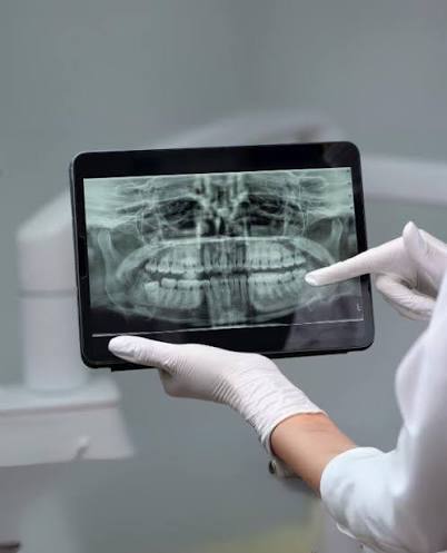
Ankara Panoramic X-Ray
In modern dentistry, successful and sustainable treatment planning begins not by focusing only on a single tooth that hurts, but by being able to interpret—on a single screen—the complex biomechanical relationship among the lower and upper jawbones, the sinus cavities, the jaw joints (TMJ), and the entire dental alignment. A panoramic X-ray, referred to in medical literature as an “Orthopantomogram (OPG),” is a fundamental “Macroscopic Screening” system that transforms the U-shaped human jaw skeleton into a two-dimensional wide-angle map by flattening it through a special scanning technology. Redefining Ankara panoramic X-ray standards with the clarity of the “Focal Corridor” (Focal Trough) and the vision of “Extraoral Comfort,” Panorama Ankara provides a professional diagnostic infrastructure that delivers a general radiograph of the entire oral flora and bony framework within seconds—without placing any foreign object inside the mouth. The systems used in our clinic are a reliable clinical radar discipline that detects impacted wisdom teeth patients may not even be aware of, silently growing jaw cysts, and generalized bone loss (periodontal) patterns before symptoms appear.
The true scientific success behind panoramic imaging technology lies in the precise trajectory the device traces around the patient’s head—known in medical literature as “Synchronized Rotation Physics.” In the advanced diagnostic unit of Panorama Ankara, once the patient is positioned in the device, the X-ray tube and the digital sensor move simultaneously in opposite directions. During this motion, only the tissues located within the horseshoe-shaped focal zone called the “Sharpness Channel” (Focal Trough) are captured clearly, while anatomical structures positioned in front of or behind the jaw (such as the spine) are intentionally blurred and effectively removed from the image. Thanks to this remarkable physical filtering, the developmental harmony of the right and left halves of the jaw (Bilateral Symmetry), the position of the jaw joint (condyle) within its socket, and the general distance of tooth roots to nerve canals can be analyzed at a glance. This vision elevates diagnostic practice in Ankara from a narrow perspective to an advanced “Holistic Medicine” center that plans the treatment pathway while considering all anatomical relationships.
Clinical Classification: Macroscopic Examination Areas and Diagnostic Algorithm
The clinical value of a panoramic X-ray lies not only in showing cavities, but in providing an immense “Road Map” for all subspecialties of dentistry (orthodontics, surgery, periodontology). While localized (periapical) X-rays show the trees, a panoramic X-ray lets us observe the entire forest from a bird’s-eye view. To help patients clearly understand the importance of this wide-angle imaging system, Panorama Ankara presents the diagnostic methodology used in our clinic within the scientific framework below.
| Diagnostic Focus | Biomechanical Analysis and Imaging Objective | Panorama Ankara Clinical Approach |
|---|---|---|
| Asymptomatic Focal Infections | Detecting cysts (radiolucent lesions) that grow deep within the jawbone without causing pain or swelling. | Stopping silent infections early through timely surgical intervention before they lead to tooth extraction or bone loss. |
| Impacted Teeth and Eruption Dynamics | Evaluating the angulation (Horizontal/Angled/Vertical) of wisdom teeth or canines within the jawbone. | Mapping in advance the potential pressure on adjacent tooth roots for orthodontic or surgical planning. |
| Periodontal (Bone) Level Analysis | Assessing the general pattern of resorption in the alveolar bone crests supporting the teeth and identifying asymmetric tissue loss. | Documenting the rate and extent of bone destruction with a wide-angle view in full-mouth gum disease (Periodontitis) treatments. |
| Orthodontic Development (Mixed Dentition) | In pediatric patients, identifying the number, position, and missing permanent tooth buds (agenesis) developing beneath primary teeth. | Determining skeletal growth direction and initiating preventive/interceptive orthodontic treatments on time. |
Extraoral Comfort and Low-Dose Radiology Discipline
The discomfort caused by small X-ray plates (sensors) placed inside the mouth can turn the diagnostic process into a nightmare—especially for young children or patients with a strong gag reflex. One of the greatest advantages of panoramic imaging is that it is an “Extraoral” (outside-the-mouth) system: the device rotates around the patient without touching the face or lips. At Panorama Ankara, these patient-friendly systems are also managed with a firm discipline in radiation safety. In this process—where holistic diagnostic power meets biological safety—our clinic safeguards radiology protocols through the following two fundamental principles:
- Because the imaging sensors scan from outside the mouth, the entire jaw anatomy can be reproduced within seconds—without trauma or physical strain—even in patients with severe nausea reflex, jaw locking (trismus), or limited mouth opening.
- Compared to capturing the whole mouth with many small individual films, the total radiation dose delivered by digital panoramic devices is dramatically lower; the use of a lead apron during the procedure is standard, and the medical ethical principle of ALARA (As Low As Reasonably Achievable) is followed at the cellular level.
Focus on the Big Picture of Your Health with Panorama Ankara
When you sit in the dental chair, settling for only the visible part of your mouth—or neglecting painless regions radiologically—is an incomplete approach that can invite future health crises. In expert hands, mapping your entire jaw framework with wide-angle digital sensors is a professional process that safeguards your health. In your search for Ankara panoramic X-rays, Panorama Ankara combines “Bilateral Symmetry Analysis” and “Macroscopic Screening” protocols to free you with scientific elegance from cysts discovered too late, incorrect treatment planning, and unexpected tooth loss. We respect the unique curved architecture of your jaw skeleton 100% and read every detail nature hides with you on our high-resolution panoramic screens.
To scan the overall health status of all your teeth, root connections, and jaw joints within seconds using our extraoral sensors; to map the most suitable “Preventive” or “Restorative” pathway without missing any detail; and to experience that holistic sense of safety every time you look in the mirror, you can schedule a detailed first examination and diagnostic consultation appointment at Panorama Ankara and entrust your health to the safe hands of experience and modern technology.