Ankara Periapical X-Ray
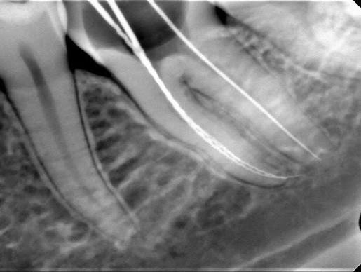
In modern dentistry, the foundation of successful treatment relies on a flawless radiological vision capable of detecting cellular-level destruction long before it becomes visible to the human eye or general screening radiographs. The system known in medical literature as “Periapical Radiography” is an isolated imaging technology that, unlike panoramic films showing the entire mouth from a distance, maps the complete anatomical boundaries of a single suspected tooth—from the crown (top) to the root tip (apex) deep within the jawbone—with millimetric resolution. Redefining Ankara periapical X-ray standards through the vision of “RVG (Radiovisiography) Sensors” and “Apical Tissue Resolution,” Panorama Ankara provides patients with a professional diagnostic infrastructure that displays the nerve canals inside the tooth and the thin bone wall surrounding the root (lamina dura) with exceptional clarity. The systems used in our clinic represent a reliable clinical navigation discipline that detects hidden cavities within the tooth or silent cysts within the bone before it is too late for treatment.
The real scientific strength behind periapical imaging technology lies in the digital traces left by X-rays as they pass through tissues of varying densities—a principle known in medical literature as “Radiolucency and Radiopacity Analysis.” In the advanced diagnostic unit of Panorama Ankara, ergonomic digital sensors placed inside the patient’s mouth scan the tooth in a fraction of a second and instantly transmit the image to high-resolution screens. Unlike traditional films that require chemical processing, these digital systems immediately reveal enamel thickness inside the tooth, dentin deterioration, and the dark “radiolucent” spaces of bone loss caused by inflammation (granuloma) at the root tip. Operating under the “ALARA” principle (As Low As Reasonably Achievable), this technology reduces the radiation dose received by the patient to a level even lower than natural daily background radiation. This vision transforms X-ray procedures in Ankara from a simple “film-taking” process into a “Micro-Diagnostic” center that determines the biological fate of the tooth and prevents unnecessary surgical interventions.
Clinical Classification: Dental Radiology Systems and Focused Diagnostic Depth
The key to success in dental diagnosis is selecting the correct imaging modality according to the patient’s complaint and the location of the pathology (disease) being investigated. Planning for jaw surgery and monitoring a single tooth during root canal therapy require completely different radiological perspectives. To help patients clearly understand these advanced diagnostic tools and why a periapical X-ray is necessary, Panorama Ankara presents the methodology used in our clinic within the scientific framework below.
| Type of Radiological Imaging | Biomechanical Scope and Diagnostic Focus | Panorama Ankara Clinical Application Goal |
|---|---|---|
| Periapical X-Ray (Isolated Target) | A radiograph that covers only 2–3 teeth and displays the root tip and the space connecting the tooth to bone with the highest level of detail. | The gold standard for root canal measurements, detection of apical cysts, and monitoring bone levels around implants. |
| Bitewing X-Ray | A radiograph showing the crowns (chewing surfaces) of upper and lower posterior teeth while the patient bites down. | Used to detect hidden “interproximal cavities” between teeth that toothbrushes cannot reach. |
| Panoramic X-Ray (General Screening) | A wide-angle scan displaying the entire upper and lower jaws, sinus cavities, and jaw joints in a single image. | Provides a roadmap for initial examination, evaluation of impacted wisdom teeth, and overall bone loss assessment. |
| Dental Tomography (3D CBCT) | A device that displays teeth and jawbone in three-dimensional slices (coronal, sagittal, axial). | Used to measure bone volume for implant surgery and calculate the distance to nerve canals with millimetric precision. |
Apical Tissue Resolution: Detecting Hidden Cysts
A tooth that appears healthy and free of decay from the outside does not necessarily mean there is no significant bone destruction at its root tip. Trauma or silently progressing infections can secretly kill the tooth’s nerve (pulp), forming an inflammatory mass at the apex. This condition, known in medical literature as “Apical Periodontitis,” may remain hidden within the broad perspective of panoramic X-rays but can be detected within seconds through the sharp and focused “apical tissue resolution” of a periapical X-ray. In the endodontic (root canal) protocols applied at Panorama Ankara, these targeted micro-images are taken before the procedure, during measurement of canal length, and at the completion of treatment. They confirm with enormous optical precision whether the canal filling ends exactly at the root tip. This represents the highest level of medical practice, where treatment is not left to chance and biological boundaries are secured with radiological evidence.
Radiological Safety and Post-Diagnostic Maintenance Discipline
Next-generation RVG periapical X-ray systems used in dentistry emit up to 90% less radiation compared to older film-based systems, protecting human health at the cellular level. Transforming this powerful digital navigation into clinical success depends on disciplined management of the process. The Panorama Ankara radiology and diagnostic board presents the following two fundamental rules as a “safety constitution” to ensure long-lasting success of applied treatments:
- If root canal treatment or surgical intervention has been performed due to a lesion (cyst) detected at the root tip, a “control periapical X-ray” should be taken 6 months and 1 year after treatment to document the regeneration process (osteogenesis) of the previously infected bone tissue.
- Although the radiation dose is even lower than the natural radiation from daily sunlight, according to the medical ethics principle of “ALARA” (As Low As Reasonably Achievable), thyroid-protective lead aprons provided by the dentist must always be used during X-ray procedures—especially for patients with suspected pregnancy.
Illuminate the Invisible with Science at Panorama Ankara
The mild sensitivity in your tooth whose source you cannot identify, the sudden pain when drinking hot tea, or the feeling of pressure in your jawbone are not threats that should remain hidden and grow over time. On the contrary, in expert hands, they are a professional process that can be instantly illuminated through digital RVG sensors and micro-diagnostic technologies. In your search for Ankara periapical X-ray services, Panorama Ankara combines “Isolated Root Analysis” and “Lamina Dura Isolation” protocols to free you with scientific elegance from misdiagnoses, unnecessary treatments, and unexpected tooth loss. We respect the hidden world of your tooth within the bone 100% and illuminate nature’s darkest regions with the power of digital pixels to secure your oral health.
To map the cellular activity at the root tip of the tooth that is causing pain or concern using our radiological sensors, determine the most appropriate “endodontic” or “restorative” treatment path before damage progresses, and achieve the reassuring feeling of certainty every time you look in the mirror, you can schedule a detailed diagnostic consultation at Panorama Ankara and entrust your health to the safe hands of experience and modern diagnostic science.