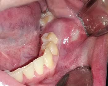
Ankara Dental Cyst Surgery
Due to their anatomical structure, the oral, dental, and jaw tissues are among the most common regions in the human body where pathological formations—especially cystic lesions—are encountered. Defined in medical literature as pathological cavities filled with fluid, semi-fluid material, or gas and surrounded by an epithelial membrane, cysts are known as the silent and insidious destroyers of the jawbones. In the early stages of development, dental cysts can often continue to grow slowly within the depths of bone tissue for months or even years without causing any pain, swelling, or discomfort. This asymptomatic progression may remain hidden until the cyst is detected incidentally during a routine radiological examination, or until the cyst volume weakens the jawbone and creates a visibly noticeable facial asymmetry. Adopting a multidisciplinary approach in Ankara dental cyst surgery, Panorama Ankara applies modern maxillofacial surgery protocols with microscopic precision to remove these insidious lesions from the area without harming surrounding tissues, healthy tooth roots, or nerve networks. The primary goal of the surgical process is not only the physical removal of the cyst, but also biomechanical restoration of jaw integrity by refilling the cavity created in the bone with healthy new bone tissue.
The origin of cystic formations in the jawbones is most often associated with infected, decayed teeth that have been left untreated in the mouth for a long time. In this process, which begins when the tooth’s nerve tissue (pulp) loses vitality, bacteria and toxins accumulating at the root tip engage in a battle with the body’s defensive cells within the bone, first forming a “granuloma” (a small inflamed tissue). In order to prevent the infection from spreading into healthy bone marrow, the body’s own defense mechanism starts to wall off the inflamed area with an epithelial cell barrier. Over time, tissue fluids enter this balloon-like enclosed structure due to osmotic pressure differences, and the lesion begins to swell and expand. As the cyst grows, it exerts hydrostatic pressure on the surrounding healthy bone tissue and causes bone resorption. To stop this destructive potential, Panorama Ankara’s maxillofacial surgery specialists analyze the patient’s general medical condition and the lesion’s detailed map within the jaw, then meticulously determine the most appropriate surgical intervention strategy based on the type of pathology.
Classification of Jaw Cysts: Radicular and Dentigerous Cysts
Jaw cysts are classified across a wide spectrum in medicine according to their cellular origin and developmental mechanisms. Correctly diagnosing the nature of the lesion is vital in determining the treatment protocol. The most commonly encountered cyst types in maxillofacial surgery clinics are tooth-origin (odontogenic) cysts. Their behavior patterns, growth rates, and recurrence potentials differ significantly from one another. Based on our patients’ radiological findings, you can review the anatomical and clinical features of the two principal odontogenic cyst types most frequently managed at Panorama Ankara—each requiring different surgical planning—in the comparative table below.
| Clinical Characteristics | Radicular Cysts (Apical/Root Tip Cysts) | Dentigerous Cysts (Follicular Cysts) |
|---|---|---|
| Cause (Etiology) | Chronic infections at the root tips of teeth with necrotic pulp due to deep decay or trauma. | Expansion of the developmental sac (follicle) surrounding an impacted tooth (especially wisdom teeth) as it fills with fluid. |
| Status of the Associated Tooth | The tooth has lost vitality (non-vital). There is often a history of failed or incomplete root canal treatment. | The tooth is fully vital but trapped within the jawbone (has not erupted). |
| Location Within the Jaw | Most frequently localized at the root tips (periapical region) of anterior or posterior teeth. | Typically surrounds the crown regions of mandibular wisdom teeth or impacted maxillary canines. |
| Surgical Treatment Approach | Removed together with the cyst lining. The related tooth may require root canal retreatment or apical surgery (apical resection). | While the cyst epithelium is completely removed, the impacted tooth causing the cyst is usually extracted surgically along with it. |
3D Volumetric Diagnosis and Boundary Mapping at Panorama Ankara
The greatest anatomical challenge in cyst surgeries is clearly determining the boundaries of the lesion and protecting vital surrounding structures (nerves, vessels, adjacent tooth roots, and the maxillary sinus membrane). While standard two-dimensional dental X-rays can show only the width and height of a cyst, they may provide misleading information about its depth within the vertical thickness of the jawbone (ridge width). A cyst that appears to be resorbing the root of a neighboring tooth on a 2D image may, in fact, pass at a much greater distance from the tooth in three-dimensional space. To eliminate such optical misinterpretations and minimize surgical risks, Panorama Ankara positions advanced 3D Volumetric Dental Tomography (CBCT) systems as a standard diagnostic tool before all cyst operations. In this way, the jawbone is examined layer by layer in a virtual environment, the cyst’s three-dimensional volume is calculated, and the millimetric surgical strategy is established long before the patient sits in the surgical chair.
Large cysts located in the upper jaw may, over time, extend toward anatomical spaces known as the maxillary sinuses and push the sinus lining (Schneiderian membrane) upward or thin it. In the lower jaw, the inferior borders of a cyst may form a dangerous proximity to the inferior alveolar nerve canal, which provides sensation to the lower lip and chin. With tomography-supported virtual navigation, the surgeon knows exactly how close they can safely approach these critical regions. When necessary, instead of rotating instruments (burs) for bone cutting and cyst curettage, ultrasonic “Piezoelectric Surgery” devices are used—tools that act only on bone but do not harm soft tissues (nerves and membranes) upon contact. This integration of technological equipment greatly increases the safety margin of cyst operations and reduces the theoretical risk of postoperative numbness or sinus complications to minimal levels.
Modern Protocols in Cyst Surgery: Enucleation and Marsupialization
In the treatment of jaw cysts, there is no medical “wait and see” approach; every detected pathological cavitation must eventually be surgically removed, as it will sooner or later lead to bone destruction. In Panorama Ankara’s clinical practice, two main surgical methods are preferred depending on the cyst’s size, its relationship with adjacent structures, and the patient’s anatomical characteristics: Enucleation and Marsupialization. “Enucleation” is a procedure used for small to medium-sized cysts, where the cyst is removed in one piece—without disrupting its lining—much like a walnut separated from its shell, by carefully stripping it from its bony bed. Thorough curettage of the cyst capsule with specialized surgical curettes to ensure no epithelial remnants remain is the most critical step in preventing recurrence in that region. All removed cystic tissues are routinely sent to pathology laboratories for histopathological examination to confirm the lesion’s nature at the cellular level.
However, in some neglected cases, a cyst may occupy such a large portion of the jawbone that removing it in a single session (via enucleation) could cause a pathological fracture of the paper-thin bone walls or damage major nerves in the area. In such massive cysts, Panorama Ankara specialists perform a far more controlled, two-stage conservative surgical method called “Marsupialization” (Decompression). In this method, a small window is first created in the roof of the cyst to drain the high-pressure fluid inside, and the cyst lining is sutured to the oral mucosa to keep the window open. Once the internal fluid pressure is eliminated, the cyst begins to shrink gradually over months, contract, and move away from the bone. When the cyst reaches a safe, manageable size, it is then completely removed in a second and final procedure (enucleation). In this way, jaw integrity and vital structures are protected from excessive surgical trauma.
Post-Surgical Tissue Regeneration and Cavity Management
After successful cyst surgery, a cavity remains in the bone corresponding to the volume occupied by the cyst. Thanks to the body’s remarkable healing capacity, small cavities gradually fill with a blood clot and enter a natural ossification process on their own. However, it may not be biologically possible—or it may take many years—for very large voids left after extensive cysts to completely fill with bone tissue. To prevent these voids from filling with soft tissue and leaving that region of the jaw weakened, Panorama Ankara utilizes advanced tissue engineering techniques. The cavity created by the removed cyst is filled and covered with highly biocompatible, sterile bone grafts (bone powders) and barrier membranes placed over the graft material. This grafting process accelerates new and robust bone formation (osteoinduction), helps the jaw regain its former strength, and prepares a solid biomechanical foundation so that implants can be safely placed in that region in the future.
For patient comfort and for the grafted area to complete cellular healing without infection, the patient’s active participation in postoperative care is essential. During the first few days after surgery, mild swelling (edema) and minimal sensitivity are expected as part of the natural tissue repair process. To ensure this period is as smooth and uncomplicated as possible, it is essential that our patients take prescribed antimicrobial and analgesic medications completely and at the times specified by the physician. In addition, applying intermittent cold compresses (ice) externally over the wound area for the first 48 hours, avoiding hard and excessively hot foods that may irritate the surgical site, and strictly abstaining from tobacco products that impair tissue oxygenation and can lead to graft failure are considered the gold standards of the healing period.
Transparent and Reliable Approaches to Your Oral Health with Panorama Ankara
Cystic lesions hidden within the jawbone can naturally appear frightening and anxiety-inducing for patients. However, at the point modern medical science has reached, Ankara dental cyst surgery processes have become highly predictable, safe, and comfortable procedures thanks to advanced imaging technologies, microsurgical instruments, and high-quality biomaterials. With its patient-centered vision, Panorama Ankara not only treats the disease but also alleviates concerns by communicating every stage of the process with digital visuals and a transparent, easy-to-understand language. The goal is to regenerate tissues affected by bone loss and preserve the functional integrity of the oral structure for many years.
Considering the insidious progression of cysts, the most effective way to protect oral health is to undergo regular radiographic check-ups even if you have no complaints. Especially if you experience mild sensitivity in teeth that previously underwent root canal treatment, small pimple-like swellings on the gums, or a sense of asymmetric fullness in the jaw area, it is crucial to consult an expert maxillofacial surgeon without delay. To secure the health of your jawbone, detect hidden pathologies before they grow, and receive detailed information about the modern surgical treatment protocols you may need, you can schedule a comprehensive tomography and consultation appointment at Panorama Ankara clinic.