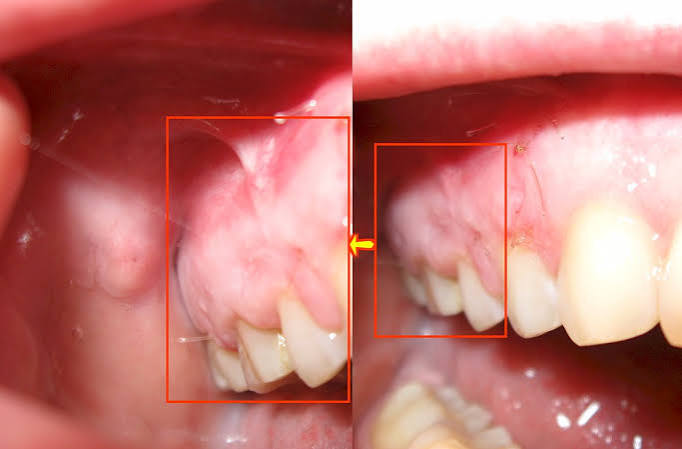
Ankara Intraoral Cyst Surgery
The oral cavity is not merely a mechanical grinding center that houses the teeth and jawbones; it is also an extraordinarily dynamic, continuously renewing, secretion-producing soft-tissue (mucosal) ecosystem that lines the inner cheeks, lips, sublingual area, and palate. Just beneath this rich ecosystem lie hundreds of minor salivary glands that keep the mouth constantly moist and help initiate digestion. However, this flawlessly functioning mechanism can sometimes be disrupted by involuntary biting of the lips and cheeks during speaking or chewing, chronic friction, micro-traumas caused by orthodontic wires, or blockage of salivary ducts. Biologically, the vast majority of cysts that develop in the soft tissues of the mouth are completely different from destructive tumoral cysts within bone; they are generally benign sacs filled with fluid or semi-fluid content, formed when an injured salivary gland cannot discharge its secretion into the mouth and instead accumulates (pools) beneath the tissue. Embracing advanced microsurgical protocols in the field of Ankara intraoral cyst surgery and soft-tissue surgery, Panorama Ankara successfully removes these mucosal pathologies—which can instantly reduce eating, drinking, and speaking comfort to zero—using atraumatic (tissue-respecting) incisions that maximize preservation of tissue integrity, while causing no harm to major salivary ducts or neural networks in the surrounding tissues. What appears alarming to our patients becomes a highly safe, rapid-healing medical intervention—often resolved within minutes in the hands of our specialist oral and maxillofacial surgeons.
In medical literature, intraoral cysts are named differently according to their clinical appearance and the specific anatomical region in which they develop. The most common pathology—and one of the leading reasons for clinical visits—is the “Mucocele” lesion, which typically appears on the inner surface of the lower lip like a bluish, translucent pearl. In individuals with a habit of biting or eating the lip, the delicate duct of a minor salivary gland in the lower lip may rupture or become obstructed. The gland continues producing saliva (mucin), but because the fluid cannot drain outward, it leaks into surrounding tissues (the Extravasation Phenomenon) and becomes trapped there. This trapped fluid forms a cystic lining (a pseudo-wall), gradually enlarges, ruptures, and then swells again. This vicious cycle can persist for months unless the cyst is completely removed surgically. Another—and larger—type of cyst is the “Ranula,” which develops under the tongue in the floor of the mouth and swells like a frog’s belly. Ranulas are broader fluid accumulations caused by injury or obstruction in the ducts of the sublingual salivary gland, which is a major (not minor) gland. When they enlarge, they may push the tongue upward, restricting movement and making swallowing difficult. Thanks to Panorama Ankara’s advanced maxillofacial expertise, these major soft-tissue cysts are treated safely by working within millimetric proximity to vital sublingual nerves (the lingual nerve) and blood vessels.
Microsurgical Protocols: Excisional Biopsy and Pathological Examination
Surgical treatment of soft-tissue cysts is not a simple drainage procedure such as emptying the fluid or popping the cyst with a needle. If the fluid is only aspirated with a needle, the damaged minor salivary gland that caused the cyst will remain alive under the tissue, and the cyst will recur within a few days—often larger than before (high recurrence rate). To prevent this medical failure, Panorama Ankara applies the “Excisional Biopsy” protocol. Under local anesthesia, after complete numbness of the lip or cheek area, a fine, aesthetic elliptical (almond-shaped) incision is made directly over the cyst. The surgeon gently removes not only the fluid-filled cystic sac, but also the tiny, cauliflower-like lobules of the problematic minor salivary gland located at the base of the cyst (the connected damaged tissues) together with the cyst wall, as a single piece (en bloc).
This excisional surgical approach is the only definitive (radical) solution that ensures the cyst will not regrow in that area. After the procedure, the mucosal tissue is closed with very fine, aesthetic micro-sutures that typically dissolve (resorb) on their own when exposed to saliva and do not leave a scar. The oral mucosa is among the most highly vascularized tissues in the body and has a remarkable rate of cellular renewal (mitosis); the delicate incision line in the lip or cheek usually heals within 7 to 10 days, returning to its former smooth texture with virtually no visible mark. In accordance with procedural standards and universal medical rules, the removed cystic tissue is always sent to pathology laboratories for histopathological examination to establish a definitive cellular diagnosis and to formally confirm its benign nature under the microscope.
Clinical Classification: Intraoral Soft-Tissue Cysts and Lesions
Not every swelling in the mouth arises from the same anatomical source. Understanding the nature of these lumps—felt with the tongue or fingers—helps patients avoid unnecessary panic. To provide transparent clinical awareness, Panorama Ankara details the most commonly encountered mucosal (soft-tissue) cyst types in the oral cavity and their specific surgical dynamics in the table below.
| Cyst Type (Pathological Classification) | Anatomical Site & Clinical Appearance | Surgical Strategy & Recurrence Risk |
|---|---|---|
| Mucocele (Mucocele / Extravasation Cyst) | Painless, bluish, translucent fluid-filled bubbles typically seen on the inner lower lip or cheek mucosa. | Removed with the underlying damaged minor salivary gland (Excisional Biopsy). If the gland is completely removed, it does not recur. |
| Ranula (Major Sublingual Cyst) | Large, tense cystic enlargements in the floor of the mouth beneath the tongue, resembling a frog’s belly. | Treated with marsupialization (opening and converting it into a pouch) or removal of the sublingual gland, depending on size. |
| Eruption Cyst | Purple-blue swellings on the gum directly over an erupting (emerging) tooth, especially in children. | Often ruptures spontaneously; if not, a small incision is made under local anesthesia to open the eruption pathway. |
| Dermoid / Epidermoid Cysts | Doughy-feeling swellings of developmental (embryological) origin located deeper in the tissues of the floor of the mouth. | Contain dense tissue rather than fluid. Complete one-piece removal with the capsule (enucleation) is essential. |
Marsupialization Technique: Pressure-Reduction Engineering in Giant Cysts
Cysts in the floor of the mouth or palate may sometimes grow to such massive sizes (for example, a giant ranula) that attempting to remove the cyst as a single piece could increase the risk of damaging vital surrounding structures, such as major salivary ducts (Wharton’s duct) or lingual nerves. To avoid harming adjacent tissues in such extreme cases, Panorama Ankara’s advanced maxillofacial specialists apply an exceptional tissue-engineering technique known in the literature as “Marsupialization.” The goal of this technique is not to rip the cyst out of its site, but to permanently eliminate the relentless internal pressure within the cyst.
The surgeon creates a round or oval “window” incision in the roof of the cyst and completely evacuates its dense fluid content. Then, by suturing the edges of the cyst lining (inner wall) to the edges of the oral mucosa (outer wall) with micro-sutures, the cyst is transformed from a closed balloon into a permanently open small “pouch” that communicates with the oral cavity. Thanks to this open window, the cyst lining gradually converts into normal oral mucosa; any fluid that accumulates drains immediately into the mouth, and the cyst slowly shrinks and disappears over months. This atraumatic approach is one of the most elegant, protective jaw-surgery protocols for eliminating massive cysts through biological remodeling—without causing any harm to vital anatomy.
Post-Operative Mucosal Healing and Patient Discipline
When intraoral cyst surgeries are performed with advanced surgical instruments and laser-assisted scalpels, they do not create externally visible swelling (edema) or bruising on the cheek or lip. However, because the oral mucosa—especially under the tongue or on highly mobile lip areas—is constantly exposed to saliva, stretching, and chewing forces, maintaining suture stability becomes an important patient responsibility. To keep the incision line sterile and allow mucosal cells to unite uninterruptedly, the Panorama Ankara surgical team recommends that patients adopt the following two basic rules as a disciplined routine:
- During the first 7 to 10 days of mucosal healing, to prevent mechanical trauma (tearing) on the sutures and to avoid acid/spice irritation, completely avoid crunchy foods, extremely hot soups, acidic beverages, and hot-pepper/spicy foods; strictly follow the “Warm and Soft Diet” regimen prescribed by your clinician.
- To stop bacterial colonization (infection) at the incision line, beginning the day after surgery, support oral hygiene carefully with medical mouth rinses prescribed by the clinician—typically containing “Chlorhexidine” or “Hydrogen Peroxide”—twice daily; strictly avoid touching the surgical area with the tongue and avoid any reflexive pulling at the sutures.
Regain Your Intraoral Comfort with Panorama Ankara
Those small fluid sacs caused by biting your lips or inner cheeks—bursting with every meal and swelling again the next day, wearing down your psychology—are not an anatomical destiny you must tolerate for life. Acting with an advanced microsurgical vision in Ankara intraoral cyst surgery and soft-tissue pathologies, Panorama Ankara permanently removes these mucosal lesions from your life within minutes—painlessly—with either laser-assisted suture-free approaches or minimally incisional techniques. Our post-excisional biopsy pathology reporting documents your surgical safety at the highest medical standards.
If you have noticed a suspicious swelling under your tongue that is gradually enlarging and making swallowing difficult, or if you want to completely escape the vicious cycle of a stubborn mucocele on your lip, you can schedule a detailed soft-tissue (cyst) surgery appointment at Panorama Ankara to determine the lesion’s diameter and anatomical boundaries and to establish the most elegant microsurgical plan while protecting your tongue/lip nerves—so you can enjoy eating and speaking freely, without anxiety.