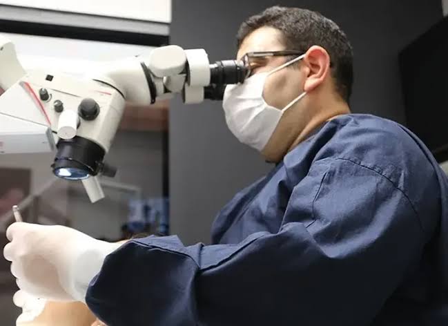
Ankara Root Canal Treatment Under a Microscope
In modern dentistry, root canal treatment has long since ceased to be a simple mechanical procedure in which the painful nerve is removed and the space is filled. Tooth roots, like the roots of a tree, advance deep into the jawbone and form a complex three-dimensional labyrinth containing microscopic branches (ramifications), curvatures, and additional canals that are impossible to see with the naked eye. In traditional methods, this procedure is performed “in the dark,” relying only on the clinician’s tactile sense and two-dimensional radiographs. At Panorama Ankara—where Ankara microscope-assisted root canal treatment standards are redefined through the vision of the “Dental Operating Microscope (DOM)” and “Coaxial Illumination”—this treatment becomes a scientific illumination. Offering patients a field of view that can reach the deepest cellular layers of the tooth under up to 25x optical magnification, our clinic is a professional endodontic rehabilitation center that preserves teeth that were said to require extraction, fail to heal despite infection, or are anatomically extremely challenging—through unwavering clinical engineering and microsurgical precision.
The true scientific achievement behind micro-endodontic technology lies in the principle known in the medical literature as “Complete Chemo-Mechanical Preparation”: the near-total removal of organic tissue remnants and anaerobic bacteria inside the root canal system. In Panorama Ankara’s advanced endodontics unit, under the microscope’s massive magnification and shadow-free light, fourth or fifth canals that the human eye can easily miss (especially the famous MB2 canal in upper molars) are identified with millimetric accuracy. Narrowed (calcified) canals are opened under the microscope using specialized ultrasonic tips—without perforating the tooth. This atraumatic approach preserves the remaining healthy dentin structure (root walls) at the maximum level, while biologically minimizing the risk of the infection forming a cyst at the root tip or the treatment failing years later (recurrence). This vision transforms root canal therapy in Ankara from a guesswork trial-and-error process into an advanced “Tissue-Saving” discipline in which every millimeter is clearly controlled by the clinician.
Clinical Classification: Micro-Endodontic Indications and Analysis of Anatomical Challenges
The success of microscopic root canal therapy (Micro-Endodontics) depends on accurately resolving anatomical variations within the root or errors from prior procedures through “Optical Mapping.” The microscope is indispensable not only for locating canals, but also as a navigation device for repairing cellular-level damage within the root. To help patients clearly understand this advanced technological process, Panorama Ankara presents the micro-intervention methodology used in our clinic in the scientific framework below.
| Intra-Canal Complication | Biomechanical Challenge and Optical Target | Panorama Ankara Microscopic Solution |
|---|---|---|
| Hidden and Accessory Canals (MB2) | Accessory nerve pathways that cannot be seen with the naked eye and, if left untreated, can lead to near-certain treatment failure. | Detection of these canals under 25x optical magnification and elimination of infection using specialized micro-files. |
| Calcified (Obstructed) Canals | Closure of the root canal by dentin deposition due to aging or trauma. | Overcoming the obstruction micron by micron with ultrasonic tips—without accidental root perforation. |
| Removal of a Separated Instrument (Retreatment) | Fragments of nickel-titanium files broken and left inside the canal during a previous failed root canal treatment. | Microscopic troughing around the separated fragment and retrieval from the canal using specialized micro-lasso instruments. |
| Repair of Perforation (Root Perforation) | Iatrogenic (clinician-induced) defects created when the root is accidentally opened toward the jawbone due to improper bur use. | Microscopic identification of the defect and cellular-level sealing with biocompatible materials such as bioceramics (MTA). |
Apical Delta Cleaning and Three-Dimensional Obturation (Filling)
The heart of root canal treatment is the “Apical Third,” the most terminal area where the tooth root meets the jawbone. In this region, the main canal branches like a river delta (Apical Delta) and opens into the bone. In the micro-endodontic protocols applied at Panorama Ankara, this delta area is irrigated by foaming and activation using laser and sonic activation devices (Irrigation). After verifying under the microscope that the canal is completely sterile and dry, three-dimensional obturation (filling) is performed with “Warm Gutta-Percha” (bio-rubber that becomes flowable when heated) techniques. The flowable material, under microscopic pressure, penetrates even into the capillary-like lateral canals beyond the main canal, locking the tooth from the inside out with a powerful seal (hermetic obturation). This is the most refined form of endodontic engineering, blocking bacteria from reaching the bone for a lifetime.
Biomechanical Maintenance and Restorative Discipline After Micro-Endodontics
A tooth whose nerve network and vascular bundle have been meticulously cleaned with microscopic precision may be completely free of infection; however, because it has lost vitality (non-vital tissue), it gradually loses flexibility over time and becomes more fragile. The Panorama Ankara restorative dentistry board presents the following two fundamental rules as a “life constitution” to ensure that this precious tooth—saved under the microscope—remains in the mouth with unwavering resistance against chewing forces:
- Between root canal sessions or immediately after the treatment is completed, until the final coronal restoration (porcelain inlay/onlay or crown) is finished, avoid chewing hard foods such as hazelnuts, roasted chickpeas, or crusty bread on the treated side to prevent vertical fractures.
- Because the cellular regeneration of an existing cystic lesion (bone loss) at the root tip back into bone by osteoblasts takes time, follow-up appointments at 6 months and 1 year should be maintained without interruption to confirm complete healing radiographically (X-ray).
Discover the Light in the Depths of Your Tooth with Panorama Ankara
The throbbing night pains you feel in your tooth, or the diagnosis that “this tooth must be extracted, the canals are blocked,” is not an irreversible fate that means you must lose that tooth. On the contrary, in expert hands, it is a professional process that can be repaired with a Dental Operating Microscope (DOM) and microsurgical instruments. In your search for Ankara microscope-assisted root canal treatment, Panorama Ankara combines “Coaxial Illumination” and “Apical Isolation” protocols to free you with scientific elegance from traditional “blind” treatments, recurring abscesses, and unnecessary tooth extractions. We respect the anatomical labyrinths of your teeth 100%, illuminating infected tissues left in darkness by nature with the microscope’s powerful light—restoring your roots to health.
To map the depth of infection or calcification in your tooth roots with our digital tomography (CBCT), design the most suitable “Micro-Endodontic Retreatment” or “Laser-Assisted Irrigation” route for you, and regain that healthy chewing sensation completely free of pain, you can schedule a detailed endodontic consultation appointment at Panorama Ankara and entrust your health to the safe hands of experience and modern science.