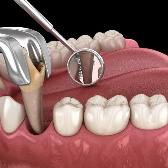
Ankara Surgical Tooth Extraction
In dental practice, removing a tooth that has been damaged beyond repair from the oral environment may not always be a standard and simple procedure (forceps extraction). Although “tooth extraction” is generally perceived by the public as merely gripping the tooth with an instrument and rocking it out of its socket, this basic approach becomes completely insufficient—and even dangerous—in many clinical scenarios where the tooth’s anatomy, the depth of decay, and the biological relationship between the tooth roots and the jawbone have been compromised. Teeth whose crowns (upper portions) have completely fractured due to decay, leaving only roots inside the bone; structures that have broken after root canal treatment and dropped below the gumline; or teeth that have become fused to the jawbone (ankylosed) after years of chronic infection—like glass “welding” to a frame—cannot be grasped with standard extraction instruments. When forced, they may cause serious fractures of the jawbone, extensive soft-tissue tearing, and severe pain that can last for weeks. In the field of Ankara surgical tooth extraction, Panorama Ankara applies advanced maxillofacial surgery principles to resolve such complex and challenging cases using “Atraumatic Microsurgery” techniques. This approach not only removes the problematic tooth but also microscopically protects the jawbone (alveolar ridge) and gum tissue—structures that are vital for future implant treatment.
Surgical tooth extraction is essentially the process of creating a controlled, millimetric surgical access window to reach the tooth or tooth root when it is not directly accessible. Tooth roots typically do not extend straight within the jawbone; they may have multi-root anatomies that are hook-shaped and curved, converge toward each other like needle-nose pliers, or diverge dramatically. If a multi-rooted molar is locked within bone and is forced out as a single unit, the roots may tear out a large portion of the surrounding jawbone as well (alveolar fracture). This major bone loss can nearly eliminate the patient’s chance of having an implant in that region in the future or may necessitate costly bone grafting surgeries that take months. To prevent these destructive biomechanical errors, Panorama Ankara’s expert oral and maxillofacial surgery team identifies the anatomical “lock points” of the roots with 3D Volumetric Tomography (CBCT) before separating the tooth from bone, and designs the surgical strategy entirely around the principle of “sacrificing the tooth—not the bone.”
Mucoperiosteal Flap and Osteotomy: Safe Access to the Surgical Field
A complicated surgical extraction procedure begins under local anesthesia with the clinician gently separating the gum tissue around the problematic tooth from the bone (raising a mucoperiosteal flap). Once the gum is reflected like a curtain, the underlying jawbone and the roots that are trapped inside the bone become fully visible. If the tooth has fractured far below the bony margin and there is no point of purchase to reach the root, our surgeons use special bone-friendly burs or piezosurgery devices that work with ultrasonic vibrations to create only a millimetric groove around the root (osteotomy). This microscopic bone removal creates the critical physical space needed for the root to mobilize and for surgical instruments (elevators) to engage.
In some multi-rooted teeth (two- or three-rooted), the root junction area (bifurcation/trifurcation) can become so integrated with bone that luxating (mobilizing) the tooth as a single unit is impossible. In such cases, Panorama Ankara’s advanced surgical protocol known as “Odontotomy” (sectioning the tooth) is employed. The clinician splits the tooth body and roots into two or three parts using high-speed surgical handpieces. Once separated, each root can be removed independently—like a small single-rooted tooth—through its own exit pathway, without applying even the slightest pressure to the surrounding bone, and can be lifted from the socket like a pearl within seconds. This bone-respecting micro-sectioning (atraumatic approach) miraculously minimizes postoperative cheek swelling, soft-tissue bleeding (hematoma), and pain compared to standard extractions.
Clinical Comparison: Simple Tooth Extraction vs. Complicated Surgical Extraction
Before patients sit in the treatment chair, understanding the biological scope of the procedure and the healing dynamics is valuable for managing postoperative expectations. The visible integrity of a tooth does not always mean the extraction will be simple; the determining factor is the root’s condition inside the bone. To provide transparent clinical information, Panorama Ankara details the anatomical and operational differences between simple (routine) extractions and advanced surgical extractions in the table below.
| Clinical and Operational Parameters | Simple (Routine) Tooth Extraction | Complicated Surgical Tooth Extraction |
|---|---|---|
| Anatomical Condition and Accessibility | The crown (upper part) is intact and easily visible in the mouth; it can be grasped with forceps. | The tooth is fractured at or below the gumline, completely crumbled due to decay, or ankylosed (fused) to bone. |
| Soft-Tissue Intervention (Incision and Sutures) | No scalpel incision is made in the gum; sutures are generally not needed afterward. | Raising a surgical flap is required, and the tissues must be closed with sutures at the end. |
| Approach to Bone and Odontotomy | No intervention to the jawbone; the tooth is mobilized and removed as a single unit from the socket. | Bone may be shaved to access the roots (osteotomy), and the tooth may be sectioned into multiple parts (odontotomy) for removal. |
| Postoperative Recovery | Patients usually return to normal eating and drinking the same day; pain and swelling are minimal. | Swelling control and cold compress are typically needed for the first 48 hours; complete cellular closure takes 7–10 days. |
Socket Preservation: Protecting the Implant Foundation on the Operating Chair
The most important concept that transforms surgical extraction from a standard “tooth removal” procedure into forward-looking tissue engineering is “Socket Preservation.” After extraction, a large void remains within the jawbone where the roots once were (the alveolar socket). The body gradually fills this space with a blood clot and converts it into new bone; however, during natural healing, the surrounding jawbone can undergo dramatic resorption and shrinkage—approximately 30% to 50% in width and height. Particularly in the anterior aesthetic zone or in posterior regions planned for implants, this bone collapse may make future implant placement physically impossible.
In Panorama Ankara’s visionary surgical protocols, immediately after the roots are removed atraumatically, the emptied socket is promptly filled by the clinician with sterile bone grafts (bone particles) and PRF (Platelet-Rich Fibrin) membranes derived from the patient’s own blood. This biological scaffold is then tightly closed with sutures. This process is called socket preservation. This small yet remarkable intervention performed simultaneously with the extraction blocks the jawbone’s natural resorption cycle. The three-dimensional bone volume—its height and thickness—is preserved as on day one. Months later, if implant placement is planned, the surgeon encounters bone of excellent thickness and quality. In this way, the patient avoids much larger, more painful, and more costly jawbone augmentation surgeries in the future through a single-session micro-intervention.
Postoperative Clot Stabilization and Alveolitis Management
The success of a surgical tooth extraction depends not only on the clinician’s technical excellence, but also on the patient’s informed approach during the first 48 hours of tissue healing (recovery) at home. The initial blood clot forming inside the extraction socket is essentially the biological “womb” for new bone and gum formation. When the clot remains firmly stabilized, the surgical site heals rapidly. However, if the clot is dislodged or disrupted, the bone becomes exposed and a painful inflammatory condition known as “Alveolar Osteitis” (Dry Socket / Alveolitis) can begin—often causing severe, throbbing pain that can prevent sleep. To prevent this painful complication and accelerate cellular healing, Panorama Ankara specialists recommend the following critical rules:
- To avoid “negative pressure” (vacuum) effects that can dislodge the vital blood clot, do not use a straw for the first 72 hours after surgery, avoid forceful rinsing/spitting, and strictly refrain from smoking, which immediately impairs capillary circulation and halts healing at the wound site.
- To suppress physiological edema (cheek swelling) that may occur in areas where a flap (incision) was performed, begin intermittent cold compress (ice) applications from the outside of the cheek immediately after the procedure, in 15-minute intervals, for the first 24 hours; and consume only lukewarm, soft foods (puree-like consistency) for the first two days.
Turn Difficult Goodbyes into Comfortable New Beginnings with Panorama Ankara
A tooth with only roots remaining, surrounded by chronic inflammation, or locked into bone is a “time bomb” that reduces your quality of life both aesthetically and functionally, and can turn into an acute abscess attack at any moment. In Ankara surgical tooth extraction services, Panorama Ankara combines advanced maxillofacial surgical experience with atraumatic, tissue-friendly technologies to remove even teeth deemed “unextractable” or “extremely difficult” without harming surrounding tissues—completely painlessly and safely.
Do not allow a broken root or an ankylosed tooth to erode your jawbone further from within. To map your bone structure with 3D tomography, analyze your root anatomies, and plan socket preservation that will create a solid foundation for future implant treatments, you can schedule a detailed surgical consultation appointment at Panorama Ankara and leave these challenging obstacles behind with expert care and high comfort.