Ankara Bone Grafting Before Implants: Alveolar Augmentation and Building a Biological Foundation with Panorama Ankara
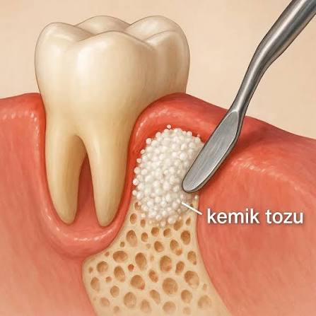
Dental implants—considered the gold standard of modern medicine for replacing missing teeth—are micro-engineered titanium roots placed into the jawbone to withstand chewing forces in the most natural way possible. However, for an implant to function healthily for years in the mouth, it requires a thick and dense volume of jawbone capable of absorbing tons of chewing pressure and gripping the implant tightly in three dimensions (both in width and in height). As a fundamental rule of human physiology, the body does not preserve tissue that is not used. When a tooth is extracted and the area is left empty for a long time, mechanical stimulation (chewing pressure) disappears in that region, and the jawbone begins to resorb and shrink rapidly within months (alveolar ridge atrophy). Past episodes of severe gum inflammation (periodontitis), traumatic and difficult extractions, or long-term use of removable dentures (palatal plates) can also accelerate this bone loss dramatically. In clinical situations where bone volume has become too thin or too short to host an implant, bone grafting (bone particulate) procedures before implants in Ankara come into play. Guided by advanced maxillofacial surgery principles, Panorama Ankara reconstructs the resorbed jawbone before implant placement using “Tissue Engineering” and “Guided Bone Regeneration” (GBR) protocols—creating a rock-solid, biological, brand-new foundation for your implants.
Bone grafting (augmentation) is not simply sprinkling a synthetic powder into the jaw; it is triggering the body’s own self-healing mechanisms (cellular repair capacity) with specialized biomaterials. Bone grafts (particulates or blocks) placed by Panorama Ankara’s expert maxillofacial surgeons into resorbed and insufficient jawbone regions work through three fundamental biological principles to generate living bone. The first is Osteoconduction: the graft particles serve as a micro-porous structural scaffold into which blood vessels and bone-forming cells (osteoblasts) can infiltrate and grow. The second is Osteoinduction: specific proteins within the graft stimulate inactive stem cells in the area, converting them into active bone-producing cells. The third is Osteogenesis: the graft directly contains living bone cells that initiate new bone formation immediately. Thanks to these advanced surgical protocols, many challenging cases that were once labeled “Implants cannot be placed here” or “Your bone is paper-thin” can be rehabilitated by expanding the jawbone both horizontally and vertically, preparing it perfectly for a standard and safe implant surgery.
Biomaterial Classification: Which Graft Type Is Right for You?
In bone augmentation procedures prior to implants, the choice of material is strategically determined by the clinician based on the size and shape of the defect (void) in the jawbone and the patient’s biological healing capacity. In medical literature, bone grafts are classified according to their source, and each type has a completely different biochemical remodeling rate. To ensure pharmacological transparency in the tissue engineering applied to patients, Panorama Ankara details the main graft types used in clinical practice in the table below.
| Graft Source & Biological Type | How It Is Obtained & Material Characteristics | Clinical Indication & Biomechanical Effect |
|---|---|---|
| Autogenous Graft (Patient’s Own Bone) | Living bone blocks or chips harvested from another area of the patient’s jaw (e.g., behind the wisdom tooth region). | The gold standard. The body recognizes it 100% (no rejection). Provides both a scaffold and immediate living cells (osteogenesis). Used for large defects. |
| Xenograft (Animal-Derived Grafts) | Bovine (cow) or porcine (pig) bone processed (sterilized) to remove all proteins, leaving only the mineral structure. | The most commonly used particulate grafts. Acts as a very strong osteoconductive scaffold, resorbs slowly, and preserves volume for a long time. |
| Allograft (Human-Derived Grafts) | Bone particulates sourced from human tissue banks, fully sterilized and stripped of antigenic properties. | Provides a scaffold and also encourages the patient’s own cells to form bone (osteoinductive). Healing is relatively fast. |
| Alloplast (Synthetic Grafts) | Completely artificial particles produced in laboratories from calcium phosphate, hydroxyapatite, or bioactive glass derivatives. | Contains no biological living components or organic residues. Supportive for filling small intra-bony voids (sockets). |
Maxillary Sinus Lifting: Where Breathing and Implantology Intersect in the Upper Jaw
In the posterior regions of the upper jaw (where the molars are), there is a highly specific anatomical challenge that can make bone grafting mandatory before implants: the Maxillary Sinuses. These air cavities located on either side of the nose above the upper jawbone help warm the air we breathe and lighten the skull. When upper posterior molars are extracted, the mechanical support that the tooth roots provided to the sinus floor disappears. As a result, the sinus membrane (the Schneiderian membrane) can sag downward into the jawbone due to gravity and air pressure changes (pneumatization). This sagging may thin the upper jawbone down to just 1–2 millimeters. Placing an implant into such thin bone is physically impossible; the implant would fall directly into the sinus cavity.
Panorama Ankara’s advanced surgical team overcomes this anatomical barrier by using Sinus Lifting (sinus floor elevation) procedures. In this microsurgical technique—performed via open or closed approaches—a very small window is created in the jawbone to access the sinus membrane, which is gently elevated upward (back to its original position) without tearing. The space created between the lifted membrane and the bone is then filled with bone graft particulates enriched with healing cells derived from the patient’s own blood (PRF). Over the following months, these graft particles transform entirely into the patient’s own living jawbone, building a substantial bony column capable of securely supporting a long, thick implant.
GBR (Guided Bone Regeneration) and the Role of Collagen Membranes
The greatest biological drawback of bone particulates placed before implants is that gum cells tend to proliferate much faster than bone cells. If the graft material is left uncovered, rapidly multiplying gingival cells infiltrate between the graft particles, preventing the site from turning into hard bone and instead creating a soft, spongy connective tissue (fibrosis). The technique developed in medical literature to ensure bone cells win this cellular race is called Guided Bone Regeneration (GBR).
At Panorama Ankara, after graft particles are placed into the resorbed area, they are tightly covered with barrier collagen membranes—or, in complex cases, with titanium meshes. These membranes function like a microscopic filter: they block gingival cells from entering from the outside while ensuring that the graft particles inside can quietly and uninterruptedly convert into true bone. At the end of this cellular maturation period—lasting for months (typically 4 to 6 months depending on the size of the case)—the previously resorbed and weakened ridge becomes a granite-hard biological foundation capable of supporting an implant for a lifetime.
Cellular Maturation After Grafting and the Discipline of Healing
Bone grafting (augmentation) is an extremely delicate microsurgical project that stimulates the body to produce a brand-new tissue in that region. For the graft particles placed during surgery to become nourished by blood vessels (angiogenesis) and to consolidate into a hard bone block without dispersing, the patient’s post-operative cellular care is just as critical as the surgery itself. To protect graft integration and complete the bone-forming process at maximum speed, the Panorama Ankara maxillofacial surgery board recommends that patients make the following two essential rules a part of their daily discipline:
- It is vital that the placed graft materials (particulates and membranes) do not move even by a millimeter and that no physical pressure is applied to the area; therefore, the surgical site should not be touched with fingers or the tongue, hard foods should not be chewed on that side, and if the patient uses a temporary removable denture (palatal plate) over the area, any pressure sores or contact on the graft must be strictly prevented until the clinician allows it.
- The most fundamental fuel bone-building cells need is oxygen; smoking, which constricts capillaries and dramatically reduces oxygenation at the surgical site—causing graft particles to fail before they transform into living bone (leading to necrosis or graft failure/rejection)—must be completely stopped during the most critical initial weeks and months of healing.
Safe Interventions for Insufficient Bone Anatomy with Panorama Ankara
Resorption in your jawbone is not a hopeless fate that forces you to remain without implants for years or to endure the exhausting sore spots of removable dentures. By bringing the latest surgical protocols of tissue engineering into clinical practice for bone grafting before implants in Ankara, Panorama Ankara rebuilds your missing anatomical foundations with patience and biological precision. Vertical insufficiencies caused by sinus descent and horizontal thinning due to extractions are transformed once again into strong bony blocks through the synergy of your body’s healing power and the expertise of our surgeons.
If you have been told that your jawbone is insufficient for implant treatment, you can schedule a detailed oral and maxillofacial surgery consultation at Panorama Ankara to analyze your current bone volume millimetrically with 3D volumetric tomography (CBCT), evaluate your sinus floors, and safely plan your personalized GBR (bone grafting) procedure—so you can begin laying the unshakable foundations of your functional smile today.